Follicular dendritic cell sarcoma

Last updated
Follicular dendritic cell sarcoma
Follicular dendritic cell sarcoma -- very high mag.jpg
Micrograph showing a follicular dendritic cell sarcoma. The cancer cells are larger and have pale staining nuclei. The smaller (benign) interspersed lymphocytes (darker blue) are common and suggest the diagnosis. H&E stain.
Specialty Oncology   OOjs UI icon edit-ltr-progressive.svg

Follicular dendritic cell sarcoma (FDCS) is an extremely rare neoplasm. While the existence of FDC tumors was predicted by Lennert in 1978, the tumor wasn't fully recognized as its own cancer until 1986 after characterization by Monda et al. [1] [2] It accounts for only 0.4% of soft tissue sarcomas, but has significant recurrent and metastatic potential and is considered an intermediate grade malignancy. [3] The major hurdle in treating FDCS has been misdiagnosis. It is a newly characterized cancer, and because of its similarities in presentation and markers to lymphoma, both Hodgkin and Non-Hodgkin subtypes, diagnosis of FDCS can be difficult. [4] With recent advancements in cancer biology better diagnostic assays and chemotherapeutic agents have been made to more accurately diagnose and treat FDCS.[ citation needed ]

Contents

Signs and symptoms

Follicular dendritic cells are localized in germinal centers of lymphoid follicles and have an integral role in regulation of the germinal center reaction and present antigens to B cells. [5] [6] Most cases of FDCS develop in the lymph nodes, but about 30% develop in extranodal sites. In 1998 the largest study on the disease was a retrospective review with fifty-one patients. [7] Of these fifty-one patients, no conclusive pattern was found in regard to age, sex, race or presentation. The median patient age was 41 (range 14–76), and while most cases presented with cervical and axillary lymphadenopathy, 17 presented in extranodal sites including the liver, spleen, bowel and pancreas. [7] With such a range of patient histories no definitive cause has been linked to FDCS. There has, however, been some evidence that previous exposure to the Epstein–Barr virus (EBV) or diagnosis of Castleman's disease can increase the risk of developing FDCSmedical literature in 2000 reported approximately 12% of all cases of FDC tumors are associated with EBV, with variance in different organs, but the role of EBV remains unclear in FDC tumor pathogenesis; and EBV does not appear to play a role in the transformation process of Castleman's disease to FDC sarcoma because all cases the report found associated with Castleman's disease were EBV negative. [8] [9] [10]

Symptoms of FDCS vary, and are largely dependent on the part of the body in which the tumor develops. The most common symptom is painless swelling in lymph nodes. This symptom alone, however, is nonconclusive, as it is associated with many other diseases, including the common cold. Other symptoms include cough, sore throat, difficulty swallowing, weight loss and tiredness. In cases that present in extranodal sites outside of the head and neck region, organ specific symptoms are observed.[ citation needed ]

Diagnosis

Staining

Proliferation of FDC cells is characteristic of many neoplastic conditions including follicular hyperplasia, follicular lymphoma, nodular lymphocyte predominate Hodgkin’s disease and angioimmunoblastic T-cell lymphoma. [11] Despite finally being recognized as its own disease in 1986, diagnosis of FDCS is still difficult. FDC cells are large, contain two nuclei, and form clusters with lymphocytes making them difficult to distinguish in staining. These cells are best visualized with immunostaining using the FDC markers CD21, CD35, R4/23, clusterin, and KiM4p. [11] Marker analysis has also led to debate over the origin of the cell type; it coexpresses CD45, a leukocyte common antigen, and CD15, a monocyte common antigen. [7] Because of the debate and difficulty of staining, pathologic diagnosis often requires morphologic, cytochemical and electron microscope analysis as well. [7]

Cellular mutations

Cellular abnormalities found within the FDCS tumor have been exploited for diagnostic purposes. Characteristically, FDCS have mircotubuloreticular structures (MTRS) and increased levels of intracellular clusterin. [12] [13] MTRS contribute to microtubule formation of many structures, including the mitotic spindle, during cell division. This contributes to many of the hallmarks of cancer, including proliferative signaling, growth activation, and replicative immortality. [14] Clusterin is a heterodimeric protein that aids in the clearance of cellular debris and is involved with apoptosis. Clusterin can be stained to help distinguish FDCS and is involved in the many important cancer hallmarks, including resistance to cell death and evading growth suppressors. [14]

Treatment

CHOP

At the time of the follicular dendritic cell sarcoma discovery, information on the effect of chemotherapy and radiation on it was nonexistent. The best physicians could do was try existing chemotherapeutic agents. With no evidence of the clinical benefit of chemotherapy, many of the first cases were treated solely with complete resection and/or radiation. However, 12 of 31 patients who had surgery alone as primary treatment relapsed. [7] Of the patients who received surgery and radiation, 2 of 8 relapsed. [7] It became apparent that better treatment options were necessary. Being so similar to lymphomas, physicians began using a common leukemia and non-Hodgkin’s lymphoma chemotherapy regimen on FDCS patients: CHOP.[ citation needed ]

Chemical structure of cyclophosphamide Cyclophosphamide-3D-balls.png
Chemical structure of cyclophosphamide

The CHOP regimen consists of Cyclophosphamide, Doxorubicin, Oncovin, and Prednisone (CHOP). They all exploit different pathways common in cancer cells. Cyclophosphamide slows or stops cell growth. It targets cells that are rapidly dividing, which include cancer cells that are self-sufficient in growth signals and insensitive to antigrowth signals. More importantly, the biological actions of cyclophosphamide are dose-dependent. [15] At high doses it is very cytotoxic; its metabolite phosphoromide adds an alkyl group to the N7 position on guanine resulting in arrested growth and cell death. The metabolite is only formed in cells with low levels of cytoplasmic aldehyde dehydrogenase (ALDH), resulting in relatively low chemotherapeutic toxicity in other non-cancer cells like bone marrow. It is also an immunosuppressant and decreases the inflammatory response. At low doses, while it is less cytotoxic, it shows some anti-angiogenic properties. The mechanism is not fully understood; it is thought that it interferes with the VEGF growth factors produced in and around the tumor microenvironment. [16]

Doxorubicin interferes with cell growth and replication by intercalating in DNA. This stops topoisomerase II from relaxing the DNA strands and inhibits transcription. Recent studies have also shown that doxorubicin may be involved in the PI3K/AKT/mTOR pathway. [17] An important hallmark of cancer, Akt is part of the cell survival pathways by inhibiting apoptosis. There is also evidence that Akt is involved in angiogenesis and vascular maturation. [18] Activation of the PI3K/AKT/mTOR pathway mediates VEGF production in cells. [18] Therefore, doxorubicin has a dual role in cancer treatment: it inhibits cell survival (causes apoptosis), and decreases angiogenesis.[ citation needed ]

Oncovin, more commonly known as vincristine, is a mitotic inhibitor. It binds to tubulin dimers, inhibiting the assembly of microtubule structures like the cytoskeleton and mitotic spindle. Although this drug still cannot strictly target cancer cells, cancer cells have a higher average turnover of microtubules making them more susceptible to the cytotoxicity of oncovin. Prednisone, the last drug in the CHOP combination therapy is a corticosteroid that acts as an immunosuppressant.[ citation needed ]

Although some results were seen in FDCS patients treated with CHOP, they were far from consistent. Using a chemotherapy regimen designed for another cancer is an archaic “guess-and-check” way of treating a disease. In 2008 the largest review of FDCS was published as a retrospective analysis on 98 patients and the authors recommended that surgery with no adjuvant treatment be the standard for FDCS treatment. [19] Patients treated with surgery alone had a recurrence rate of 40% and those treated with adjuvant therapy after surgery did not have a significantly different recurrence rate. [19] Radiation and/or chemotherapy had no significant effect in improving patients’ disease-free survival. With developments in our understanding of the hallmarks of cancer, however, novel approaches to specifically targeting and treating FDCS are being developed.[ citation needed ]

(PEG)-liposomal doxorubicin

One such development is in the delivery of doxorubicin. While it is an effective inducer of apoptosis, doxorubicin is quickly filtered out of the body. By loading a PEG-liposome with doxorubicin the circulation time and localization to tumors greatly increases. [3] Cancerous tumors characteristically have extensive angiogenesis and leaky vasculatures, which causes the PEG-liposomes to naturally accumulate in the tumor. This also allows for patients to receive lower and fewer doses of the drug and experience fewer side effects.

Taxotere and gemcitabine

Chemical structure of Gemcitabine Gemcitabine.svg
Chemical structure of Gemcitabine
Chemical structure of Taxotere Docetaxel.png
Chemical structure of Taxotere

Newer cases are also starting to be treated by taxotere and gemcitabine. Taxotere is similar to Oncovin used in CHOP; it irreversibly binds beta tubulin halting formation of microtubules. Taxotere has an added benefit though; it also phosphorylates bcl-2 to halt the anti-apoptotic pathway. [20] The dual effect of taxotere on integral cancer pathways makes it a more potent drug than Oncovin. Gemcitabene is a nucleoside analog and when incorporated into DNA during replication leads to apoptosis; the fluorine on the 2’ carbon atom stops other nucleosides from attaching. [21] The most important part of this combination therapy, however, is the synergism between the drugs. While researchers are not entirely sure of the mechanism, there is evidence of synergistic effects of taxotere and gemcitabine when used in combination. [22] [23] This allows for decreased dosages of each single agent with an increased apoptotic response.[ citation needed ]

Research

All advances in the understanding and treatment of FDCS come from advances made in other cancers. Funding for research is hard to come by and being such a rare cancer FDCS does not receive monetary priority. CHOP, Gemcitabine, and Taxotere were all initially developed for other cancers, but mutually mutated pathways allow for its use in FDCS. The hallmarks of cancer have helped physicians realize that there are biological commonalities between seemingly very different cancer types that can be exploited to develop new and better treatment plans. [14]

Related Research Articles

<span class="mw-page-title-main">Lymphoma</span> Hematologic cancer that affects lymphocytes

Lymphoma is a group of blood and lymph tumors that develop from lymphocytes. In current usage the name usually refers to just the cancerous versions rather than all such tumours. Signs and symptoms may include enlarged lymph nodes, fever, drenching sweats, unintended weight loss, itching, and constantly feeling tired. The enlarged lymph nodes are usually painless. The sweats are most common at night.

<span class="mw-page-title-main">Burkitt lymphoma</span> Medical condition

Burkitt lymphoma is a cancer of the lymphatic system, particularly B lymphocytes found in the germinal center. It is named after Denis Parsons Burkitt, the Irish surgeon who first described the disease in 1958 while working in equatorial Africa. The overall cure rate for Burkitt lymphoma in developed countries is about 90%, and it is worse in low-income countries. Burkitt lymphoma is uncommon in adults, in whom it has a worse prognosis.

<span class="mw-page-title-main">Tumors of the hematopoietic and lymphoid tissues</span> Medical condition

Tumors of the hematopoietic and lymphoid tissues or tumours of the haematopoietic and lymphoid tissues are tumors that affect the blood, bone marrow, lymph, and lymphatic system. Because these tissues are all intimately connected through both the circulatory system and the immune system, a disease affecting one will often affect the others as well, making myeloproliferation and lymphoproliferation closely related and often overlapping problems. While uncommon in solid tumors, chromosomal translocations are a common cause of these diseases. This commonly leads to a different approach in diagnosis and treatment of hematological malignancies. Hematological malignancies are malignant neoplasms ("cancer"), and they are generally treated by specialists in hematology and/or oncology. In some centers "hematology/oncology" is a single subspecialty of internal medicine while in others they are considered separate divisions. Not all hematological disorders are malignant ("cancerous"); these other blood conditions may also be managed by a hematologist.

<span class="mw-page-title-main">Anaplastic large-cell lymphoma</span> Medical condition

Anaplastic large cell lymphoma (ALCL) refers to a group of non-Hodgkin lymphomas in which aberrant T cells proliferate uncontrollably. Considered as a single entity, ALCL is the most common type of peripheral lymphoma and represents ~10% of all peripheral lymphomas in children. The incidence of ALCL is estimated to be 0.25 cases per 100,000 people in the United States of America. There are four distinct types of anaplastic large cell lymphomas that on microscopic examination share certain key histopathological features and tumor marker proteins. However, the four types have very different clinical presentations, gene abnormalities, prognoses, and/or treatments.

<span class="mw-page-title-main">Doxorubicin</span> Chemotherapy medication

Doxorubicin, sold under the brand name Adriamycin among others, is a chemotherapy medication used to treat cancer. This includes breast cancer, bladder cancer, Kaposi's sarcoma, lymphoma, and acute lymphocytic leukemia. It is often used together with other chemotherapy agents. Doxorubicin is given by injection into a vein.

CHOP is the acronym for a chemotherapy regimen used in the treatment of non-Hodgkin lymphoma. CHOP consists of:

<span class="mw-page-title-main">Follicular lymphoma</span> Medical condition

Follicular lymphoma (FL) is a cancer that involves certain types of white blood cells known as lymphocytes. The cancer originates from the uncontrolled division of specific types of B-cells known as centrocytes and centroblasts. These cells normally occupy the follicles in the germinal centers of lymphoid tissues such as lymph nodes. The cancerous cells in FL typically form follicular or follicle-like structures in the tissues they invade. These structures are usually the dominant histological feature of this cancer.

<span class="mw-page-title-main">Primary effusion lymphoma</span> Medical condition

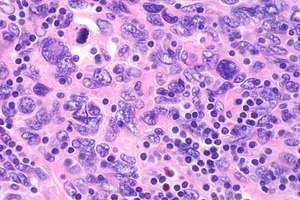
Primary effusion lymphoma (PEL) is classified as a diffuse large B cell lymphoma. It is a rare malignancy of plasmablastic cells that occurs in individuals that are infected with the Kaposi's sarcoma-associated herpesvirus. Plasmablasts are immature plasma cells, i.e. lymphocytes of the B-cell type that have differentiated into plasmablasts but because of their malignant nature do not differentiate into mature plasma cells but rather proliferate excessively and thereby cause life-threatening disease. In PEL, the proliferating plasmablastoid cells commonly accumulate within body cavities to produce effusions, primarily in the pleural, pericardial, or peritoneal cavities, without forming a contiguous tumor mass. In rare cases of these cavitary forms of PEL, the effusions develop in joints, the epidural space surrounding the brain and spinal cord, and underneath the capsule which forms around breast implants. Less frequently, individuals present with extracavitary primary effusion lymphomas, i.e., solid tumor masses not accompanied by effusions. The extracavitary tumors may develop in lymph nodes, bone, bone marrow, the gastrointestinal tract, skin, spleen, liver, lungs, central nervous system, testes, paranasal sinuses, muscle, and, rarely, inside the vasculature and sinuses of lymph nodes. As their disease progresses, however, individuals with the classical effusion-form of PEL may develop extracavitary tumors and individuals with extracavitary PEL may develop cavitary effusions.

<span class="mw-page-title-main">T-cell lymphoma</span> Medical condition

T-cell lymphoma is a rare form of cancerous lymphoma affecting T-cells. Lymphoma arises mainly from the uncontrolled proliferation of T-cells and can become cancerous.

<span class="mw-page-title-main">Diffuse large B-cell lymphoma</span> Type of blood cancer

Diffuse large B-cell lymphoma (DLBCL) is a cancer of B cells, a type of lymphocyte that is responsible for producing antibodies. It is the most common form of non-Hodgkin lymphoma among adults, with an annual incidence of 7–8 cases per 100,000 people per year in the US and UK. This cancer occurs primarily in older individuals, with a median age of diagnosis at ~70 years, although it can occur in young adults and, in rare cases, children. DLBCL can arise in virtually any part of the body and, depending on various factors, is often a very aggressive malignancy. The first sign of this illness is typically the observation of a rapidly growing mass or tissue infiltration that is sometimes associated with systemic B symptoms, e.g. fever, weight loss, and night sweats.

Angioimmunoblastic T-cell lymphoma is a mature T-cell lymphoma of blood or lymph vessel immunoblasts characterized by a polymorphous lymph node infiltrate showing a marked increase in follicular dendritic cells (FDCs) and high endothelial venules (HEVs) and systemic involvement.

<span class="mw-page-title-main">Breast cancer chemotherapy</span>

Breast cancer chemotherapy refers to the use of cytotoxic drugs (chemotherapy) in the treatment of breast cancer.

<span class="mw-page-title-main">Aggressive lymphoma</span> Medical condition

Aggressive lymphoma, also known as high-grade lymphoma, is a group of fast growing non-Hodgkin lymphoma.

<span class="mw-page-title-main">Langerhans cell sarcoma</span> Medical condition

Langerhans cell sarcoma (LCS) is a rare form of malignant histiocytosis. It should not be confused with Langerhans cell histiocytosis, which is cytologically benign. It can present most commonly in the skin and lymphatic tissue, but may also present in the lung, liver, and bone marrow. Treatment is most commonly with surgery or chemotherapy.

Extranodal NK/T-cell lymphoma, nasal type (ENKTCL-NT) is a rare type of lymphoma that commonly involves midline areas of the nasal cavity, oral cavity, and/or pharynx At these sites, the disease often takes the form of massive, necrotic, and extremely disfiguring lesions. However, ENKTCL-NT can also involve the eye, larynx, lung, gastrointestinal tract, skin, and various other tissues. ENKTCL-NT mainly affects adults; it is relatively common in Asia and to lesser extents Mexico, Central America, and South America but is rare in Europe and North America. In Korea, ENKTCL-NT often involves the skin and is reported to be the most common form of cutaneous lymphoma after mycosis fungoides.

<span class="mw-page-title-main">Plasmablastic lymphoma</span> Type of large B-cell lymphoma

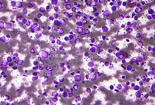
Plasmablastic lymphoma (PBL) is a type of large B-cell lymphoma recognized by the World Health Organization (WHO) in 2017 as belonging to a subgroup of lymphomas termed lymphoid neoplasms with plasmablastic differentiation. The other lymphoid neoplasms within this subgroup are: plasmablastic plasma cell lymphoma ; primary effusion lymphoma that is Kaposi's sarcoma-associated herpesvirus positive or Karposi's sarcoma-associated Herpesvirus negative; anaplastic lymphoma kinase-positive large B-cell lymphoma; and human herpesvirus 8-positive diffuse large B-cell lymphoma, not otherwise specified. All of these lymphomas are malignancies of plasmablasts, i.e. B-cells that have differentiated into plasmablasts but because of their malignant nature: fail to differentiate further into mature plasma cells; proliferate excessively; and accumulate in and injure various tissues and organs.

Epstein–Barr virus–associated lymphoproliferative diseases are a group of disorders in which one or more types of lymphoid cells, i.e. B cells, T cells, NK cells, and histiocytic-dendritic cells, are infected with the Epstein–Barr virus (EBV). This causes the infected cells to divide excessively, and is associated with the development of various non-cancerous, pre-cancerous, and cancerous lymphoproliferative disorders (LPDs). These LPDs include the well-known disorder occurring during the initial infection with the EBV, infectious mononucleosis, and the large number of subsequent disorders that may occur thereafter. The virus is usually involved in the development and/or progression of these LPDs although in some cases it may be an "innocent" bystander, i.e. present in, but not contributing to, the disease.

Primary testicular diffuse large B-cell lymphoma (PT-DLBCL), also termed testicular diffuse large B-cell lymphoma and diffuse large B-cell lymphoma of the testes, is a variant of the diffuse large B-cell lymphomas (DLBCL). DLBCL are a large and diverse group of B-cell malignancies with the great majority (-85%) being typed as diffuse large B-cell lymphoma, not otherwise specified. PT-DLBCL is a variant of DLBCL, NOS that involves one or, in uncommon cases, both testicles. Other variants and subtypes of DLBCL may involve the testes by spreading to them from their primary sites of origin in other tissues. PT-DLBCL differs from these other DLBCL in that it begins in the testes and then may spread to other sites.

Diffuse large B-cell lymphoma associated with chronic inflammation (DLBCL-CI) is a subtype of the Diffuse large B-cell lymphomas and a rare form of the Epstein–Barr virus-associated lymphoproliferative diseases, i.e. conditions in which lymphocytes infected with the Epstein-Barr virus (EBV) proliferate excessively in one or more tissues. EBV infects ~95% of the world's population to cause no symptoms, minor non-specific symptoms, or infectious mononucleosis. The virus then enters a latency phase in which the infected individual becomes a lifetime asymptomatic carrier of the virus. Some weeks, months, years, or decades thereafter, a very small fraction of these carriers, particularly those with an immunodeficiency, develop any one of various EBV-associated benign or malignant diseases.

Mature T-cell lymphoma, also called peripheral T-cell lymphoma, is a group of rare, aggressive lymphomas that develop from mature white blood cells and originate from lymphoid tissues outside of the bone marrow. Mature T-cell lymphoma is under the category of non-Hodgkin lymphoma. Mature T-cell lymphomas account for 10% to 15% of all lymphomas and is more common in Asia than in Europe and America. Its common subtypes include angioimmunoblastic T-cell lymphoma, anaplastic large cell lymphoma and peripheral T-cell lymphoma not otherwise specified. While different subtypes have variable symptoms, common symptoms include enlarged painless lymph nodes, fever, weight loss, rash and night sweats.

References

  1. Lennert, Karl (1978). Malignant lymphomas other than Hodgkin's disease, histology, cytology, ultrastructure, immunology. Berlin: Springer-Verlag. pp. 59–64. ISBN   978-0-387-08020-8.
  2. Monda, Lauren; Warnke, Roger; Rosai, Juan (1986). "A primary lymph node malignancy with features suggestive of dendritic reticulum cell differentiation. A report of 4 cases". The American Journal of Pathology. 122 (3): 562–72. PMC   1888214 . PMID   2420185.
  3. 1 2 Sharpe, Miriam; Easthope, Stephanie E.; Keating, Gillian M.; Lamb, Harriet M. (2002). "Polyethylene glycol-liposomal doxorubicin: a review of its use in the management of solid and haematological malignancies and AIDS-related Kaposi's sarcoma". Drugs. 62 (14): 2089–126. doi:10.2165/00003495-200262140-00012. PMID   12269857. S2CID   46974328.
  4. Fonseca, Rafael; Tefferi, Ayalew; Strickler, John G. (1997). "Follicular dendritic cell sarcoma mimicking diffuse large cell lymphoma: A case report". American Journal of Hematology. 55 (3): 148–55. doi: 10.1002/(SICI)1096-8652(199707)55:3<148::AID-AJH6>3.0.CO;2-S . PMID   9256295.
  5. Kosco, Marie H.; Gray, David (1992). "Signals Involved in Germinal Center Reactions". Immunological Reviews. 126: 63–76. doi:10.1111/j.1600-065X.1992.tb00631.x. PMID   1597321. S2CID   6172404.
  6. Tew, John G.; Kosco, Marie H.; Burton, Gregory F.; Szakal, Andras K. (1990). "Follicular Dendritic Cells as Accessory Cells". Immunological Reviews. 117: 185–211. doi:10.1111/j.1600-065X.1990.tb00573.x. PMID   2258191. S2CID   5556197.
  7. 1 2 3 4 5 6 Fonseca, R.; Yamakawa, M.; Nakamura, S.; Van Heerde, P.; Miettinen, M.; Shek, T.W. H.; Jensen, O. Myhre; Rousselet, M. C.; Tefferi, A. (1998). "Follicular dendritic cell sarcoma and interdigitating reticulum cell sarcoma: A review". American Journal of Hematology. 59 (2): 161–7. doi: 10.1002/(SICI)1096-8652(199810)59:2<161::AID-AJH10>3.0.CO;2-C . PMID   9766802.
  8. Biddle, David A.; Ro, Jae Y.; Yoon, Gil S.; Yong, Yap-Whang H.; Ayala, Alberto G.; Ordonez, Nelson G.; Ro, J (2002). "Extranodal Follicular Dendritic Cell Sarcoma of the Head and Neck Region: Three New Cases, with a Review of the Literature". Modern Pathology. 15 (1): 50–8. doi: 10.1038/modpathol.3880489 . PMID   11796841.
  9. Chen Tse-Ching; Kuo Tseng-tong; Ng Kwai-Fong (2001). "Follicular Dendritic Cell Tumor of the Liver: A Clinicopathologic and Epstein-Barr Virus Study of Two Cases". Modern Pathology. 14 (4): 354–360. doi: 10.1038/modpathol.3880315 . PMID   11301353.
  10. Horiguchi H.; Matsui-Horiguchi M.; Sakata H.; Ichinose M.; Yamamoto T.; Fujiwara M.; Ohse H. (February 2004). "Inflammatory pseudotumor-like follicular dendritic cell tumor of the spleen". Pathology International. 54 (2): 124–131. doi:10.1111/j.1440-1827.2004.01589.x. PMID   14720144. S2CID   8651824.
  11. 1 2 Chan, John K. C.; Fletcher, Christopher D. M.; Nayler, Simon J.; Cooper, Kum (1997). "Follicular dendritic cell sarcoma". Cancer. 79 (2): 294–313. doi: 10.1002/(SICI)1097-0142(19970115)79:2<294::AID-CNCR13>3.0.CO;2-W . PMID   9010103.
  12. Ono, Yuri; Terashima, Kazuo; Liu, Aimin; Yokoyama, Munehiro; Yokoshima, Kazuhiro; Mizukami, Miki; Watanabe, Ken; Mochimaru, Yoko; et al. (2009). "Follicular dendritic cell sarcoma with microtubuloreticular structure and virus-like particle productionin vitro". Pathology International. 59 (5): 332–44. doi:10.1111/j.1440-1827.2009.02375.x. PMID   19432677. S2CID   41080132.
  13. Grogg, Karen L; Macon, William R; Kurtin, Paul J; Nascimento, Antonio G (2004). "A survey of clusterin and fascin expression in sarcomas and spindle cell neoplasms: strong clusterin immunostaining is highly specific for follicular dendritic cell tumor". Modern Pathology. 18 (2): 260–6. doi: 10.1038/modpathol.3800294 . PMID   15467709.
  14. 1 2 3 Hanahan, Douglas; Weinberg, Robert A. (2011). "Hallmarks of Cancer: The Next Generation". Cell. 144 (5): 646–74. doi: 10.1016/j.cell.2011.02.013 . PMID   21376230.
  15. Nicolini, A; Mancini, P; Ferrari, P; Anselmi, L; Tartarelli, G; Bonazzi, V; Carpi, A; Giardino, R (2004). "Oral low-dose cyclophosphamide in metastatic hormone refractory prostate cancer (MHRPC)". Biomedicine & Pharmacotherapy. 58 (8): 447–50. doi:10.1016/j.biopha.2004.08.006. PMID   15464874.
  16. Nelius, Thomas; Klatte, Tobias; Riese, Werner; Haynes, Allan; Filleur, Stephanie (2009). "Clinical outcome of patients with docetaxel-resistant hormone-refractory prostate cancer treated with second-line cyclophosphamide-based metronomic chemotherapy". Medical Oncology. 27 (2): 363–7. doi:10.1007/s12032-009-9218-8. PMID   19365737. S2CID   27539332.
  17. Wendel, Hans-Guido; Stanchina, Elisa de; Fridman, Jordan S.; Malina, Abba; Ray, Sagarika; Kogan, Scott; Cordon-Cardo, Carlos; Pelletier, Jerry; Lowe, Scott W. (2004). "Survival signalling by Akt and eIF4E in oncogenesis and cancer therapy". Nature. 428 (6980): 332–7. Bibcode:2004Natur.428..332W. doi:10.1038/nature02369. PMID   15029198. S2CID   4426215.
  18. 1 2 Chen, Juhua; Somanath, Payaningal R; Razorenova, Olga; Chen, William S; Hay, Nissim; Bornstein, Paul; Byzova, Tatiana V (2005). "Akt1 regulates pathological angiogenesis, vascular maturation and permeability in vivo". Nature Medicine. 11 (11): 1188–96. doi:10.1038/nm1307. PMC   2277080 . PMID   16227992.
  19. 1 2 Depas, T; Spitaleri, G; Pruneri, G; Curigliano, G; Noberasco, C; Luini, A; Andreoni, B; Testori, A; Debraud, F (2008). "Dendritic cell sarcoma: An analytic overview of the literature and presentation of original five cases". Critical Reviews in Oncology/Hematology. 65 (1): 1–7. doi:10.1016/j.critrevonc.2007.06.003. PMID   17658269.
  20. Pathan, N; Aime-Sempe, C; Kitada, S; Basu, A; Haldar, S; Reed, JC (2001). "Microtubule-Targeting Drugs Induce Bcl-2 Phosphorylation and Association with Pin1". Neoplasia. 3 (6): 550–9. doi:10.1038/sj.neo.7900213. PMC   1506558 . PMID   11774038.
  21. Mini, E.; Nobili, S; Caciagli, B; Landini, I; Mazzei, T (2006). "Cellular pharmacology of gemcitabine". Annals of Oncology. 17: v7–12. doi: 10.1093/annonc/mdj941 . PMID   16807468.
  22. Leu, K. M.; Ostruszka, LJ; Shewach, D; Zalupski, M; Sondak, V; Biermann, JS; Lee, JS; Couwlier, C; et al. (2004). "Laboratory and Clinical Evidence of Synergistic Cytotoxicity of Sequential Treament With Gemcitabine Followed by Docetaxel in the Treatment of Sarcoma". Journal of Clinical Oncology. 22 (9): 1706–12. doi:10.1200/JCO.2004.08.043. PMID   15117993.
  23. Bay, Jacques-Olivier; Ray-Coquard, Isabelle; Fayette, Jérôme; Leyvraz, Serge; Cherix, Stephane; Piperno-Neumann, Sophie; Chevreau, Christine; Isambert, Nicolas; et al. (2006). "Docetaxel and gemcitabine combination in 133 advanced soft-tissue sarcomas: A retrospective analysis". International Journal of Cancer. 119 (3): 706–11. doi: 10.1002/ijc.21867 . PMID   16496406.