Protein kinase B

Last updated
AKT1
Crystal structure of Akt-1-inhibitor complexes.png
Ribbon Representation of crystal structure of Akt-1-inhibitor complexes. [1]
Identifiers
Symbol AKT1
NCBI gene 207
HGNC 391
OMIM 164730
RefSeq NM_005163
UniProt P31749
Other data
Locus Chr. 14 q32.32-32.33
Search for
Structures Swiss-model
Domains InterPro
AKT2
3D0E Ribbon.png
Crystal structure of Akt-2-inhibitor complexes. [2]
Identifiers
Symbol AKT2
NCBI gene 208
HGNC 392
OMIM 164731
RefSeq NM_001626
UniProt P31751
Other data
Locus Chr. 19 q13.1-13.2
Search for
Structures Swiss-model
Domains InterPro
AKT3
Identifiers
Symbol AKT3
NCBI gene 10000
HGNC 393
OMIM 611223
RefSeq NM_181690
UniProt Q9Y243
Other data
Locus Chr. 1 q43-44
Search for
Structures Swiss-model
Domains InterPro

Protein kinase B (PKB), also known as Akt, is the collective name of a set of three serine/threonine-specific protein kinases that play key roles in multiple cellular processes such as glucose metabolism, apoptosis, cell proliferation, transcription, and cell migration.

Contents

Family members - Isoforms

There are three different genes that encode isoforms of protein kinase B. These three genes are referred to as AKT1 , AKT2 , and AKT3 and encode the RAC alpha, beta, and gamma serine/threonine protein kinases respectively. The terms PKB and Akt may refer to the products of all three genes collectively, but sometimes are used to refer to PKB alpha and Akt1 alone.[ citation needed ]

Akt1 is involved in cellular survival pathways, by inhibiting apoptotic processes. Akt1 is also able to induce protein synthesis pathways, and is therefore a key signaling protein in the cellular pathways that lead to skeletal muscle hypertrophy and general tissue growth. A mouse model with complete deletion of the Akt1 gene manifests growth retardation and increased spontaneous apoptosis in tissues such as testes and thymus. [3] Since it can block apoptosis and thereby promote cell survival, Akt1 has been implicated as a major factor in many types of cancer. Akt1 was originally identified as the oncogene in the transforming retrovirus, AKT8. [4]

Akt2 is an important signaling molecule in the insulin signaling pathway. It is required to induce glucose transport. In a mouse which is null for Akt1 but normal for Akt2, glucose homeostasis is unperturbed, but the animals are smaller, consistent with a role for Akt1 in growth. In contrast, mice which do not have Akt2, but have normal Akt1, have mild growth deficiency and display a diabetic phenotype (insulin resistance), again consistent with the idea that Akt2 is more specific for the insulin receptor signaling pathway. [5]

The role of Akt3 is less clear, though it appears to be predominantly expressed in the brain. It has been reported that mice lacking Akt3 have small brains. [6]

Akt isoforms are overexpressed in a variety of human tumors, and, at the genomic level, are amplified in gastric adenocarcinomas (Akt1), ovarian (Akt2), pancreatic (Akt2) and breast (Akt2) cancers. [7] [8]

Name

The name Akt does not refer to its function. The "Ak" in Akt refers to the AKR mouse strain that develops spontaneous thymic lymphomas. The "t" stands for 'thymoma'; the letter was added when a transforming retrovirus was isolated from the Ak mouse strain, which was termed "Akt-8". The authors state, "Stock A Strain k AKR mouse originally inbred in the laboratory of Dr. C. P. Rhoads by K. B. Rhoads at the Rockefeller Institute." When the oncogene encoded in this virus was discovered, it was termed v-Akt. Thus, the more recently identified human analogs were named accordingly. [9]

Regulation

Akt1 is involved in the PI3K/AKT/mTOR pathway and other signaling pathways. [10] [ citation needed ]

Binding phospholipids

The Akt proteins possess a protein domain known as a PH domain, or pleckstrin homology domain, named after pleckstrin, the protein in which it was first discovered. This domain binds to phosphoinositides with high affinity. In the case of the PH domain of the Akt proteins, it binds either PIP3 (phosphatidylinositol (3,4,5)-trisphosphate, PtdIns(3,4,5)P3) or PIP2 (phosphatidylinositol (3,4)-bisphosphate, PtdIns(3,4)P2). [11] This is useful for control of cellular signaling because the di-phosphorylated phosphoinositide PIP2 is only phosphorylated by the family of enzymes, PI 3-kinases (phosphoinositide 3-kinase or PI3-K), and only upon receipt of chemical messengers which tell the cell to begin the growth process. For example, PI 3-kinases may be activated by a G protein coupled receptor or receptor tyrosine kinase such as the insulin receptor. Once activated, PI 3-kinase phosphorylates PIP2 to form PIP3.

Phosphorylation

Once correctly positioned at the membrane via binding of PIP3, Akt can then be phosphorylated by its activating kinases, phosphoinositide-dependent kinase-1 (PDPK1 at threonine 308 in Akt1 and threonine 309 in Akt2) and the mammalian target of rapamycin complex 2 (mTORC2 at serine 473 (Akt1) and 474 (Akt2)) which is found at high levels in the fed state, [12] [13] first by mTORC2. mTORC2 therefore functionally acts as the long-sought PDK2 molecule, although other molecules, including integrin-linked kinase (ILK) and mitogen-activated protein kinase-activated protein kinase-2 (MAPKAPK2) can also serve as PDK2. Phosphorylation by mTORC2 stimulates the subsequent phosphorylation of Akt isoforms by PDPK1.

Activated Akt isoforms can then go on to activate or deactivate their myriad substrates (e.g. mTOR) via their kinase activity.

Besides being a downstream effector of PI 3-kinases, Akt isoforms can also be activated in a PI 3-kinase-independent manner. [14] ACK1 or TNK2, a non-receptor tyrosine kinase, phosphorylates Akt at its tyrosine 176 residue, leading to its activation in PI 3-kinase-independent manner. [14] Studies have suggested that cAMP-elevating agents could also activate Akt through protein kinase A (PKA) in the presence of insulin. [15]

O-GlcNAcylation

Akt can be O-GlcNAcylated by OGT. O-GlcNAcylation of Akt is associated with a decrease in T308 phosphorylation. [16]

Ubiquitination

Akt1 is normally phosphorylated at position T450 in the turn motif when Akt1 is translated. If Akt1 is not phosphorylated at this position, Akt1 does not fold in the right way. The T450-non-phosphorylated misfolded Akt1 is ubiquitinated and degraded by the proteasome. Akt1 is also phosphorylated at T308 and S473 during IGF-1 response, and the resulting polyphosphorylated Akt is ubiquitinated partly by E3 ligase NEDD4. Most of the ubiquitinated-phosphorylated-Akt1 is degraded by the proteasome, while a small amount of phosphorylated-Akt1 translocates to the nucleus in a ubiquitination-dependent way to phosphorylate its substrate. A cancer-derived mutant Akt1 (E17K) is more readily ubiquitinated and phosphorylated than the wild type Akt1. The ubiquitinated-phosphorylated-Akt1 (E17K) translocates more efficiently to the nucleus than the wild type Akt1. This mechanism may contribute to E17K-Akt1-induced cancer in humans. [17]

Lipid phosphatases and PIP3

PI3K-dependent Akt1 activation can be regulated through the tumor suppressor PTEN, which works essentially as the opposite of PI3K mentioned above. [18] PTEN acts as a phosphatase to dephosphorylate PIP3 back to PIP2. This removes the membrane-localization factor from the Akt signaling pathway. Without this localization, the rate of Akt1 activation decreases significantly, as do all of the downstream pathways that depend on Akt1 for activation.

PIP3 can also be de-phosphorylated at the "5" position by the SHIP family of inositol phosphatases, SHIP1 and SHIP2. These poly-phosphate inositol phosphatases dephosphorylate PIP3 to form PIP2.

Protein phosphatases

The phosphatases in the PHLPP family, PHLPP1 and PHLPP2 have been shown to directly de-phosphorylate, and therefore inactivate, distinct Akt isoforms. PHLPP2 dephosphorylates Akt1 and Akt3, whereas PHLPP1 is specific for Akt2 and Akt3.[ citation needed ]

Function

The Akt kinases regulate cellular survival [19] and metabolism by binding and regulating many downstream effectors, e.g. Nuclear Factor-κB, Bcl-2 family proteins, master lysosomal regulator TFEB and murine double minute 2 (MDM2).

Cell survival

Overview of signal transduction pathways involved in apoptosis. Signal transduction pathways.svg
Overview of signal transduction pathways involved in apoptosis.

Akt kinases can promote growth factor-mediated cell survival both directly and indirectly. BAD is a pro-apoptotic protein of the Bcl-2 family. Akt1 can phosphorylate BAD on Ser136, [20] which makes BAD dissociate from the Bcl-2/Bcl-X complex and lose the pro-apoptotic function. [21] Akt1 can also activate NF-κB via regulating IκB kinase (IKK), thus result in transcription of pro-survival genes. [22]

Cell cycle

The Akt isoforms are known to play a role in the cell cycle. Under various circumstances, activation of Akt1 was shown to overcome cell cycle arrest in G1 [23] and G2 [24] phases. Moreover, activated Akt1 may enable proliferation and survival of cells that have sustained a potentially mutagenic impact and, therefore, may contribute to acquisition of mutations in other genes.

Metabolism

Akt2 is required for the insulin-induced translocation of glucose transporter 4 (GLUT4) to the plasma membrane. Glycogen synthase kinase 3 (GSK-3) could be inhibited upon phosphorylation by Akt, which results in increase of glycogen synthesis. GSK3 is also involved in Wnt signaling cascade, so Akt might be also implicated in the Wnt pathway. Its role in HCV induced steatosis is unknown.[ citation needed ]

Lysosomal biogenesis and autophagy

Akt1 regulates TFEB, a master controller of lysosomal biogenesis, [25] by direct phosphorylation at serine 467. [26] Phosphorylated TFEB is excluded from the nucleus and less active. [26] Pharmacological inhibition of Akt promotes nuclear translocation of TFEB, lysosomal biogenesis and autophagy. [26]

Angiogenesis

Akt1 has also been implicated in angiogenesis and tumor development. Although deficiency of Akt1 in mice inhibited physiological angiogenesis, it enhanced pathological angiogenesis and tumor growth associated with matrix abnormalities in skin and blood vessels. [27] [28]

Clinical relevance

Akt proteins are associated with tumor cell survival, proliferation, and invasiveness. The activation of Akt is also one of the most frequent alterations observed in human cancer and tumor cells. Tumor cells that have constantly active Akt may depend on Akt for survival. [29] Therefore, understanding the Akt proteins and their pathways is important for the creation of better therapies to treat cancer and tumor cells. A mosaic-activating mutation (c. 49G→A, p.Glu17Lys) in Akt1 is associated with the Proteus Syndrome, which causes overgrowth of skin, connective tissue, brain and other tissues. [30]

Akt inhibitors

Akt inhibitors may treat cancers such as neuroblastoma. Some Akt inhibitors have undergone clinical trials. In 2007 VQD-002 had a phase I trial. [31] In 2010 Perifosine reached phase II. [32] but it failed phase III in 2012.

Miltefosine is approved for leishmaniasis and under investigation for other indications including HIV.

Akt1 is now thought to be the "key" for cell entry by HSV-1 and HSV-2 (herpes virus: oral and genital, respectively). Intracellular calcium release by the cell allows for entry by the herpes virus; the virus activates Akt1, which in turn causes the release of calcium. Treating the cells with Akt inhibitors before virus exposure leads to a significantly lower rate of infection. [33]

MK-2206 reported phase 1 results for advanced solid tumors in 2011, [34] and subsequently has undergone numerous phase II studies for a wide variety of cancer types. [35]

In 2013 AZD5363 reported phase I results regarding solid tumors. [36] with a study of AZD5363 with olaparib reporting in 2016. [37]

Ipatasertib is in phase II trials for breast cancer. [38]

Decreased Akt isoforms can cause deleterious effects

Akt isoform activation is associated with many malignancies; however, a research group from Massachusetts General Hospital and Harvard University unexpectedly observed a converse role for Akt and one of its downstream effector FOXOs in acute myeloid leukemia (AML). They claimed that low levels of Akt activity associated with elevated levels of FOXOs are required to maintain the function and immature state of leukemia-initiating cells (LICs). FOXOs are active, implying reduced Akt activity, in ~40% of AML patient samples regardless of genetic subtype; and either activation of Akt or compound deletion of FoxO1/3/4 reduced leukemic cell growth in a mouse model. [39]

Hyperactivation of Akt1 can cause deleterious effects

Two studies show that Akt1 is involved in Juvenile Granulosa Cell tumors (JGCT). In-frame duplications in the pleckstrin-homology domain (PHD) of the protein were found in more than 60% of JGCTs occurring in girls under 15 years of age. The JGCTs without duplications carried point mutations affecting highly conserved residues. The mutated proteins carrying the duplications displayed a non-wild-type subcellular distribution, with a marked enrichment at the plasma membrane. This led to a striking degree of Akt1 activation demonstrated by a strong phosphorylation level and corroborated by reporter assays. [40]

Analysis by RNA-Seq pinpointed a series of differentially expressed genes, involved in cytokine and hormone signaling and cell division-related processes. Further analyses pointed to a possible dedifferentiation process and suggested that most of the transcriptomic dysregulations might be mediated by a limited set of transcription factors perturbed by Akt1 activation. These results incriminate somatic mutations of Akt1 as major probably driver events in the pathogenesis of JGCTs. [41]

See also

Related Research Articles

<span class="mw-page-title-main">Insulin receptor</span> Mammalian protein found in Homo sapiens

The insulin receptor (IR) is a transmembrane receptor that is activated by insulin, IGF-I, IGF-II and belongs to the large class of receptor tyrosine kinase. Metabolically, the insulin receptor plays a key role in the regulation of glucose homeostasis; a functional process that under degenerate conditions may result in a range of clinical manifestations including diabetes and cancer. Insulin signalling controls access to blood glucose in body cells. When insulin falls, especially in those with high insulin sensitivity, body cells begin only to have access to lipids that do not require transport across the membrane. So, in this way, insulin is the key regulator of fat metabolism as well. Biochemically, the insulin receptor is encoded by a single gene INSR, from which alternate splicing during transcription results in either IR-A or IR-B isoforms. Downstream post-translational events of either isoform result in the formation of a proteolytically cleaved α and β subunit, which upon combination are ultimately capable of homo or hetero-dimerisation to produce the ≈320 kDa disulfide-linked transmembrane insulin receptor.

<span class="mw-page-title-main">GSK-3</span> Class of enzymes

Glycogen synthase kinase 3 (GSK-3) is a serine/threonine protein kinase that mediates the addition of phosphate molecules onto serine and threonine amino acid residues. First discovered in 1980 as a regulatory kinase for its namesake, glycogen synthase (GS), GSK-3 has since been identified as a protein kinase for over 100 different proteins in a variety of different pathways. In mammals, including humans, GSK-3 exists in two isozymes encoded by two homologous genes GSK-3α (GSK3A) and GSK-3β (GSK3B). GSK-3 has been the subject of much research since it has been implicated in a number of diseases, including type 2 diabetes, Alzheimer's disease, inflammation, cancer, addiction and bipolar disorder.

mTOR Mammalian protein found in humans

The mammalian target of rapamycin (mTOR), also referred to as the mechanistic target of rapamycin, and sometimes called FK506-binding protein 12-rapamycin-associated protein 1 (FRAP1), is a kinase that in humans is encoded by the MTOR gene. mTOR is a member of the phosphatidylinositol 3-kinase-related kinase family of protein kinases.

<span class="mw-page-title-main">Phosphatidylinositol (3,4,5)-trisphosphate</span> Chemical compound

Phosphatidylinositol (3,4,5)-trisphosphate (PtdIns(3,4,5)P3), abbreviated PIP3, is the product of the class I phosphoinositide 3-kinases' (PI 3-kinases) phosphorylation of phosphatidylinositol (4,5)-bisphosphate (PIP2). It is a phospholipid that resides on the plasma membrane.

<span class="mw-page-title-main">Phosphoinositide 3-kinase</span> Class of enzymes

Phosphoinositide 3-kinases (PI3Ks), also called phosphatidylinositol 3-kinases, are a family of enzymes involved in cellular functions such as cell growth, proliferation, differentiation, motility, survival and intracellular trafficking, which in turn are involved in cancer.

<span class="mw-page-title-main">Platelet-derived growth factor receptor</span> Cell surface receptors

Platelet-derived growth factor receptors (PDGF-R) are cell surface tyrosine kinase receptors for members of the platelet-derived growth factor (PDGF) family. PDGF subunits -A and -B are important factors regulating cell proliferation, cellular differentiation, cell growth, development and many diseases including cancer. There are two forms of the PDGF-R, alpha and beta each encoded by a different gene. Depending on which growth factor is bound, PDGF-R homo- or heterodimerizes.

The PHLPP isoforms are a pair of protein phosphatases, PHLPP1 and PHLPP2, that are important regulators of Akt serine-threonine kinases and conventional/novel protein kinase C (PKC) isoforms. PHLPP may act as a tumor suppressor in several types of cancer due to its ability to block growth factor-induced signaling in cancer cells.

<span class="mw-page-title-main">AKT1</span> Protein-coding gene in the species Homo sapiens

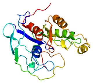
RAC(Rho family)-alpha serine/threonine-protein kinase is an enzyme that in humans is encoded by the AKT1 gene. This enzyme belongs to the AKT subfamily of serine/threonine kinases that contain SH2 protein domains. It is commonly referred to as PKB, or by both names as "Akt/PKB".

<span class="mw-page-title-main">Insulin receptor substrate 1</span> Protein-coding gene in the species Homo sapiens

Insulin receptor substrate 1(IRS-1) is a signaling adapter protein that in humans is encoded by the IRS1 gene. It is a 131 kDa protein with amino acid sequence of 1242 residues. It contains a single pleckstrin homology (PH) domain at the N-terminus and a PTB domain ca. 40 residues downstream of this, followed by a poorly conserved C-terminus tail. Together with IRS2, IRS3 (pseudogene) and IRS4, it is homologous to the Drosophila protein chico, whose disruption extends the median lifespan of flies up to 48%. Similarly, Irs1 mutant mice experience moderate life extension and delayed age-related pathologies.

<span class="mw-page-title-main">AKT2</span> Protein-coding gene in the species Homo sapiens

AKT2, also known as RAC-beta serine/threonine-protein kinase, is an enzyme that in humans is encoded by the AKT2 gene. It influences metabolite storage as part of the insulin signal transduction pathway.

<span class="mw-page-title-main">AKT3</span> Protein-coding gene in the species Homo sapiens

RAC-gamma serine/threonine-protein kinase is an enzyme that in humans is encoded by the AKT3 gene.

<span class="mw-page-title-main">TNK2</span> Protein-coding gene in the species Homo sapiens

Activated CDC42 kinase 1, also known as ACK1, is an enzyme that in humans is encoded by the TNK2 gene. TNK2 gene encodes a non-receptor tyrosine kinase, ACK1, that binds to multiple receptor tyrosine kinases e.g. EGFR, MERTK, AXL, HER2 and insulin receptor (IR). ACK1 also interacts with Cdc42Hs in its GTP-bound form and inhibits both the intrinsic and GTPase-activating protein (GAP)-stimulated GTPase activity of Cdc42Hs. This binding is mediated by a unique sequence of 47 amino acids C-terminal to an SH3 domain. The protein may be involved in a regulatory mechanism that sustains the GTP-bound active form of Cdc42Hs and which is directly linked to a tyrosine phosphorylation signal transduction pathway. Several alternatively spliced transcript variants have been identified from this gene, but the full-length nature of only two transcript variants has been determined.

<span class="mw-page-title-main">RPTOR</span> Protein-coding gene in humans

Regulatory-associated protein of mTOR also known as raptor or KIAA1303 is an adapter protein that is encoded in humans by the RPTOR gene. Two mRNAs from the gene have been identified that encode proteins of 1335 and 1177 amino acids long.

<span class="mw-page-title-main">RICTOR</span> Protein-coding gene in the species Homo sapiens

Rapamycin-insensitive companion of mammalian target of rapamycin (RICTOR) is a protein that in humans is encoded by the RICTOR gene.

The Akt signaling pathway or PI3K-Akt signaling pathway is a signal transduction pathway that promotes survival and growth in response to extracellular signals. Key proteins involved are PI3K and Akt.

<span class="mw-page-title-main">Forkhead box protein O1</span> Protein

Forkhead box protein O1 (FOXO1), also known as forkhead in rhabdomyosarcoma (FKHR), is a protein that in humans is encoded by the FOXO1 gene. FOXO1 is a transcription factor that plays important roles in regulation of gluconeogenesis and glycogenolysis by insulin signaling, and is also central to the decision for a preadipocyte to commit to adipogenesis. It is primarily regulated through phosphorylation on multiple residues; its transcriptional activity is dependent on its phosphorylation state.

<span class="mw-page-title-main">PI3K/AKT/mTOR pathway</span> Cell cycle regulation pathway

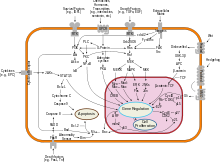
The PI3K/AKT/mTOR pathway is an intracellular signaling pathway important in regulating the cell cycle. Therefore, it is directly related to cellular quiescence, proliferation, cancer, and longevity. PI3K activation phosphorylates and activates AKT, localizing it in the plasma membrane. AKT can have a number of downstream effects such as activating CREB, inhibiting p27, localizing FOXO in the cytoplasm, activating PtdIns-3ps, and activating mTOR which can affect transcription of p70 or 4EBP1. There are many known factors that enhance the PI3K/AKT pathway including EGF, shh, IGF-1, insulin, and CaM. Both leptin and insulin recruit PI3K signalling for metabolic regulation. The pathway is antagonized by various factors including PTEN, GSK3B, and HB9.

mTOR inhibitors Class of pharmaceutical drugs

mTOR inhibitors are a class of drugs used to treat several human diseases, including cancer, autoimmune diseases, and neurodegeneration. They function by inhibiting the mammalian target of rapamycin (mTOR), which is a serine/threonine-specific protein kinase that belongs to the family of phosphatidylinositol-3 kinase (PI3K) related kinases (PIKKs). mTOR regulates cellular metabolism, growth, and proliferation by forming and signaling through two protein complexes, mTORC1 and mTORC2. The most established mTOR inhibitors are so-called rapalogs, which have shown tumor responses in clinical trials against various tumor types.

mTORC1 Protein complex

mTORC1, also known as mammalian target of rapamycin complex 1 or mechanistic target of rapamycin complex 1, is a protein complex that functions as a nutrient/energy/redox sensor and controls protein synthesis.

mTOR Complex 2 (mTORC2) is an acutely rapamycin-insensitive protein complex formed by serine/threonine kinase mTOR that regulates cell proliferation and survival, cell migration and cytoskeletal remodeling. The complex itself is rather large, consisting of seven protein subunits. The catalytic mTOR subunit, DEP domain containing mTOR-interacting protein (DEPTOR), mammalian lethal with sec-13 protein 8, and TTI1/TEL2 complex are shared by both mTORC2 and mTORC1. Rapamycin-insensitive companion of mTOR (RICTOR), mammalian stress-activated protein kinase interacting protein 1 (mSIN1), and protein observed with rictor 1 and 2 (Protor1/2) can only be found in mTORC2. Rictor has been shown to be the scaffold protein for substrate binding to mTORC2.

References

  1. PDB: 3MV5 ; Freeman-Cook KD, Autry C, Borzillo G, Gordon D, Barbacci-Tobin E, Bernardo V, et al. (June 2010). "Design of selective, ATP-competitive inhibitors of Akt". Journal of Medicinal Chemistry. 53 (12): 4615–22. doi:10.1021/jm1003842. PMID   20481595.
  2. PDB: 3D0E ; Heerding DA, Rhodes N, Leber JD, Clark TJ, Keenan RM, Lafrance LV, et al. (September 2008). "Identification of 4-(2-(4-amino-1,2,5-oxadiazol-3-yl)-1-ethyl-7-{[(3S)-3-piperidinylmethyl]oxy}-1H-imidazo[4,5-c]pyridin-4-yl)-2-methyl-3-butyn-2-ol (GSK690693), a novel inhibitor of AKT kinase". Journal of Medicinal Chemistry. 51 (18): 5663–79. doi:10.1021/jm8004527. PMID   18800763.
  3. Chen WS, Xu PZ, Gottlob K, Chen ML, Sokol K, Shiyanova T, et al. (September 2001). "Growth retardation and increased apoptosis in mice with homozygous disruption of the Akt1 gene". Genes & Development. 15 (17): 2203–8. doi:10.1101/gad.913901. PMC   312770 . PMID   11544177.
  4. Staal SP, Hartley JW, Rowe WP (July 1977). "Isolation of transforming murine leukemia viruses from mice with a high incidence of spontaneous lymphoma". Proceedings of the National Academy of Sciences of the United States of America. 74 (7): 3065–7. Bibcode:1977PNAS...74.3065S. doi: 10.1073/pnas.74.7.3065 . PMC   431413 . PMID   197531.
  5. Garofalo RS, Orena SJ, Rafidi K, Torchia AJ, Stock JL, Hildebrandt AL, et al. (July 2003). "Severe diabetes, age-dependent loss of adipose tissue, and mild growth deficiency in mice lacking Akt2/PKB beta". The Journal of Clinical Investigation. 112 (2): 197–208. doi:10.1172/JCI16885. PMC   164287 . PMID   12843127.
  6. Yang ZZ, Tschopp O, Baudry A, Dümmler B, Hynx D, Hemmings BA (April 2004). "Physiological functions of protein kinase B/Akt". Biochemical Society Transactions. 32 (Pt 2): 350–4. doi:10.1042/BST0320350. PMID   15046607.
  7. Hill MM, Hemmings BA (2002). "Inhibition of protein kinase B/Akt. implications for cancer therapy". Pharmacology & Therapeutics. 93 (2–3): 243–51. doi:10.1016/S0163-7258(02)00193-6. PMID   12191616.
  8. Mitsiades CS, Mitsiades N, Koutsilieris M (May 2004). "The Akt pathway: molecular targets for anti-cancer drug development". Current Cancer Drug Targets. 4 (3): 235–56. doi:10.2174/1568009043333032. PMID   15134532.
  9. Xie, J; Weiskirchen, R (2020). "What Does the "AKT" Stand for in the Name "AKT Kinase"? Some Historical Comments". Frontiers in Oncology. 10: 1329. doi: 10.3389/fonc.2020.01329 . PMC   7431881 . PMID   32850422.
  10. Hsu, P.P. et al. The mTOR-regulated phosphoproteome reveals a mechanism of mTORC1-mediated inhibition of growth factor signaling. Science 332, 1317–1322 (2011). DOI: 10.1126/science.1199498
  11. Franke TF, Kaplan DR, Cantley LC, Toker A (January 1997). "Direct regulation of the Akt proto-oncogene product by phosphatidylinositol-3,4-bisphosphate". Science. 275 (5300): 665–8. doi:10.1126/science.275.5300.665. PMID   9005852. S2CID   31186873.
  12. Sarbassov DD, Guertin DA, Ali SM, Sabatini DM (February 2005). "Phosphorylation and regulation of Akt/PKB by the rictor-mTOR complex". Science. 307 (5712): 1098–101. Bibcode:2005Sci...307.1098S. doi:10.1126/science.1106148. PMID   15718470. S2CID   45837814.
  13. Jacinto E, Facchinetti V, Liu D, Soto N, Wei S, Jung SY, et al. (October 2006). "SIN1/MIP1 maintains rictor-mTOR complex integrity and regulates Akt phosphorylation and substrate specificity". Cell. 127 (1): 125–37. doi: 10.1016/j.cell.2006.08.033 . PMID   16962653. S2CID   230319.
  14. 1 2 Mahajan K, Coppola D, Challa S, Fang B, Chen YA, Zhu W, et al. (March 2010). "Ack1 mediated AKT/PKB tyrosine 176 phosphorylation regulates its activation". PLOS ONE. 5 (3): e9646. Bibcode:2010PLoSO...5.9646M. doi: 10.1371/journal.pone.0009646 . PMC   2841635 . PMID   20333297.
  15. Stuenaes JT, Bolling A, Ingvaldsen A, Rommundstad C, Sudar E, Lin FC, et al. (May 2010). "Beta-adrenoceptor stimulation potentiates insulin-stimulated PKB phosphorylation in rat cardiomyocytes via cAMP and PKA". British Journal of Pharmacology. 160 (1): 116–29. doi:10.1111/j.1476-5381.2010.00677.x. PMC   2860212 . PMID   20412069.
  16. Yang X, Ongusaha PP, Miles PD, Havstad JC, Zhang F, So WV, et al. (February 2008). "Phosphoinositide signalling links O-GlcNAc transferase to insulin resistance". Nature. 451 (7181): 964–9. Bibcode:2008Natur.451..964Y. doi:10.1038/nature06668. PMID   18288188. S2CID   18459576.
  17. Fan CD, Lum MA, Xu C, Black JD, Wang X (January 2013). "Ubiquitin-dependent regulation of phospho-AKT dynamics by the ubiquitin E3 ligase, NEDD4-1, in the insulin-like growth factor-1 response". The Journal of Biological Chemistry. 288 (3): 1674–84. doi: 10.1074/jbc.M112.416339 . PMC   3548477 . PMID   23195959.
  18. Cooper GM (2000). "Figure 15.37: PTEN and PI3K". The cell: a molecular approach. Washington, D.C: ASM Press. ISBN   978-0-87893-106-4.
  19. Song G, Ouyang G, Bao S (2005). "The activation of Akt/PKB signaling pathway and cell survival". Journal of Cellular and Molecular Medicine. 9 (1): 59–71. doi:10.1111/j.1582-4934.2005.tb00337.x. PMC   6741304 . PMID   15784165.
  20. Alberts B, Johnson A, Lewis J, Raff M, Roberts K, Walter P (2002). "Figure 15-60: BAD phosphorylation by Akt". Molecular biology of the cell. New York: Garland Science. ISBN   978-0-8153-3218-3.
  21. Lodish H, Berk A, Zipursky LS, Matsudaira P, Baltimore D, Darnell J (1999). "Figure 23-50: BAD interaction with Bcl-2". Molecular cell biology. New York: Scientific American Books. ISBN   978-0-7167-3136-8.
  22. Faissner A, Heck N, Dobbertin A, Garwood J (2006). "DSD-1-Proteoglycan/Phosphacan and Receptor Protein Tyrosine Phosphatase-Beta Isoforms during Development and Regeneration of Neural Tissues". Brain Repair. Advances in Experimental Medicine and Biology. Vol. 557. pp. 25–53, Figure 2: regulation of NF–κB. doi:10.1007/0-387-30128-3_3. ISBN   978-0-306-47859-8. PMID   16955703.
  23. Ramaswamy S, Nakamura N, Vazquez F, Batt DB, Perera S, Roberts TM, Sellers WR (March 1999). "Regulation of G1 progression by the PTEN tumor suppressor protein is linked to inhibition of the phosphatidylinositol 3-kinase/Akt pathway". Proceedings of the National Academy of Sciences of the United States of America. 96 (5): 2110–5. Bibcode:1999PNAS...96.2110R. doi: 10.1073/pnas.96.5.2110 . PMC   26745 . PMID   10051603.
  24. Kandel ES, Skeen J, Majewski N, Di Cristofano A, Pandolfi PP, Feliciano CS, et al. (November 2002). "Activation of Akt/protein kinase B overcomes a G(2)/m cell cycle checkpoint induced by DNA damage". Molecular and Cellular Biology. 22 (22): 7831–41. doi:10.1128/MCB.22.22.7831-7841.2002. PMC   134727 . PMID   12391152.
  25. Sardiello M, Palmieri M, di Ronza A, Medina DL, Valenza M, Gennarino VA, et al. (July 2009). "A gene network regulating lysosomal biogenesis and function". Science. 325 (5939): 473–7. Bibcode:2009Sci...325..473S. doi: 10.1126/science.1174447 . PMID   19556463. S2CID   20353685.
  26. 1 2 3 Palmieri M, Pal R, Nelvagal HR, Lotfi P, Stinnett GR, Seymour ML, et al. (February 2017). "mTORC1-independent TFEB activation via Akt inhibition promotes cellular clearance in neurodegenerative storage diseases". Nature Communications. 8: 14338. Bibcode:2017NatCo...814338P. doi:10.1038/ncomms14338. PMC   5303831 . PMID   28165011.
  27. Chen J, Somanath PR, Razorenova O, Chen WS, Hay N, Bornstein P, Byzova TV (November 2005). "Akt1 regulates pathological angiogenesis, vascular maturation and permeability in vivo". Nature Medicine. 11 (11): 1188–96. doi:10.1038/nm1307. PMC   2277080 . PMID   16227992.
  28. Somanath PR, Razorenova OV, Chen J, Byzova TV (March 2006). "Akt1 in endothelial cell and angiogenesis". Cell Cycle. 5 (5): 512–8. doi:10.4161/cc.5.5.2538. PMC   1569947 . PMID   16552185.
  29. "Tumor Genetics; AKT Function and Oncogenic Activity" (PDF). Scientific Report. Fox Chase Cancer Center. 2005. Archived from the original (PDF) on 2010-06-04. Retrieved 2013-01-23.
  30. Lindhurst MJ, Sapp JC, Teer JK, Johnston JJ, Finn EM, Peters K, et al. (August 2011). "A mosaic activating mutation in AKT1 associated with the Proteus syndrome". The New England Journal of Medicine. 365 (7): 611–9. doi:10.1056/NEJMoa1104017. PMC   3170413 . PMID   21793738.
  31. "VioQuest Pharmaceuticals Announces Phase I/IIa Trial For Akt Inhibitor VQD-002". Apr 2007.
  32. Ghobrial IM, Roccaro A, Hong F, Weller E, Rubin N, Leduc R, et al. (February 2010). "Clinical and translational studies of a phase II trial of the novel oral Akt inhibitor perifosine in relapsed or relapsed/refractory Waldenstrom's macroglobulinemia". Clinical Cancer Research. 16 (3): 1033–41. doi:10.1158/1078-0432.CCR-09-1837. PMC   2885252 . PMID   20103671.
  33. Cheshenko N, Trepanier JB, Stefanidou M, Buckley N, Gonzalez P, Jacobs W, Herold BC (July 2013). "HSV activates Akt to trigger calcium release and promote viral entry: novel candidate target for treatment and suppression". FASEB Journal. 27 (7): 2584–99. doi: 10.1096/fj.12-220285 . PMC   3688744 . PMID   23507869.
  34. Yap TA, Yan L, Patnaik A, Fearen I, Olmos D, Papadopoulos K, et al. (December 2011). "First-in-man clinical trial of the oral pan-AKT inhibitor MK-2206 in patients with advanced solid tumors". Journal of Clinical Oncology. 29 (35): 4688–95. doi:10.1200/JCO.2011.35.5263. PMID   22025163.
  35. MK-2206 phase-2 trials
  36. AKT inhibitor AZD5363 well tolerated, yielded partial response in patients with advanced solid tumors
  37. "PARP/AKT Inhibitor Combination Active in Multiple Tumor Types. April 2016". Archived from the original on 2016-05-07. Retrieved 2016-04-20.
  38. Jabbarzadeh Kaboli P, Salimian F, Aghapour S, Xiang S, Zhao Q, Li M, et al. (June 2020). "Akt-targeted therapy as a promising strategy to overcome drug resistance in breast cancer - A comprehensive review from chemotherapy to immunotherapy". Pharmacological Research. 156: 104806. doi:10.1016/j.phrs.2020.104806. PMID   32294525. S2CID   215793444.
  39. Sykes SM, Lane SW, Bullinger L, Kalaitzidis D, Yusuf R, Saez B, et al. (September 2011). "AKT/FOXO signaling enforces reversible differentiation blockade in myeloid leukemias". Cell. 146 (5): 697–708. doi:10.1016/j.cell.2011.07.032. PMC   3826540 . PMID   21884932.
  40. Bessière L, Todeschini AL, Auguste A, Sarnacki S, Flatters D, Legois B, et al. (May 2015). "A Hot-spot of In-frame Duplications Activates the Oncoprotein AKT1 in Juvenile Granulosa Cell Tumors". eBioMedicine. 2 (5): 421–31. doi:10.1016/j.ebiom.2015.03.002. PMC   4485906 . PMID   26137586.
  41. Auguste A, Bessière L, Todeschini AL, Caburet S, Sarnacki S, Prat J, et al. (December 2015). "Molecular analyses of juvenile granulosa cell tumors bearing AKT1 mutations provide insights into tumor biology and therapeutic leads". Human Molecular Genetics. 24 (23): 6687–98. doi:10.1093/hmg/ddv373. PMID   26362254.

Further reading