Galectin-3 is a protein that in humans is encoded by the LGALS3 gene. [5] [6] Galectin-3 is a member of the lectin family, of which 14 mammalian galectins have been identified. [7] [8]
Galectin-3 is approximately 30 kDa and, like all galectins, contains a carbohydrate-recognition-binding domain (CRD) of about 130 amino acids that enable the specific binding of β-galactosides. [7] [9] [10] [11]
Galectin-3 (Gal-3) is also a member of the beta-galactoside-binding protein family that plays an important role in cell-cell adhesion, cell-matrix interactions, macrophage activation, angiogenesis, metastasis, apoptosis.
Galectin-3 is encoded by a single gene, LGALS3, located on chromosome 14, locus q21–q22. [7] [12] Galectin-3 is expressed in the nucleus, cytoplasm, mitochondrion, cell surface, and extracellular space. [7] [9] [10]
Galectin-3 has an affinity for beta-galactosides and exhibits antimicrobial activity against bacteria and fungi. [8]
This protein has been shown to be involved in the following biological processes: cell adhesion, cell activation and chemoattraction, cell growth and differentiation, cell cycle, and apoptosis. [7] Given galectin-3's broad biological functionality, it has been demonstrated to be involved in cancer, inflammation and fibrosis, heart disease, and stroke. [7] [11] [13] [14] Studies have also shown that the expression of galectin-3 is implicated in a variety of processes associated with heart failure, including myofibroblast proliferation, fibrogenesis, tissue repair, inflammation, and ventricular remodeling. [13] [15] [16]
Galectin-3 associates with the primary cilium and modulates renal cyst growth in congenital polycystic kidney disease. [17]
The functional roles of galectins in cellular response to membrane damage are rapidly expanding. [18] [19] [20] It has recently shown that Galectin-3 recruits ESCRTs to damaged lysosomes so that lysosomes can be repaired. [19]
A correlation between galectin-3 expression levels and various types of fibrosis has been found. Galectin-3 is upregulated in cases of liver fibrosis, renal fibrosis, and idiopathic pulmonary fibrosis (IPF). In several studies with mice deficient in or lacking galectin-3, conditions that caused control mice to develop IPF, renal, or liver fibrosis either induced limited fibrosis or failed to induce fibrosis entirely. [21] [22] [23] Companies have developed galectin modulators that block the binding of galectins to carbohydrate structures. The galectin-3 inhibitor, TD139 and GR-MD-02 have the potential to treat fibrosis. [23]
Elevated levels of galectin-3 have been found to be significantly associated with higher risk of death in both acute decompensated heart failure and chronic heart failure populations. [24] [25] In normal human, murine, and rat cells galectin-3 levels are low. However, as heart disease progresses, significant upregulation of galectin-3 occurs in the myocardium. [26]
Galectin-3 also may be used as a biomarker to identify at risk individuals, and predict patient response to different drugs and therapies. For instance, galectin-3 levels could be used in early detection of failure-prone hearts and lead to intervention strategies including broad spectrum anti-inflammatory agents. [13] One study concluded that individuals with systolic heart failure of ischaemic origin and elevated galectin-3 levels may benefit from statin treatment. [27] Galectin-3 has also been associated as a factor promoting ventricular remodeling following mitral valve repair, and may identify patients requiring additional therapies to obtain beneficial reverse remodeling. [28]
The wide variety of effects of galectin-3 on cancerous cells are due to the unique structure and various interaction properties of the molecule. Overexpression and changes in the localization of galectin-3 molecules affects the prognosis of the patient and targeting the actions of galectin-3 poses a promising therapeutic strategy for the development of effective therapeutic agents for cancer treatment.
Overexpression and changes in sub- and inter-cellular localization of galectin-3 are commonly seen in cancerous conditions. The many interaction and binding properties of galectin-3 influence various cell activities based on its location. Altered galectin-3 expression can affect cancer cell growth and differentiation, chemoattraction, apoptosis, immunosuppression, angiogenesis, adhesion, invasion and metastasis. [29]
Galectin-3 overexpression promotes neoplastic transformation and the maintenance of transformed phenotypes as well as enhances the tumour cell's adhesion to the extracellular matrix and increase metastatic spreading. Galectin-3 can be either an inhibitory or a promoting apoptotic depending on its sub-cellular localization. In immune regulation, galectin-3 can regulate immune cell activities and helps contribute to the tumour cell's evasion of the immune system. Galectin-3 also helps promote angiogenesis. [29]
The roles of galectins and galectin-3, in particular, in cancer have been heavily investigated. [30] Of note, galectin-3 has been suggested to play important roles in cancer metastasis. [31]
Chronic heart failure has been found to be indicated by a galectin-3 tests, using the ARCHITECT immunochemistry platform developed by BG Medicine and marketed by Abbott, helping to determine which patients are most at risk for the disease. This test is also offered on the VIDAS platform marketed by bioMérieux. [32] Pecta-Sol C binds to galectin-3 binding sites on the surfaces of cells as a preventative measure created by Isaac Eliaz in conjunction with EcoNugenics. [33]
Galectin-3 is upregulated in patients with idiopathic pulmonary fibrosis. The cells that receive galectin-3 stimulation (fibroblasts, epithelial cells, and myofibroblasts) upregulated the formation of fibrosis and collagen formation. [34] Fibrosis is necessary in many aspects of intrabody regeneration. The myocardial lining constantly undergoes necessary fibrosis, and the inhibition of galectin-3 interferes with myocardial fibrogenesis. A study concluded that pharmacological inhibition of galectin-3 attenuates cardiac fibrosis, LV dysfunction, and subsequent heart failure development. [34]
Galecto Biotech in Sweden is focused on developing drugs targeting galectin-3 to treat fibrosis, specifically idiopathic pulmonary fibrosis. [35] Galectin Therapeutics in the United States is also targeting galectins for clinical applications. Preclinical studies demonstrate that inhibition of galectin-3 significantly reduces portal hypertension and fibrosis. [36] Galectin Therapeutics galectin-3 inhibitor GR-MD-02 (belapectin) is currently in human clinical trials for nonalcoholic steatohepatitis (NASH) and for increasing the effectiveness and reducing side effects of cancer immunotherapy. [37] [38] [39]
Galectin-3 is increasingly being used as a diagnostic marker for different cancers. It can be screened for and used as a prognostic factor to predict the progression of the cancer. Galectin-3 has varying effects in different types of cancer. [40] One approach to cancers with high galectin-3 expression is to inhibit galectin-3 to enhance treatment response. [41]
LGALS3 has been shown to interact with LGALS3BP. [42] [43] [44]
In melanocytic cells LGALS3 gene expression may be regulated by MITF. [45]
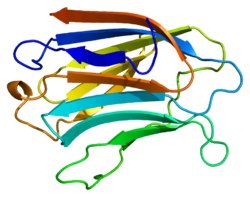
Fibronectin is a high-molecular weight glycoprotein of the extracellular matrix that binds to membrane-spanning receptor proteins called integrins. It is approved for marketing as a topical solution in India by Central Drugs Standard Control organization in 2020 under the brand name FIBREGA for chronic wounds. Fibronectin also binds to other extracellular matrix proteins such as collagen, fibrin, and heparan sulfate proteoglycans.
In biology, the extracellular matrix (ECM), also called intercellular matrix, is a network consisting of extracellular macromolecules and minerals, such as collagen, enzymes, glycoproteins and hydroxyapatite that provide structural and biochemical support to surrounding cells. Because multicellularity evolved independently in different multicellular lineages, the composition of ECM varies between multicellular structures; however, cell adhesion, cell-to-cell communication and differentiation are common functions of the ECM.
Cell adhesion is the process by which cells interact and attach to neighbouring cells through specialised molecules of the cell surface. This process can occur either through direct contact between cell surfaces such as cell junctions or indirect interaction, where cells attach to surrounding extracellular matrix, a gel-like structure containing molecules released by cells into spaces between them. Cells adhesion occurs from the interactions between cell-adhesion molecules (CAMs), transmembrane proteins located on the cell surface. Cell adhesion links cells in different ways and can be involved in signal transduction for cells to detect and respond to changes in the surroundings. Other cellular processes regulated by cell adhesion include cell migration and tissue development in multicellular organisms. Alterations in cell adhesion can disrupt important cellular processes and lead to a variety of diseases, including cancer and arthritis. Cell adhesion is also essential for infectious organisms, such as bacteria or viruses, to cause diseases.
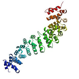
Osteopontin (OPN), also known as bone /sialoprotein I, early T-lymphocyte activation (ETA-1), secreted phosphoprotein 1 (SPP1), 2ar and Rickettsia resistance (Ric), is a protein that in humans is encoded by the SPP1 gene. The murine ortholog is Spp1. Osteopontin is a SIBLING (glycoprotein) that was first identified in 1986 in osteoblasts.
Catenin beta-1, also known as beta-catenin (β-catenin), is a protein that in humans is encoded by the CTNNB1 gene.
Mannan-binding lectin serine protease 1 also known as mannose-associated serine protease 1 (MASP-1) is an enzyme that in humans is encoded by the MASP1 gene.
Galectins are a class of proteins that bind specifically to β-galactoside sugars, such as N-acetyllactosamine, which can be bound to proteins by either N-linked or O-linked glycosylation. They are also termed S-type lectins due to their dependency on disulphide bonds for stability and carbohydrate binding. There have been about 15 galectins discovered in mammals, encoded by the LGALS genes, which are numbered in a consecutive manner. Only galectin-1, -2, -3, -4, -7, -7B, -8, -9, -9B, 9C, -10, -12, -13, -14, and -16 have been identified in humans. Galectin-5 and -6 are found in rodents, whereas galectin-11 and -15 are uniquely found in sheep and goats. Members of the galectin family have also been discovered in other mammals, birds, amphibians, fish, nematodes, sponges, and some fungi. Unlike the majority of lectins they are not membrane bound, but soluble proteins with both intra- and extracellular functions. They have distinct but overlapping distributions but found primarily in the cytosol, nucleus, extracellular matrix or in circulation. Although many galectins must be secreted, they do not have a typical signal peptide required for classical secretion. The mechanism and reason for this non-classical secretion pathway is unknown.
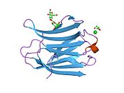
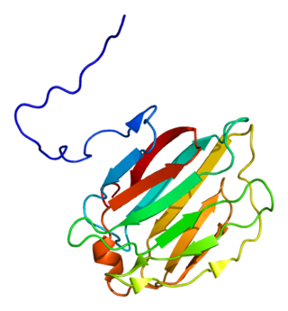
Galectin-1 is a protein that in humans is encoded by the LGALS1 gene.
Integrin beta-6 is a protein that in humans is encoded by the ITGB6 gene. It is the β6 subunit of the integrin αvβ6. Integrins are αβ heterodimeric glycoproteins which span the cell’s membrane, integrating the outside and inside of the cell. Integrins bind to specific extracellular proteins in the extracellular matrix or on other cells and subsequently transduce signals intracellularly to affect cell behaviour. One α and one β subunit associate non-covalently to form 24 unique integrins found in mammals. While some β integrin subunits partner with multiple α subunits, β6 associates exclusively with the αv subunit. Thus, the function of ITGB6 is entirely associated with the integrin αvβ6.
Galectin-3-binding protein is a protein that in humans is encoded by the LGALS3BP gene.
Galectin-10 is an enzyme that in humans is encoded by the CLC gene.
Galectin-8 is a protein of the galectin family that in humans is encoded by the LGALS8 gene.
Galectin-9 was first isolated from mouse embryonic kidney in 1997 as a 36 kDa beta-galactoside lectin protein. Human galectin-9 is encoded by the LGALS9 gene.
Galectin-2 is a protein that in humans is encoded by the LGALS2 gene.
Hepatitis A virus cellular receptor 2 (HAVCR2), also known as T-cell immunoglobulin and mucin-domain containing-3 (TIM-3), is a protein that in humans is encoded by the HAVCR2 (TIM-3)gene. HAVCR2 was first described in 2002 as a cell surface molecule expressed on IFNγ producing CD4+ Th1 and CD8+ Tc1 cells. Later, the expression was detected in Th17 cells, regulatory T-cells, and innate immune cells. HAVCR2 receptor is a regulator of the immune response.
Galectin-7 is a protein that in humans is encoded by the LGALS7 gene.
Placental protein 13 (PP13) is a protein that in humans is encoded by the LGALS13 gene.
Galectin-4 is a protein that in humans is encoded by the LGALS4 gene.
Cenderitide is a natriuretic peptide developed by the Mayo Clinic as a potential treatment for heart failure. Cenderitide is created by the fusion of the 15 amino acid C-terminus of dendroaspis natriuretic peptide (DNP) with the full C-type natriuretic peptide (CNP) structure both peptide which are endogenous to humans. This peptide chimera is a dual activator of the natriuretic peptide receptors NPR-A and NPR-B and therefore exhibits the natriuretic and diuretic properties of DNP, as well as the antiproliferative and antifibrotic properties of CNP.
MFAP4 is an extracellular matrix protein encoded by the MFAP4 gene. It is part of the MFAP family of proteoglycans, which are involved in cell adhesion, intercellular interactions and the assembly and/or maintenance of elastic fibres.
{{cite journal}}
: Cite journal requires |journal=
(help)This article incorporates text from the United States National Library of Medicine, which is in the public domain.