Renal cyst

Last updated
Renal cyst
Other namesKidney cysst
Renal cyst MRI.jpg
Renal cyst of the left kidney (hyperintense area) as shown on MRI.
Specialty Urology   OOjs UI icon edit-ltr-progressive.svg
Simple renal cyst Simple renal cyst.jpg
Simple renal cyst

A renal cyst is a fluid collection in or on the kidney. There are several types based on the Bosniak classification. The majority are benign, simple cysts that can be monitored and not intervened upon. However, some are cancerous or are suspicious for cancer and are commonly removed in a surgical procedure called nephrectomy.

Contents

Numerous renal cysts are seen in the cystic kidney diseases, which include polycystic kidney disease and medullary sponge kidney.

Classification

Renal cysts are classified by malignant risk using the Bosniak classification system. The system was created by Morton Bosniak (1929–2016), a faculty member at the New York University Langone Medical Center in New York City. [1]

The Bosniak classification categorizes renal cysts into five groups. [2]

Category I

Benign simple cyst with thin wall without septa, calcifications, or solid components, and has a density of 0–20 Hounsfield units (HU) [3] (about equal to that of water). In such cases, a CT scan without intravenous contrast is enough for classification. [4] Still, if a contrast CT is performed, a category I cyst should not show significant enhancement, [4] which can be regarded as an increase of less than 10HU. [5]

Category II

Benign cyst with a few thin septa, which may contain fine calcifications or a small segment of mildly thickened calcification. This includes homogenous, high-attenuation (60–70 Hounsfield units [3] ) lesions less than 3 cm with sharp margins but without enhancement. Hyperdense cysts must be exophytic with at least 75 percent of its wall outside the kidney to allow for appropriate assessment of margins, otherwise they are categorized as IIF. [6]

Category IIF

A Bosniak category IIF cyst. This one is 3 cm wide, with calcifications within its wall, seen as very radiodense (white in this presentation) areas in its margins. There is also a septation which is calcified. Yet, the cyst does not show enhancement (uptake of contrast). Bosniak 2F renal cyst.jpg
A Bosniak category IIF cyst. This one is 3 cm wide, with calcifications within its wall, seen as very radiodense (white in this presentation) areas in its margins. There is also a septation which is calcified. Yet, the cyst does not show enhancement (uptake of contrast).

This category includes renal cysts with multiple thin septa, a septum thicker than hairline, slightly thick wall, or with calcification, which may be thick. It also includes intrarenal cysts larger than 3 centimetres (1.2 inches) if:

Category IIF cysts have a 5–10% risk of being kidney cancer, and therefore follow-up is recommended. However, there is no consensus recommendation on the appropriate interval of follow up. [7]

Category III

Indeterminate cystic masses with thickened, irregular or smooth walls or septa with measurable enhancement. Approximately 40 to 60% of these lesions are ultimately found to be malignant, most commonly in the forms of cystic renal cell carcinoma and its multiloculated variant. [8] [9] [10] The remaining lesions are benign and include hemorrhagic cysts, chronic infected cysts, and multiloculated cystic nephromas.

Category IV

Malignant cystic masses with all the characteristics of category III lesions but also with enhancing soft tissue components independent of but adjacent to the septa. Approximately 85 to 100% of these lesions are malignant. [8] [11]

The presence of measurable contrast enhancement of the lesion is the most important characteristic in distinguishing between high-risk cysts (classifications III and IV) from the typically benign, low-risk Bosniak I, II, and IIF cysts. [8] Such contrast enhancement should be at least 10 to 15 Hounsfield units higher when compared with unenhanced images.

Bosniak category
IIIIIFIIIIV
Attenuation [3] 0–20 HU 60–70 Hounsfield units
Walls [3] Thin and smoothSmall and fine calcificationsNodular or irregular calcificationsThick, heterogeneous. Gross calcifications with enhancement
Solid components [3] NoYes

Diagnosis

The complex cyst can be further evaluated with doppler ultrasonography, and for Bosniak classification and follow-up of complex cysts, either contrast-enhanced ultrasound (CEUS) or contrast CT is used. [12]

Histopathology of the lining of a simple cyst of the kidney, incidentally found on autopsy, with inconspicuous nuclei. They usually have a single layer of cuboidal, flattened or atrophic epithelium, but this case has a somewhat thicker fibrous layer. H&E stain. Histopathology of simple cyst of the kidney.jpg
Histopathology of the lining of a simple cyst of the kidney, incidentally found on autopsy, with inconspicuous nuclei. They usually have a single layer of cuboidal, flattened or atrophic epithelium, but this case has a somewhat thicker fibrous layer. H&E stain.

Treatment

This system is more directly focused on the most appropriate management. These alternatives are broadly to ignore the cyst, schedule follow-up or perform a surgical excision of it. When a cyst shows discrepancy in severity across categories, it is the most worrisome feature that is used in deciding about management. There is no established rule regarding the follow-up frequency, but one possibility is after 6 months, which can later be doubled if unchanged. [5]

Recommended management [5]
IgnoreFollowExcise
Calcification
  • Small, smooth and liquid (moves to lowest point when changing position)
Thick, nodular
If radiodensity > 20 HU without radiocontrast
  • Sharp margin, < 3 cm, not completely intrarenal and homogenous
  • Must also be clearly cystic if seen on ultrasound
  • totally intrarenal
  • >3 cm
SeptationsThin and smoothSlightly greater than hairline
  • thick
  • irregular
  • nodular
Enhancement (increase with radiocontrast)< 10 HU 10–15 HU> 15 HU
MultilocularIf infectionAll others
Nodularity [5] Very small and nonenhancingAll others
Wall thickening [5] If infectionAll others

Peripelvic versus parapelvic cysts

Non-contrast CT (at left) showing peripelvic fluid accumulations, which may be hydronephrosis. CT urography (at right) reveals non-dilated calyces and pelvises. The fluid accumulations are thus peripelvic cysts. CT of peripelvic cysts with non-contrast and urography.jpg
Non-contrast CT (at left) showing peripelvic fluid accumulations, which may be hydronephrosis. CT urography (at right) reveals non-dilated calyces and pelvises. The fluid accumulations are thus peripelvic cysts.

Parapelvic cysts originate from around the kidney at the adjacent renal parenchyma, and plunge into the renal sinus. Peripelvic cysts are contained entirely within the renal sinus, possibly related to dilated lymphatic channels. When viewed on CT in absence of contrast, they can mimic hydronephrosis. [14] If symptomatic, they can be laparoscopically decorticated - removal of the outer layer or cortex. [15]

Epidemiology

Up to 27 percent of individuals older than 50 years may have simple renal cysts that cause no symptoms. [16]

See also

Related Research Articles

<span class="mw-page-title-main">Medical ultrasound</span> Diagnostic and therapeutic technique

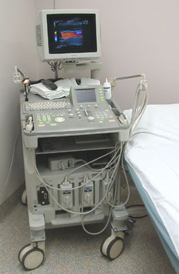
Medical ultrasound includes diagnostic techniques using ultrasound, as well as therapeutic applications of ultrasound. In diagnosis, it is used to create an image of internal body structures such as tendons, muscles, joints, blood vessels, and internal organs, to measure some characteristics or to generate an informative audible sound. The usage of ultrasound to produce visual images for medicine is called medical ultrasonography or simply sonography, or echography. The practice of examining pregnant women using ultrasound is called obstetric ultrasonography, and was an early development of clinical ultrasonography. The machine used is called an ultrasound machine, a sonograph or an echograph. The visual image formed using this technique is called an ultrasonogram, a sonogram or an echogram.

<span class="mw-page-title-main">Cyst</span> Closed sac growth on the body

A cyst, also traditionally known from Old English as a wen, is a closed sac, having a distinct envelope and division compared with the nearby tissue. Hence, it is a cluster of cells that have grouped together to form a sac ; however, the distinguishing aspect of a cyst is that the cells forming the "shell" of such a sac are distinctly abnormal when compared with all surrounding cells for that given location. A cyst may contain air, fluids, or semi-solid material. A collection of pus is called an abscess, not a cyst. Once formed, a cyst may resolve on its own. When a cyst fails to resolve, it may need to be removed surgically, but that would depend upon its type and location.

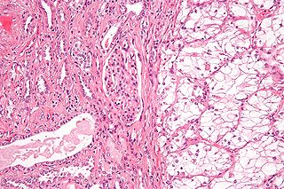
Liver tumors are abnormal growth of liver cells on or in the liver. Several distinct types of tumors can develop in the liver because the liver is made up of various cell types. Liver tumors can be classified as benign (non-cancerous) or malignant (cancerous) growths. They may be discovered on medical imaging, and the diagnosis is often confirmed with liver biopsy. Signs and symptoms of liver masses vary from being asymptomatic to patients presenting with an abdominal mass, hepatomegaly, abdominal pain, jaundice, or some other liver dysfunction. Treatment varies and is highly specific to the type of liver tumor.

<span class="mw-page-title-main">Renal cell carcinoma</span> Medical condition

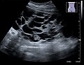
Renal cell carcinoma (RCC) is a kidney cancer that originates in the lining of the proximal convoluted tubule, a part of the very small tubes in the kidney that transport primary urine. RCC is the most common type of kidney cancer in adults, responsible for approximately 90–95% of cases. RCC occurrence shows a male predominance over women with a ratio of 1.5:1. RCC most commonly occurs between 6th and 7th decade of life.

<span class="mw-page-title-main">Kidney cancer</span> Medical condition

Kidney cancer, also known as renal cancer, is a group of cancers that starts in the kidney. Symptoms may include blood in the urine, lump in the abdomen, or back pain. Fever, weight loss, and tiredness may also occur. Complications can include spread to the lungs or brain.

In medical or research imaging, an incidental imaging finding is an unanticipated finding which is not related to the original diagnostic inquiry. As with other types of incidental medical findings, they may represent a diagnostic, ethical, and philosophical dilemma because their significance is unclear. While some coincidental findings may lead to beneficial diagnoses, others may lead to overdiagnosis that results in unnecessary testing and treatment, sometimes called the "cascade effect".

<span class="mw-page-title-main">Giant-cell tumor of bone</span> Medical condition

Giant-cell tumor of the bone (GCTOB), is a relatively uncommon tumor of the bone. It is characterized by the presence of multinucleated giant cells. Malignancy in giant-cell tumor is uncommon and occurs in about 2% of all cases. However, if malignant degeneration does occur, it is likely to metastasize to the lungs. Giant-cell tumors are normally benign, with unpredictable behavior. It is a heterogeneous tumor composed of three different cell populations. The giant-cell tumour stromal cells (GCTSC) constitute the neoplastic cells, which are from an osteoblastic origin and are classified based on expression of osteoblast cell markers such as alkaline phosphatase and osteocalcin. In contrast, the mononuclear histiocytic cells (MNHC) and multinucleated giant cell (MNGC) fractions are secondarily recruited and comprise the non-neoplastic cell population. They are derived from an osteoclast-monocyte lineage determined primarily by expression of CD68, a marker for monocytic precursor cells. In most patients, the tumors are slow to develop, but may recur locally in as many as 50% of cases.

<span class="mw-page-title-main">Angiomyolipoma</span> Medical condition

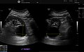
Angiomyolipomas are the most common benign tumour of the kidney. Although regarded as benign, angiomyolipomas may grow such that kidney function is impaired or the blood vessels may dilate and burst, leading to bleeding.

<span class="mw-page-title-main">Abdominal ultrasonography</span>

Abdominal ultrasonography is a form of medical ultrasonography to visualise abdominal anatomical structures. It uses transmission and reflection of ultrasound waves to visualise internal organs through the abdominal wall. For this reason, the procedure is also called a transabdominal ultrasound, in contrast to endoscopic ultrasound, the latter combining ultrasound with endoscopy through visualize internal structures from within hollow organs.

<span class="mw-page-title-main">Thyroid nodule</span> Medical condition

Thyroid nodules are nodules which commonly arise within an otherwise normal thyroid gland. They may be hyperplastic or tumorous, but only a small percentage of thyroid tumors are malignant. Small, asymptomatic nodules are common, and often go unnoticed. Nodules that grow larger or produce symptoms may eventually need medical care. A goitre may have one nodule – uninodular, multiple nodules – multinodular, or be diffuse.

<span class="mw-page-title-main">Lung nodule</span> Medical condition

A lung nodule or pulmonary nodule is a relatively small focal density in the lung. A solitary pulmonary nodule (SPN) or coin lesion, is a mass in the lung smaller than three centimeters in diameter. A pulmonary micronodule has a diameter of less than three millimetres. There may also be multiple nodules.

<span class="mw-page-title-main">Kidney tumour</span> Medical condition

Kidney tumours are tumours, or growths, on or in the kidney. These growths can be benign or malignant.

<span class="mw-page-title-main">Diktyoma</span> Type of eye tumor

Diktyoma, or ciliary body medulloepithelioma, or teratoneuroma, is a rare tumor arising from primitive medullary epithelium in the ciliary body of the eye. Almost all diktyomas arise in the ciliary body, although, rarely, they may arise from the optic nerve head or retina.

Theca lutein cyst is a type of bilateral functional ovarian cyst filled with clear, straw-colored fluid. These cysts result from exaggerated physiological stimulation due to elevated levels of beta-human chorionic gonadotropin (beta-hCG) or hypersensitivity to beta-hCG. On ultrasound and MRI, theca lutein cysts appear in multiples on ovaries that are enlarged.

<span class="mw-page-title-main">Computed tomography of the abdomen and pelvis</span>

Computed tomography of the abdomen and pelvis is an application of computed tomography (CT) and is a sensitive method for diagnosis of abdominal diseases. It is used frequently to determine stage of cancer and to follow progress. It is also a useful test to investigate acute abdominal pain. Renal stones, appendicitis, pancreatitis, diverticulitis, abdominal aortic aneurysm, and bowel obstruction are conditions that are readily diagnosed and assessed with CT. CT is also the first line for detecting solid organ injury after trauma.

<span class="mw-page-title-main">Scrotal ultrasound</span> Medical ultrasound examination of the scrotum.

Scrotalultrasound is a medical ultrasound examination of the scrotum. It is used in the evaluation of testicular pain, and can help identify solid masses.

<span class="mw-page-title-main">Renal ultrasonography</span> Examination of one or both kidneys using medical ultrasound

Renal ultrasonography is the examination of one or both kidneys using medical ultrasound.

Ultrasonography of liver tumors involves two stages: detection and characterization.

In CT scan of the thyroid, focal and diffuse thyroid abnormalities are commonly encountered. These findings can often lead to a diagnostic dilemma, as the CT reflects nonspecific appearances. Ultrasound (US) examination has a superior spatial resolution and is considered the modality of choice for thyroid evaluation. Nevertheless, CT detects incidental thyroid nodules (ITNs) and plays an important role in the evaluation of thyroid cancer.

<span class="mw-page-title-main">Emilio Quaia</span> Italian radiologist, academic, and author

Emilio Quaia is an Italian radiologist, academic, and author. He is a professor of Radiology and the Director of the Radiology Department at the University of Padova.

References

  1. "Education & Research at NYU Langone Health".
  2. Curry NS, Cochran ST, Bissada NK (August 2000). "Cystic renal masses: accurate Bosniak classification requires adequate renal CT". American Journal of Roentgenology. 175 (2): 339–42. doi:10.2214/ajr.175.2.1750339. PMID   10915671.
  3. 1 2 3 4 5 6 Muglia, Valdair F.; Westphal en, Antonio Carlos (2014). "Classificação de Bosniak para cistos renais complexos: histórico e análise crítica". Radiologia Brasileiro. 47 (6): 368–373. doi:10.1590/0100-3984.2013.1797. ISSN   0100-3984. PMC   4341374 . PMID   25741120.
  4. 1 2 Karen Garfield and Stephen W. Leslie (2019-03-14). Simple Renal Cyst. StatPearls. PMID   29763075.{{cite book}}: |website= ignored (help)
  5. 1 2 3 4 5 David S. Hartman, MD and Ileana Chesaru, MD. "Kidney – Cystic masses". Radiology Assistant . Retrieved 2017-10-20.{{cite web}}: CS1 maint: multiple names: authors list (link)
  6. De Miranda, C. M.; Maranhão, C. P.; Dos Santos, C. J.; Padilha, I. G.; De Farias Lde, P; Da Rocha, M. 's. (2014). "Bosniak classification of renal cystic lesions according to multidetector computed tomography findings". Radiologia Brasileiro. 47 (2): 115–21. doi:10.1590/S0100-39842014000200015. PMC   4337166 . PMID   25741060.
  7. 1 2 "Bosniak classification of renal cystic disease". GPnotebook . Retrieved 2017-05-17.
  8. 1 2 3 Israel, Gary M.; Bosniak, Morton A. (September 2005). "An update of the Bosniak renal cyst classification system". Urology. 66 (3): 484–488. doi:10.1016/j.urology.2005.04.003. PMID   16140062.
  9. Curry, Nancy S.; Cochran, Sachiko T.; Bissada, Nabil K. (August 2000). "Cystic Renal Masses: Accurate Bosniak Classification Requires Adequate Renal CT". American Journal of Roentgenology. 175 (2): 339–342. doi:10.2214/ajr.175.2.1750339. ISSN   0361-803X. PMID   10915671.
  10. Harisinghani, Mukesh G.; Maher, Michael M.; Gervais, Debra A.; McGovern, Francis; Hahn, Peter; Jhaveri, Kartik; Varghese, Jose; Mueller, Peter R. (March 2003). "Incidence of Malignancy in Complex Cystic Renal Masses (Bosniak Category III): Should Imaging-Guided Biopsy Precede Surgery?". American Journal of Roentgenology. 180 (3): 755–758. doi:10.2214/ajr.180.3.1800755. ISSN   0361-803X. PMID   12591691.
  11. Bosniak, M A (May 1991). "The small (less than or equal to 3.0 cm) renal parenchymal tumor: detection, diagnosis, and controversies". Radiology. 179 (2): 307–317. doi:10.1148/radiology.179.2.2014269. ISSN   0033-8419. PMID   2014269.
  12. 1 2 Hansen, Kristoffer; Nielsen, Michael; Ewertsen, Caroline (2015). "Ultrasonography of the Kidney: A Pictorial Review". Diagnostics. 6 (1): 2. doi: 10.3390/diagnostics6010002 . ISSN   2075-4418. PMC   4808817 . PMID   26838799. (CC-BY 4.0)
  13. Mandolin S. Ziadie, M.D. "Simple cysts". Pathology Outlines. Topic Completed: 1 November 2011. Minor changes: 1 October 2019
  14. Zinn, H. L.; Becker, J. A. (1997). "Peripelvic cysts simulating hydronephrosis". Abdominal Imaging. 22 (3): 346–7. doi:10.1007/s002619900205. PMID   9107666. S2CID   9514583.
  15. Shiraishi, K; Eguchi, S; Mohri, J; Kamiryo, Y (2006). "Laparoscopic decortication of symptomatic simple renal cysts: 10-year experience from one institution". BJU International. 98 (2): 405–8. doi:10.1111/j.1464-410X.2006.06249.x. PMID   16879687.
  16. Tada S, Yamagishi J, Kobayashi H, Hata Y, Kobari T (July 1983). "The incidence of simple renal cyst by computed tomography". Clinical Radiology. 34 (4): 437–9. doi:10.1016/S0009-9260(83)80238-4. PMID   6872451.