Genotoxicity

Last updated

Genotoxicity is the property of chemical agents that damage the genetic information within a cell causing mutations, which may lead to cancer. While genotoxicity is often confused with mutagenicity, all mutagens are genotoxic, but some genotoxic substances are not mutagenic. The alteration can have direct or indirect effects on the DNA: the induction of mutations, mistimed event activation, and direct DNA damage leading to mutations. The permanent, heritable changes can affect either somatic cells of the organism or germ cells to be passed on to future generations. [1] Cells prevent expression of the genotoxic mutation by either DNA repair or apoptosis; however, the damage may not always be fixed leading to mutagenesis.

Contents

To assay for genotoxic molecules, researchers assay for DNA damage in cells exposed to the toxic substrates. This DNA damage can be in the form of single- and double-strand breaks, loss of excision repair, cross-linking, alkali-labile sites, point mutations, and structural and numerical chromosomal aberrations. [2] The compromised integrity of the genetic material has been known to cause cancer. As a consequence, many sophisticated techniques including Ames Assay, in vitro and in vivo Toxicology Tests, and Comet Assay have been developed to assess the chemicals' potential to cause DNA damage that may lead to cancer.

Mechanism

Definition of transitions and transversions. They are a common mutation caused by genotoxic compounds. Transitions and transversions.svg
Definition of transitions and transversions. They are a common mutation caused by genotoxic compounds.

Genotoxic substances induce damage to the genetic material in the cells through interactions with the DNA sequence and structure. For example, the transition metal chromium interacts with DNA in its high-valent oxidation state, incurring DNA lesions which lead to carcinogenesis. The metastable oxidation state Cr(V) is achieved through reductive activation. Researchers performed an experiment to study the interaction between DNA with the carcinogenic chromium by using a Cr(V)-Salen complex at the specific oxidation state. [3] The interaction was specific to the guanine nucleotide in the genetic sequence. In order to narrow the interaction between the Cr(V)-Salen complex with the guanine base, the researchers modified the bases to 8-oxo-G so to have site specific oxidation. The reaction between the two molecules caused DNA lesions; the two lesions observed at the modified base site were guanidinohydantoin and spiroiminodihydantoin. To further analyze the site of lesion, it was observed that polymerase stopped at the site and adenine was inappropriately incorporated into the DNA sequence opposite of the 8-oxo-G base. Therefore, these lesions predominately contain G-->T transversions. High-valent chromium is seen to act as a carcinogen as researchers found that "the mechanism of damage and base oxidation products for the interaction between high-valent chromium and DNA... are relevant to in vivo formation of DNA damage leading to cancer in chromate-exposed human populations". [3] Consequently, it shows how high-valent chromium can act as a carcinogen with 8-oxo-G forming xenobiotics. [3]

Another example of a genotoxic substance causing DNA damage are pyrrolizidine alkaloids (PAs). These substances are found mainly in plant species and are poisonous to animals, including humans; about half of them have been identified as genotoxic and many as tumorigenic. The researchers concluded from testing that when metabolically activated, "PAs produce DNA adducts, DNA cross-linking, DNA breaks, sister chromatid exchange, micronuclei, chromosomal aberrations, gene mutations, and chromosome mutations in vivo and in vitro." [4] The most common mutation within the genes are G:C --> T:A tranversions and tandem base substitution. The pyrrolizidine alkaloids are mutagenic in vivo and in vitro and, therefore, responsible for the carcinogenesis prominently in the liver. [4] Comfrey is an example of a plant species that contains fourteen different PAs. The active metabolites interact with DNA to cause DNA damage, mutation induction, and cancer development in liver endothelial cells and hepatocytes. The researchers discovered in the end that the "comfrey is mutagenic in liver, and PA contained in comfrey appear to be responsible for comfrey-induced toxicity and tumor induction,". [5]

Test techniques

The purpose of genotoxicity testing is to determine if a substrate will influence genetic material or may cause cancer. They can be performed in bacterial, yeast, and mammalian cells. [2] With the knowledge from the tests, one can control early development of vulnerable organisms to genotoxic substances. [1]

Bacterial Reverse Mutation Assay

The Bacterial Reverse Mutation Assay, also known as the Ames Assay, is used in laboratories to test for gene mutation. The technique uses many different bacterial strains in order to compare the different changes in the genetic material. The result of the test detects the majority of genotoxic carcinogens and genetic changes; the types of mutations detected are frame shifts and base substitutions. [6]

Ames test procedure to test for gene mutations present in the various bacterial strains Ames test.svg
Ames test procedure to test for gene mutations present in the various bacterial strains

in vitro toxicology testing

The purpose of in vitro testing is to determine whether a substrate, product, or environmental factor induces genetic damage. One technique entails cytogenetic assays using different mammalian cells. [6] The types of aberrations detected in cells affected by a genotoxic substance are chromatid and chromosome gaps, chromosome breaks, chromatid deletions, fragmentation, translocation, complex rearrangements, and many more. The clastogenic or aneugenic effects from the genotoxic damage will cause an increase in frequency of structural or numerical aberrations of the genetic material. [6] This is similar to the micronucleus test and chromosome aberration assay, which detect structural and numerical chromosomal aberrations in mammalian cells. [1]

In a specific mammalian tissue, one can perform a mouse lymphoma TK+/- assay to test for changes in the genetic material. [6] Gene mutations are commonly point mutations, altering only one base within the genetic sequence to alter the ensuing transcript and amino acid sequence; these point mutations include base substitutions, deletions, frame-shifts, and rearrangements. Also, chromosomes' integrity may be altered through chromosome loss and clastogenic lesions causing multiple gene and multilocus deletions. The specific type of damage is determined by the size of the colonies, distinguishing between genetic mutations (mutagens) and chromosomal aberrations (clastogens). [6]

The SOS/umu assay test evaluates the ability of a substance to induce DNA damage; it is based on the alterations in the induction of the SOS response due to DNA damage. The benefits of this technique are that it is a fast and simple method and convenient for numerous substances. These techniques are performed on water and wastewater in the environment. [7]

Overview of the use of the SOS response for genotoxicity testing Genotoxic Damage.png
Overview of the use of the SOS response for genotoxicity testing

in vivo testing

The purpose for in vivo testing is to determine the potential of DNA damage that can affect chromosomal structure or disturb the mitotic apparatus that changes chromosome number; the factors that could influence the genotoxicity are ADME and DNA repair. It can also detect genotoxic agents missed in in vitro tests. The positive result of induced chromosomal damage is an increase in frequency of micronucelated PCEs. [6] A micronucleus is a small structure separate from the nucleus containing nuclear DNA arisen from DNA fragments or whole chromosomes that were not incorporated in the daughter cell during mitosis. Causes for this structure are mitotic loss of acentric chromosomal fragments (clastogenicity), mechanical problems from chromosomal breakage and exchange, mitotic loss of chromosomes (aneugenicity), and apoptosis. The micronucleus test in vivo is similar to the in vitro one because it tests for structural and numerical chromosomal aberrations in mammalian cells, especially in rats' blood cells. [6]

Comet assay

Comet assays are one of the most common tests for genotoxicity. The technique involves lysing cells using detergents and salts. The DNA released from the lysed cell is electrophoresed in an agarose gel under neutral pH conditions. Cells containing DNA with an increased number of double-strand breaks will migrate more quickly to the anode. This technique is advantageous in that it detects low levels of DNA damage, requires only a very small number of cells, is cheaper than many techniques, is easy to execute, and quickly displays results. However, it does not identify the mechanism underlying the genotoxic effect or the exact chemical or chemical component causing the breaks. [8]

Cancer

Genotoxic effects such as deletions, breaks and/or rearrangements can lead to cancer if the damage does not immediately lead to cell death. Regions sensitive to breakage, called fragile sites, may result from genotoxic agents (such as pesticides). Some chemicals have the ability to induce fragile sites in regions of the chromosome where oncogenes are present, which could lead to carcinogenic effects. In keeping with this finding, occupational exposure to some mixtures of pesticides are positively correlated with increased genotoxic damage in the exposed individuals. DNA damage is not uniform in its severity across populations because Individuals vary in their ability to activate or detoxify genotoxic substances, which leads to variability in the incidence of cancer among individuals. The difference in ability to detoxify certain compounds is due to individuals' inherited polymorphisms of genes involved in the metabolism of the chemical. Differences may also be attributed to individual variation in efficiency of DNA repair mechanisms [9]

The metabolism of some chemicals results in the production of reactive oxygen species (ROS), which is a possible mechanism of genotoxicity. This is seen in the metabolism of arsenic, which produces hydroxyl radicals, which are known to cause genotoxic effects. [10] Similarly, ROS have been implicated in genotoxicity caused by particles and fibers. Genotoxicity of nonfibrous and fibrous particles is characterized by high production of ROS from inflammatory cells. [11]

Genotoxins implicated in the four most common cancers worldwide

Major genotoxic agents responsible for the four most common cancers worldwide (lung, breast, colon and stomach) have been identified.

Lung cancer is the most frequent cancer in the world, both in terms of yearly cases (1.61 million cases; 12.7% of all cancer cases) and deaths (1.38 million deaths; 18.2% of all cancer deaths). [12] Tobacco smoke is the main cause of lung cancer. Risk estimates for lung cancer indicate that tobacco smoke is responsible for 90% of lung cancers in the United States. Tobacco smoke contains more than 5,300 identified chemicals. The most significant carcinogens in tobacco smoke have been determined by a "Margin of Exposure" approach. [13] By this approach, the tumorigenic compounds in tobacco smoke were, in order of importance, acrolein, formaldehyde, acrylonitrile, 1,3-butadiene, cadmium, acetaldehyde, ethylene oxide, and isoprene. In general, these compounds are genotoxic and cause DNA damage. As examples, DNA damaging effects have been reported for acrolein, [14] formaldehyde, [15] and acrylonitrile. [16]

Breast cancer is the second most frequent cancer worldwide on a yearly basis [(1.38 million cases, 10.9% of all cancer cases), and ranks 5th as cause of death (458,000, 6.1% of all cancer deaths)]. [12] Breast cancer risk is associated with persistently high blood levels of estrogen. [17] Estrogen likely contributes to breast carcinogenesis by the following three processes; (1) metabolic conversion of estrogen to genotoxic, mutagenic carcinogens, (2) stimulation of growth of tissues, and (3) repression of phase II detoxification enzymes that metabolize genotoxic reactive oxygen species, thus resulting in increased oxidative DNA damage. [18] [19] [20] The principal human estrogen, estradiol, can be metabolized to quinone derivatives that form DNA adducts. [21] These derivatives can cause the removal of bases from the phosphodiester backbone of DNA (e.g. depurination). This removal may be followed by inaccurate repair or replication of the apurinic site leading to mutation and eventually cancer.

Colorectal cancer is the third most frequent cancer worldwide [1.23 million cases (9.7% of all cancer cases), 608,000 deaths (8.0% of all cancer deaths)]. [12] In the United States, tobacco smoke may be responsible for up to 20% of colorectal cancers. [22] In addition, bile acids are implicated by substantial evidence as an important genotoxic factor in colon cancer. [23] In particular, the bile acid deoxycholic acid acid causes the production of DNA-damaging reactive oxygen species in human and rodent colon epithelial cells. [23]

Stomach cancer is the fourth most common cancer worldwide [990,000 cases (7.8% of all cancer cases), 738,000 deaths (9.7% of all cancer deaths )]. [12] Infection by Helicobacter pylori is the main causative factor in stomach cancer. Chronic inflammation due to H. pylori, if untreated, is often long-standing. H. pylori infection of gastric epithelial cells causes increased production of genotoxic reactive oxygen species (ROS). [24] [25] ROS cause oxidative damage to DNA that includes the major base alteration 8-Oxo-2'-deoxyguanosine. In a recent retrospective study it was found that use of a bile acid sequestrant was associated with a significant reduction in gastric cancer risk, suggesting that bile acids may be a contributory factor in stomach cancer. [26]

Genotoxic chemotherapy

Genotoxic chemotherapy is the treatment of cancer with the use of one or more genotoxic drugs. The treatment is traditionally part of standardized regime. By utilizing the destructive properties of genotoxins treatments aims to induce DNA damage into cancer cells. Any damage done to a cancer is passed on to descendent cancer cells as proliferation continues. If this damage is severe enough, it will induce cells to undergo apoptosis. [27]

Risks

A drawback of treatment is that many genotoxic drugs are effective on cancerous cells and normal cells alike. Selectivity of a particular drug's action is based on the sensitivity of the cells themselves. So, while rapidly dividing cancer cells are particularly sensitive to many drug treatments, often normal functioning cells are affected. [27]

Another risk of treatment is that, in addition to being genotoxic, many of the drugs are also mutagenic and cytotoxic. So the effects of these drugs are not limited to just DNA damage. In addition, some of these drugs that are meant to treat cancers are also carcinogens themselves, raising the risk of secondary cancers, such as leukemia. [27]

Different treatments

This table depicts different genotoxic-based cancer treatments along with examples. [27]

TreatmentMechanismExamples
Alkylating agents interfere with DNA replication and transcription by modifying DNA bases. Busulfan, Carmustine, Mechlorethamine
Intercalating agents interfere with DNA replication and transcription by wedging themselves into the spaces in between DNA's nucleotides Daunorubicin, Doxorubicin, Epirubicin
Enzyme inhibitors inhibit enzymes that are crucial to DNA replication Decitabine, Etoposide, Irinotecan

Kidney

Genotoxic DNA damage in the kidney has been implicated in both acute and chronic kidney injury, as well as in renal cell carcinoma [28] . Studies of humans with genetic deficiencies in DNA repair pathways have revealed that their kidneys are particularly vulnerable to DNA damages such as DNA crosslinks, DNA breaks and transcription-blocking damage [28] .

See also

Related Research Articles

<span class="mw-page-title-main">Ames test</span> Biological testing method

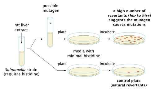
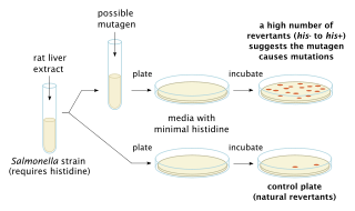
The Ames test is a widely employed method that uses bacteria to test whether a given chemical can cause mutations in the DNA of the test organism. More formally, it is a biological assay to assess the mutagenic potential of chemical compounds. A positive test indicates that the chemical is mutagenic and therefore may act as a carcinogen, because cancer is often linked to mutation. The test serves as a quick and convenient assay to estimate the carcinogenic potential of a compound because standard carcinogen assays on mice and rats are time-consuming and expensive. However, false-positives and false-negatives are known.

<span class="mw-page-title-main">Carcinogen</span> Substance, radionuclide, or radiation directly involved in causing cancer

A carcinogen is any substance, radionuclide, or radiation that promotes carcinogenesis. This may be due to the ability to damage the genome or to the disruption of cellular metabolic processes. Several radioactive substances are considered carcinogens, but their carcinogenic activity is attributed to the radiation, for example gamma rays and alpha particles, which they emit. Common examples of non-radioactive carcinogens are inhaled asbestos, certain dioxins, and tobacco smoke. Although the public generally associates carcinogenicity with synthetic chemicals, it is equally likely to arise from both natural and synthetic substances. Carcinogens are not necessarily immediately toxic; thus, their effect can be insidious.

Mutagenesis is a process by which the genetic information of an organism is changed by the production of a mutation. It may occur spontaneously in nature, or as a result of exposure to mutagens. It can also be achieved experimentally using laboratory procedures. A mutagen is a mutation-causing agent, be it chemical or physical, which results in an increased rate of mutations in an organism's genetic code. In nature mutagenesis can lead to cancer and various heritable diseases, and it is also a driving force of evolution. Mutagenesis as a science was developed based on work done by Hermann Muller, Charlotte Auerbach and J. M. Robson in the first half of the 20th century.

<span class="mw-page-title-main">Mutagen</span> Physical or chemical agent that increases the rate of genetic mutation

In genetics, a mutagen is a physical or chemical agent that permanently changes genetic material, usually DNA, in an organism and thus increases the frequency of mutations above the natural background level. As many mutations can cause cancer in animals, such mutagens can therefore be carcinogens, although not all necessarily are. All mutagens have characteristic mutational signatures with some chemicals becoming mutagenic through cellular processes.

<span class="mw-page-title-main">Methylcholanthrene</span> Chemical compound

Methylcholanthrene is a highly carcinogenic polycyclic aromatic hydrocarbon produced by burning organic compounds at very high temperatures. Methylcholanthrene is also known as 3-methylcholanthrene, 20-methylcholanthrene or the IUPAC name 3-methyl-1,2-dyhydrobenzo[j]aceanthrylene. The short notation often used is 3-MC or MCA. This compound forms pale yellow solid crystals when crystallized from benzene and ether. It has a melting point around 180 °C and its boiling point is around 280 °C at a pressure of 80 mmHg. Methylcholanthrene is used in laboratory studies of chemical carcinogenesis. It is an alkylated derivative of benz[a]anthracene and has a similar UV spectrum. The most common isomer is 3-methylcholanthrene, although the methyl group can occur in other places.

<span class="mw-page-title-main">Sudan I</span> Chemical compound

Sudan I is an organic compound, typically classified as an azo dye. It is an intensely orange-red solid that is added to colourise waxes, oils, petrol, solvents, and polishes. Sudan I has also been adopted for colouring various foodstuffs, especially curry powder and chili powder, although the use of Sudan I in foods is now banned in many countries, because Sudan I, Sudan III, and Sudan IV have been classified as category 3 carcinogens by the International Agency for Research on Cancer. Sudan I is still used in some orange-coloured smoke formulations and as a colouring for cotton refuse used in chemistry experiments.

Carcinogenesis, also called oncogenesis or tumorigenesis, is the formation of a cancer, whereby normal cells are transformed into cancer cells. The process is characterized by changes at the cellular, genetic, and epigenetic levels and abnormal cell division. Cell division is a physiological process that occurs in almost all tissues and under a variety of circumstances. Normally, the balance between proliferation and programmed cell death, in the form of apoptosis, is maintained to ensure the integrity of tissues and organs. According to the prevailing accepted theory of carcinogenesis, the somatic mutation theory, mutations in DNA and epimutations that lead to cancer disrupt these orderly processes by interfering with the programming regulating the processes, upsetting the normal balance between proliferation and cell death. This results in uncontrolled cell division and the evolution of those cells by natural selection in the body. Only certain mutations lead to cancer whereas the majority of mutations do not.

<span class="mw-page-title-main">Micronucleus</span>

Micronucleus is the name given to the small nucleus that forms whenever a chromosome or a fragment of a chromosome is not incorporated into one of the daughter nuclei during cell division. It usually is a sign of genotoxic events and chromosomal instability. Micronuclei are commonly seen in cancerous cells and may indicate genomic damage events that can increase the risk of developmental or degenerative diseases. Micronuclei form during anaphase from lagging acentric chromosome or chromatid fragments caused by incorrectly repaired or unrepaired DNA breaks or by nondisjunction of chromosomes. This incorrect segregation of chromosomes may result from hypomethylation of repeat sequences present in pericentromeric DNA, irregularities in kinetochore proteins or their assembly, dysfunctional spindle apparatus, or flawed anaphase checkpoint genes. Micronuclei can contribute to genome instability by promoting a catastrophic mutational event called chromothripsis. Many micronucleus assays have been developed to test for the presence of these structures and determine their frequency in cells exposed to certain chemicals or subjected to stressful conditions.

<span class="mw-page-title-main">Comet assay</span>

The single cell gel electrophoresis assay is an uncomplicated and sensitive technique for the detection of DNA damage at the level of the individual eukaryotic cell. It was first developed by Östling & Johansson in 1984 and later modified by Singh et al. in 1988. It has since increased in popularity as a standard technique for evaluation of DNA damage/repair, biomonitoring and genotoxicity testing. It involves the encapsulation of cells in a low-melting-point agarose suspension, lysis of the cells in neutral or alkaline (pH>13) conditions, and electrophoresis of the suspended lysed cells. The term "comet" refers to the pattern of DNA migration through the electrophoresis gel, which often resembles a comet.

<span class="mw-page-title-main">Clastogen</span> Substance that can cause breaks in chromosomes

A clastogen is a mutagenic agent that disturbs normal DNA related processes or directly causes DNA strand breakages, thus causing the deletion, insertion, or rearrangement of entire chromosome sections. These processes are a form of mutagenesis which if left unrepaired, or improperly repaired, can lead to cancer. Known clastogens include acridine yellow, benzene, ethylene oxide, arsenic, phosphine, mimosine, actinomycin D, camptothecin, methotrexate, methyl acrylate, resorcinol and 5-fluorodeoxyuridine. Additionally, 1,2-dimethylhydrazine is a known colon carcinogen and shows signs of possessing clastogenic activity. There are many clastogens not listed here and research is ongoing to discover new clastogens. Some known clastogens only exhibit clastogenic activity in certain cell types, such as caffeine which exhibits clastogenic activity in plant cells. Researchers are interested in clastogens for researching cancer, as well as for other human health concerns such as the inheritability of clastogen effected paternal germ cells that lead to fetus developmental defects.

<span class="mw-page-title-main">DNA adduct</span> Segment of DNA bound to a cancer-causing chemical

In molecular genetics, a DNA adduct is a segment of DNA bound to a cancer-causing chemical. This process could lead to the development of cancerous cells, or carcinogenesis. DNA adducts in scientific experiments are used as biomarkers of exposure. They are especially useful in quantifying an organism's exposure to a carcinogen. The presence of such an adduct indicates prior exposure to a potential carcinogen, but it does not necessarily indicate the presence of cancer in the subject animal.

<span class="mw-page-title-main">Micronucleus test</span> Test for potential genotoxic compounds

A micronucleus test is a test used in toxicological screening for potential genotoxic compounds. The assay is now recognized as one of the most successful and reliable assays for genotoxic carcinogens, i.e., carcinogens that act by causing genetic damage and is recommended by the OECD guideline for the testing of chemicals. There are two major versions of this test, one in vivo and the other in vitro.

<i>o</i>-Toluidine Aryl amine

o-Toluidine (ortho-toluidine) is an organic compound with the chemical formula CH3C6H4NH2. It is the most important of the three isomeric toluidines. It is a colorless liquid although commercial samples are often yellowish. It is a precursor to the herbicides metolachlor and acetochlor.

Arsenic biochemistry refers to biochemical processes that can use arsenic or its compounds, such as arsenate. Arsenic is a moderately abundant element in Earth's crust, and although many arsenic compounds are often considered highly toxic to most life, a wide variety of organoarsenic compounds are produced biologically and various organic and inorganic arsenic compounds are metabolized by numerous organisms. This pattern is general for other related elements, including selenium, which can exhibit both beneficial and deleterious effects. Arsenic biochemistry has become topical since many toxic arsenic compounds are found in some aquifers, potentially affecting many millions of people via biochemical processes.

Cancer is caused by genetic changes leading to uncontrolled cell growth and tumor formation. The basic cause of sporadic (non-familial) cancers is DNA damage and genomic instability. A minority of cancers are due to inherited genetic mutations. Most cancers are related to environmental, lifestyle, or behavioral exposures. Cancer is generally not contagious in humans, though it can be caused by oncoviruses and cancer bacteria. The term "environmental", as used by cancer researchers, refers to everything outside the body that interacts with humans. The environment is not limited to the biophysical environment, but also includes lifestyle and behavioral factors.

Toxicodynamics, termed pharmacodynamics in pharmacology, describes the dynamic interactions of a toxicant with a biological target and its biological effects. A biological target, also known as the site of action, can be binding proteins, ion channels, DNA, or a variety of other receptors. When a toxicant enters an organism, it can interact with these receptors and produce structural or functional alterations. The mechanism of action of the toxicant, as determined by a toxicant’s chemical properties, will determine what receptors are targeted and the overall toxic effect at the cellular level and organismal level.

Bernd Kaina, born on 7 January 1950 in Drewitz, is a German biologist and toxicologist. His research is devoted to DNA damage and repair, DNA damage response, genotoxic signaling and cell death induced by carcinogenic DNA damaging insults.

<span class="mw-page-title-main">Hydroxylation of estradiol</span>

The hydroxylation of estradiol is one of the major routes of metabolism of the estrogen steroid hormone estradiol. It is hydroxylated into the catechol estrogens 2-hydroxyestradiol and 4-hydroxyestradiol and into estriol (16α-hydroxyestradiol), reactions which are catalyzed by cytochrome P450 enzymes predominantly in the liver, but also in various other tissues.

The somatic mutation and recombination tests (SMARTs) are in vivo genotoxicity tests performed in Drosophila melanogaster (Fruit fly). These fruit fly tests are a short-term test and a non-mammalian approach for in vivo testing of putative genotoxins found in the environment. D. melanogaster has a short lifespan, which allows for fast reproductive cycles and high-throughput genotoxicity testing. D. melanogaster also has around 75% functional orthologs of human disease-related genes, making it an attractive in vivo model for human research. The tests identify loss of heterozygosity for the specified genetic markers in heterozygous or trans-heterozygous adults using phenotypically observable genetic markers in adult tissues. Although diverse events like point mutations/deletions, nondisjunction, and homologous mitotic recombination might theoretically cause this loss of heterozygosity, nondisjunction processes are generally not relevant for most of the examined chemicals. SMARTs are two different tests that use the same genetic foundation, but target different adult tissues and are named accordingly: the wing-spot test and the eye-spot test.

Diana Anderson is a British biomedical scientist who is a professor at the University of Bradford. Her research has focussed on early cancer detection and genome stability. She was appointed an Order of the British Empire in 2022 for her services to cancer detection.

References

  1. 1 2 3 Kolle S (2012-06-01). "Genotoxicity and Carcinogenicity". BASF The Chemical Company. Archived from the original on 2013-06-28. Retrieved 2013-03-16.
  2. 1 2 "Genotoxicity: Validated Non-animal Alternatives". AltTox.org. 2011-06-20. Retrieved 2013-03-16.
  3. 1 2 3 Sugden KD, Campo CK, Martin BD (September 2001). "Direct oxidation of guanine and 7,8-dihydro-8-oxoguanine in DNA by a high-valent chromium complex: a possible mechanism for chromate genotoxicity". Chemical Research in Toxicology. 14 (9): 1315–22. doi:10.1021/tx010088+. PMID   11559048.
  4. 1 2 Chen T, Mei N, Fu PP (April 2010). "Genotoxicity of pyrrolizidine alkaloids". Journal of Applied Toxicology. 30 (3): 183–96. doi:10.1002/jat.1504. PMC   6376482 . PMID   20112250.
  5. Mei N, Guo L, Fu PP, Fuscoe JC, Luan Y, Chen T (October 2010). "Metabolism, genotoxicity, and carcinogenicity of comfrey". Journal of Toxicology and Environmental Health Part B: Critical Reviews. 13 (7–8): 509–26. doi:10.1080/10937404.2010.509013. PMC   5894094 . PMID   21170807.
  6. 1 2 3 4 5 6 7 Furman G (2008-04-17). "Genotoxicity Testing for Pharmaceuticals Current and Emerging Practices" (PDF). Paracelsus, Inc. Archived from the original (PDF) on 2014-01-16. Retrieved 2013-03-16.
  7. Končar H (2011). "In Vitro Genotoxicity Testing". National Institute of Biology. Archived from the original on 2013-03-07. Retrieved 2013-03-16.
  8. Tice RR, Agurell E, Anderson D, Burlinson B, Hartmann A, Kobayashi H, et al. (2000). "Single cell gel/comet assay: guidelines for in vitro and in vivo genetic toxicology testing" (PDF). Environmental and Molecular Mutagenesis. 35 (3): 206–21. doi: 10.1002/(SICI)1098-2280(2000)35:3<206::AID-EM8>3.0.CO;2-J . PMID   10737956.
  9. Bolognesi, Claudia (June 2003). "Genotoxicity of pesticides: A review of human biomonitoring studies". Mutation Research. 543 (3): 251–272. doi:10.1016/S1383-5742(03)00015-2. PMID   12787816.
  10. Liu SX, Athar M, Lippai I, Waldren C, Hei TK (February 2001). "Induction of oxyradicals by arsenic: implication for mechanism of genotoxicity". Proceedings of the National Academy of Sciences of the United States of America. 98 (4): 1643–8. Bibcode:2001PNAS...98.1643L. doi: 10.1073/pnas.98.4.1643 . PMC   29310 . PMID   11172004.
  11. Schins RP (January 2002). "Mechanisms of genotoxicity of particles and fibers". Inhalation Toxicology. 14 (1): 57–78. doi:10.1080/089583701753338631. PMID   12122560. S2CID   24802577.
  12. 1 2 3 4 Ferlay J, Shin HR, Bray F, Forman D, Mathers C, Parkin DM (December 2010). "Estimates of worldwide burden of cancer in 2008: GLOBOCAN 2008". International Journal of Cancer. 127 (12): 2893–2917. doi:10.1002/ijc.25516. PMID 21351269. S2CID 23583962
  13. Cunningham FH, Fiebelkorn S, Johnson M, Meredith C (November 2011). "A novel application of the Margin of Exposure approach: segregation of tobacco smoke toxicants". Food and Chemical Toxicology. 49 (11): 2921–2933. doi:10.1016/j.fct.2011.07.019. PMID 21802474
  14. Liu D, Cheng Y, Tang Z, Mei X, Cao X, Liu J. Toxicity mechanism of acrolein on DNA damage and apoptosis in BEAS-2B cells: Insights from cell biology and molecular docking analyses. Toxicology. 2022 Jan 30;466:153083. doi: 10.1016/j.tox.2021.153083. Epub 2021 Dec 24. PMID 34958888
  15. Mulderrig L, Garaycoechea JI, Tuong ZK, Millington CL, Dingler FA, Ferdinand JR, Gaul L, Tadross JA, Arends MJ, O'Rahilly S, Crossan GP, Clatworthy MR, Patel KJ. Aldehyde-driven transcriptional stress triggers an anorexic DNA damage response. Nature. 2021 Dec;600(7887):158-163. doi: 10.1038/s41586-021-04133-7. Epub 2021 Nov 24. PMID 34819667
  16. Pu X, Kamendulis LM, Klaunig JE. Acrylonitrile-induced oxidative stress and oxidative DNA damage in male Sprague-Dawley rats. Toxicol Sci. 2009 Sep;111(1):64-71. doi: 10.1093/toxsci/kfp133. Epub 2009 Jun 22. PMID 19546159; PMCID: PMC2726299
  17. Yager JD, Davidson NE (January 2006). "Estrogen carcinogenesis in breast cancer". The New England Journal of Medicine. 354 (3): 270–282. doi:10.1056/NEJMra050776. PMID 16421368
  18. Ansell PJ, Espinosa-Nicholas C, Curran EM, Judy BM, Philips BJ, Hannink M, Lubahn DB (January 2004). "In vitro and in vivo regulation of antioxidant response element-dependent gene expression by estrogens". Endocrinology. 145 (1): 311–317. doi:10.1210/en.2003-0817. PMID 14551226
  19. Belous AR, Hachey DL, Dawling S, Roodi N, Parl FF (January 2007). "Cytochrome P450 1B1-mediated estrogen metabolism results in estrogen-deoxyribonucleoside adduct formation". Cancer Research. 67 (2): 812–817. doi:10.1158/0008-5472.CAN-06-2133. PMID 17234793
  20. Bolton JL, Thatcher GR (January 2008). "Potential mechanisms of estrogen quinone carcinogenesis". Chemical Research in Toxicology. 21 (1): 93–101. doi:10.1021/tx700191p. PMC 2556295. PMID 18052105
  21. Yue W, Santen RJ, Wang JP, Li Y, Verderame MF, Bocchinfuso WP, et al. (September 2003). "Genotoxic metabolites of estradiol in breast: potential mechanism of estradiol induced carcinogenesis". The Journal of Steroid Biochemistry and Molecular Biology. 86 (3–5): 477–486. doi:10.1016/s0960-0760(03)00377-7. PMID 14623547. S2CID 31885800
  22. Giovannucci E, Martínez ME (December 1996). "Tobacco, colorectal cancer, and adenomas: a review of the evidence". Journal of the National Cancer Institute. 88 (23): 1717–1730. doi:10.1093/jnci/88.23.1717. PMID 8944002
  23. 1 2 Bernstein H, Bernstein C. Bile acids as carcinogens in the colon and at other sites in the gastrointestinal system. Exp Biol Med (Maywood). 2022 Nov 19:15353702221131858. doi: 10.1177/15353702221131858. Epub ahead of print. PMID 36408538
  24. Ding SZ, Minohara Y, Fan XJ, Wang J, Reyes VE, Patel J, et al. (August 2007). "Helicobacter pylori infection induces oxidative stress and programmed cell death in human gastric epithelial cells". Infection and Immunity. 75 (8): 4030–4039. doi:10.1128/IAI.00172-07. PMC 1952011. PMID 17562777
  25. Handa O, Naito Y, Yoshikawa T (2011). "Redox biology and gastric carcinogenesis: the role of Helicobacter pylori". Redox Report. 16 (1): 1–7. doi:10.1179/174329211X12968219310756. PMC 6837368. PMID
  26. Noto JM, Piazuelo MB, Shah SC, Romero-Gallo J, Hart JL, Di C, Carmichael JD, Delgado AG, Halvorson AE, Greevy RA, Wroblewski LE, Sharma A, Newton AB, Allaman MM, Wilson KT, Washington MK, Calcutt MW, Schey KL, Cummings BP, Flynn CR, Zackular JP, Peek RM Jr. Iron deficiency linked to altered bile acid metabolism promotes Helicobacter pylori-induced inflammation-driven gastric carcinogenesis. J Clin Invest. 2022 May 16;132(10):e147822. doi: 10.1172/JCI147822. PMID 35316215; PMCID: PMC9106351
  27. 1 2 3 4 Walsh D (2011-11-18). "Genotoxic Drugs". Cancerquest.org. Archived from the original on 2013-03-02. Retrieved 2013-03-16.
  28. 1 2 Garaycoechea JI, Quinlan C, Luijsterburg MS. Pathological consequences of DNA damage in the kidney. Nat Rev Nephrol. 2023 Apr;19(4):229-243. doi: 10.1038/s41581-022-00671-z. Epub 2023 Jan 26. PMID: 36702905

Further reading