Haematoxylin

Last updated

Contents

Haematoxylin
Haematoxylin powder.jpg
Hamatoxylin.svg
Haematoxylin 3D ball.png
Names
Preferred IUPAC name
(6aS,11bR)-7,11b-Dihydroindolo[2,1-c] [1]benzopyran-3,4,6a,9,10(6H)-pentol
Other names
Hematoxylin; Natural Black 1; Hematoxyline; Hydroxybrazilin; Hydroxybrasilin; C.I. 75290
Identifiers
3D model (JSmol)
ChEMBL
ChemSpider
ECHA InfoCard 100.007.490 OOjs UI icon edit-ltr-progressive.svg
MeSH Hematoxylin
PubChem CID
UNII
  • InChI=1S/C16H14O6/c17-10-2-1-8-13-9-4-12(19)11(18)3-7(9)5-16(13,21)6-22-15(8)14(10)20/h1-4,13,17-21H,5-6H2/t13?,16-/m0/s1 Yes check.svgY
    Key: WZUVPPKBWHMQCE-VYIIXAMBSA-N Yes check.svgY
  • InChI=1/C16H14O6/c17-10-2-1-8-13-9-4-12(19)11(18)3-7(9)5-16(13,21)6-22-15(8)14(10)20/h1-4,13,17-21H,5-6H2/t13?,16-/m0/s1
    Key: WZUVPPKBWHMQCE-VYIIXAMBBJ
  • Oc2cc3C[C@]4(O)COc1c(O)c(O)ccc1C4c3cc2O
Properties
C16H14O6
Molar mass 302.282 g·mol−1
Melting point 100-120 °C
Except where otherwise noted, data are given for materials in their standard state (at 25 °C [77 °F], 100 kPa).
X mark.svgN  verify  (what is  Yes check.svgYX mark.svgN ?)

Haematoxylin or hematoxylin ( /ˌhməˈtɒksɪlɪn/ ), also called natural black 1 or C.I. 75290, is a compound extracted from heartwood of the logwood tree ( Haematoxylum campechianum ) [1] [2] with a chemical formula of C
16
H
14
O
6
. This naturally derived dye has been used as a histologic stain, as an ink [3] [4] [5] [6] and as a dye in the textile and leather industry. [7] [8] As a dye, haematoxylin has been called palo de Campeche, [8] logwood extract, [7] bluewood [9] and blackwood. [9] In histology, haematoxylin staining is commonly followed by counterstaining with eosin. [1] [10] [11] When paired, this staining procedure is known as H&E staining and is one of the most commonly used combinations in histology. [1] [7] [12] [13] [14] In addition to its use in the H&E stain, haematoxylin is also a component of the Papanicolaou stain (or Pap stain) which is widely used in the study of cytology specimens. [1] [14]

Although the stain is commonly called haematoxylin, the active colourant is the oxidized form haematein, which forms strongly coloured complexes with certain metal ions (commonly Fe(III) and Al(III) salts). [1] [7] [8] [15] [16] In its pure form, haematoxylin is a colourless and crystalline solid, [7] [17] although commercial samples are typically light to dark brown based on the level of impurities present. [2] [18]

Extraction and purification

Logwood (Haematoxylum campechianum) chips Haematoxylum campechianum wood chips.jpg
Logwood (Haematoxylum campechianum) chips

Haematoxylin has been synthesized, [19] [20] although never in commercially viable amounts. [14] [21] Historically the logwood was exported and the haematoxylin extracted in Europe. More recently extraction takes place closer to where the logwood is harvested. [18] Extraction of haematoxylin from logwood on industrial scales has been accomplished in the 'French process' by boiling the wood chips or in the 'American process' with steam and pressure. [9] [22] Once extracted, the dye can be sold as a liquid concentrate or dried and sold in a crystalline form. [9] Modern production methods use water, ether or alcohol as a solvent, at which point the extracts may be further refined to the level of purity needed. [18]

The commercial product may vary from batch to batch and between manufacturers [18] in both the level of impurities and in the ratio of haematoxylin to haematein. [23] [2] [24] For histologic use, this variability can affect the stains interaction with biological tissue samples, and is therefore of concern to histologists and pathologists. [23] [2] [18] Haematoxylin, like other biological stains, may be certified by the Biological Stain Commission, signifying that a particular batch of stain works in a standardized test, although this does not specify the dye's actual purity. [23]

Use as a histologic stain

Hematoxylin staining shown as "basophilic" at top, seen with dual staining with hematoxylin and eosin (H&E). Eosinophilic, basophilic, chromophobic and amphophilic staining.png
Hematoxylin staining shown as "basophilic" at top, seen with dual staining with hematoxylin and eosin (H&E).

Haematoxylin stain is commonly followed (or counterstained) with another histologic stain, eosin. [10] [11] [1] When paired, this staining procedure is known as H&E staining, and is one of the most commonly used combinations in histology. [1] [12] [7] [14] Haematoxylin is also a component of the Papanicolaou stain (or PAP stain) which is widely used in the study of cytology specimens, notably in the PAP test used to detect cervical cancer. [14] [1]

Principally used as a nuclear stain (to stain the cell nucleus), haematoxylin will also stain rough endoplasmic reticulum, ribosomes, collagen, myelin, elastic fibers, and acid mucins. [10] Haematoxylin alone is not an effective stain, but when oxidized to hematein, and combined with a mordant, stains chromatin in cell nuclei dark blue to black. [1] [7] [25] [10] The colour and specificity of haematoxylin stains are controlled by the chemical nature, and amount, of the mordant used, and the pH of the staining solution, thus, a variety of haematoxylin formulations have been developed. [1] [10] [15]

Stain formulations

Hematoxylin solution for staining microscopy slides. Hematoxylin solution.jpg
Hematoxylin solution for staining microscopy slides.

Haematoxylin stain formulations can be broadly classified based on how the haematoxylin is oxidized (or ripened) and by choice of the mordant used. [1] Haematoxylin stain formulations may either be natural oxidized by exposure to air and sunlight, or more commonly, especially in commercially prepared solutions, [7] chemically oxidized using sodium iodate. [1] [26] [11] Commonly only enough oxidizer is added to convert one half of the haematoxylin to haematein, allowing the remainder to naturally oxidize during use, this extends the staining solution's useful life as more haematein is produced, while some haematein is further oxidized to oxyhaematein. [13] [27] [11] Of the metallic salts used as mordants, aluminium is the most common, [11] other mordants include salts of iron, tungsten, molybdenum and lead. [1]

Depending on the formulation or staining technique, haematoxylin stains may be used in what is called a progressive manner, in which the length of time the tissue remains in contact with the staining solution is used to control the amount of colouration, or in a regressive manner, in which the tissue is over-stained, and excess stain is removed in a secondary step of the procedure. [11] [25] [1] Removal of unwanted staining, or differentiation, typically involves a solution of diluted ethanol and hydrochloric acid. [11] [1] [20]

Table of significant formulations

Formula nameReferenceMordantOxidation methodTypical use
Ehrlich's Haematoxylin [26] Ehrlich, 1886 Potassium alum NaturalNuclear stain in H&E
Delafield's Haematoxylin [26] Prudden, 1855 Ammonium alum NaturalNuclear stain in H&E
Mayer's Haematoxylin [26] Mayer, 1903Potassium or Ammonium alum Sodium iodate Nuclear stain in H&E
Harris's Haematoxylin [26] Harris, 1900 [28] Potassium alum Mercuric oxide Nuclear stain in H&E, also used in the classical versions of the Papanicolaou stain [29]
Cole's Haematoxylin [1] Cole, 1943 [30] Potassium alum Iodine Nuclear stain in H&E
Carazzi's Haematoxylin [1] Carazzi, 1911Potassium alum Potassium iodate Nuclear stain in H&E, urgent biopsy sections
Weigert's Haematoxylin [26] Weigert, 1904 Ferric chloride NaturalNuclear stain in H&E, resistant to acids
Verhoeff's Haematoxylin [1] Verhoeff, 1908Ferric chloride Iodine elastic fibers, myelin [20]
Mallory's phosphotungstic acid Haematoxylin [1] Mallory, 1897 Phosphotungstic acid Natural or chemical Fibrin, muscle striations
Gill's Haematoxylin (I, II, and III)Culling et al. 1985 [11] [27] Aluminium sulfate Sodium iodateNuclear stain in H&E

Early use as a histologic stain

In 1758, Georg Christian Reichel used haemotoxylin, without a mordant, to stain plant tissues. [31] [12] [32] John Thomas Quekett in an 1852 book, [33] suggests using "logwood" (haematoxylin) to dye translucent material for examination under the microscope. [32] [31] In 1863, Wilhelm von Waldeyer-Hartz used haematoxylin on animal tissue without a mordant (with limited success), [34] and is sometimes credited as being the first to do so, [8] [12] [35] [34] although this is not universally accepted. [35] [8] Franz Böhmer in 1865 published a haematoxylin formula using alum as a mordant, [34] [21] [12] [8] [35] [31] and in 1891, Paul Mayer published a formulation using a chemical oxidizer to convert haematoxylin into haematein. [26] [31] [12] The first use of haematoxylin with eosin as a counterstain, which is currently the most used stain combination in histology, was first suggested by A. Wissowzky in 1876. [15] [31] By the early 1900s, haematoxylin had become widely accepted as a histologic stain. [12]

Shortages and possible alternatives

During World War I, the late 1920s, World War II, the early 1970s (summer 1973 [22] ) and in 2008, there were shortages of haematoxylin due to interruptions in its extraction from logwood. [18] These shortages prompted a search for alternative nuclear stains. [22] [18] Several synthetic dyes have been recommended as replacements, notably celestine blue (CI 51050), [18] gallocyanine [7] [11] (CI 51030), gallein [18] (CI 45445) and eriochrome cyanine R [18] [11] also called chromoxane cyanine R and solochrome cyanine (CI 43820). All four have Fe(III) as the mordant. An alternative is the aluminium complex of oxidized brazilin, which differs from haematoxylin by only one hydroxyl group. A replacement stain for haematoxylin in H&E staining must also not disrupt the ability of histologists and pathologists, [14] who have spent years of training with H&E stained slides, to examine the slides and make medical diagnoses. [7] None of proposed replacement stains have been widely adopted. [14] [7]

Use as a textile dye

Haematoxylin was first used as a dye by the Mayans and Aztecs in Central America where logwood trees grow natively. [8] [9] The dye was first introduced to Europe by the Spanish, and soon after was widely adopted. [9] [8] Haematoxylin was used to produce blacks, blues and purples on various textiles, and remained an important industrial dye until the introduction of suitable replacements in the form of synthetic dyes. [9] As a blue dye (with alum as a mordant), the initial results were not as lightfast as those produced using indigo. [7] [9] In reaction to this perceived inferiority of the quality of the blue colour produced with haematoxylin, its use to dye fabric was barred in England from 1581 to 1662. [8] [9] After the introduction of synthetic black dyes in the late 19th century, haematoxylin was first replaced as a dye for cotton. [9] A 1902 German treatise on the dyeing textiles notes "...logwood in the black dyeing of cotton has suffered considerably from the competition of aniline black". [36] Haematoxylin remained important as a black dye (using copperas or chrome as a mordant) for wool until the 1920s when a black synthetic dye compatible with wool became available. [9] Contemporary usage of haematoxylin includes the dyeing of silk, leather, and sutures. [7]

Use as a writing and drawing ink

Haematoxylin has been used as the primary component of writing and drawing inks, although the timing of first use as an ink is unclear. [37] Haematoxylin was also added to some iron gall inks, which take time to fully darken when applied to paper. [4] [37] In this case the Haematoxylin provided some initial colour before the iron gall reached its full depth of colour. [4] [37] William Lewis in 1763 is credited with being the first to use haematoxylin as an additive in iron gall inks. [6] In 1848, Friedlieb Ferdinand Runge produced a heamatoxylin ink that was non-acidic, using a potassium chromate as the mordant, which had the advantage of not corroding steel pens. [6] Van Gogh is known to have used haematoxylin ink with a chrome mordant in a number of his drawings and letters. [6] [5] [37]

See also

Further reading

Related Research Articles

<span class="mw-page-title-main">Histology</span> Study of the microscopic anatomy of cells and tissues of plants and animals

Histology, also known as microscopic anatomy or microanatomy, is the branch of biology that studies the microscopic anatomy of biological tissues. Histology is the microscopic counterpart to gross anatomy, which looks at larger structures visible without a microscope. Although one may divide microscopic anatomy into organology, the study of organs, histology, the study of tissues, and cytology, the study of cells, modern usage places all of these topics under the field of histology. In medicine, histopathology is the branch of histology that includes the microscopic identification and study of diseased tissue. In the field of paleontology, the term paleohistology refers to the histology of fossil organisms.

<span class="mw-page-title-main">Eosin</span> Group of bromo derivatives of fluorescein used as red dye

Eosin is the name of several fluorescent acidic compounds which bind to and form salts with basic, or eosinophilic, compounds like proteins containing amino acid residues such as arginine and lysine, and stains them dark red or pink as a result of the actions of bromine on eosin. In addition to staining proteins in the cytoplasm, it can be used to stain collagen and muscle fibers for examination under the microscope. Structures that stain readily with eosin are termed eosinophilic. In the field of histology, Eosin Y is the form of eosin used most often as a histologic stain.

<span class="mw-page-title-main">Alizarin</span> Chemical compound and histologic stain

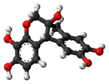
Alizarin is an organic compound with formula C
14
H
8
O
4
that has been used throughout history as a prominent red dye, principally for dyeing textile fabrics. Historically it was derived from the roots of plants of the madder genus. In 1869, it became the first natural dye to be produced synthetically.

<span class="mw-page-title-main">Romanowsky stain</span> Family of related stains for examination of blood including the detection of parasites

Romanowsky staining is a prototypical staining technique that was the forerunner of several distinct but similar stains widely used in hematology and cytopathology. Romanowsky-type stains are used to differentiate cells for microscopic examination in pathological specimens, especially blood and bone marrow films, and to detect parasites such as malaria within the blood.

<span class="mw-page-title-main">Mordant</span> Substance used for binding dyes to fabrics

A mordant or dye fixative is a substance used to set dyes on fabrics. It does this by forming a coordination complex with the dye, which then attaches to the fabric. It may be used for dyeing fabrics or for intensifying stains in cell or tissue preparations. Although mordants are still used, especially by small batch dyers, they have been largely displaced in industry by directs.

<span class="mw-page-title-main">Staining</span> Technique used to enhance visual contrast of specimens observed under a microscope

Staining is a technique used to enhance contrast in samples, generally at the microscopic level. Stains and dyes are frequently used in histology, in cytology, and in the medical fields of histopathology, hematology, and cytopathology that focus on the study and diagnoses of diseases at the microscopic level. Stains may be used to define biological tissues, cell populations, or organelles within individual cells.

<i>Haematoxylum campechianum</i> Species of plant

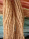
Haematoxylum campechianum is a species of flowering tree in the legume family, Fabaceae, that is native to southern Mexico,where it is known as Árbol de campeche, and introduced to the Caribbean, northern Central America, and other localities around the world. The tree was of great economic importance from the 17th century to the 19th century, when it was commonly logged and exported to Europe for use in dyeing fabrics. The modern nation of Belize developed from 17th- and 18th-century logging camps established by the English. The tree's scientific name means "bloodwood".

<span class="mw-page-title-main">Eosinophilic</span> Quality of being colored by eosin

Eosinophilic is the staining of tissues, cells, or organelles after they have been washed with eosin, a dye.

<span class="mw-page-title-main">Basophilic</span> Microscopic appearance of cells stained with a basic dye

Basophilic is a technical term used by pathologists. It describes the appearance of cells, tissues and cellular structures as seen through the microscope after a histological section has been stained with a basic dye. The most common such dye is haematoxylin.

<span class="mw-page-title-main">Crystal violet</span> Triarylmethane dye used as a histological stain and in Grams method of classifying bacteria

Crystal violet or gentian violet, also known as methyl violet 10B or hexamethyl pararosaniline chloride, is a triarylmethane dye used as a histological stain and in Gram's method of classifying bacteria. Crystal violet has antibacterial, antifungal, and anthelmintic (vermicide) properties and was formerly important as a topical antiseptic. The medical use of the dye has been largely superseded by more modern drugs, although it is still listed by the World Health Organization.

Trichrome staining is a histological staining method that uses two or more acid dyes in conjunction with a polyacid. Staining differentiates tissues by tinting them in contrasting colours. It increases the contrast of microscopic features in cells and tissues, which makes them easier to see when viewed through a microscope.

<span class="mw-page-title-main">Eosin Y</span> Chemical compound

Eosin Y, also called C.I. 45380 or C.I. Acid Red 87, is a member of the triarylmethane dyes. It is produced from fluorescein by bromination.

<span class="mw-page-title-main">Hematein</span> Chemical compound

Hematein or haematein is an oxidized derivative of haematoxylin, used in staining. Haematein should not be confused with haematin, which is a brown to black iron-containing pigment formed by decomposition of haemoglobin. In the Colour Index, haematein is called haematine.

<span class="mw-page-title-main">Papanicolaou stain</span> Histological staining method

Papanicolaou stain is a multichromatic (multicolored) cytological staining technique developed by George Papanicolaou in 1942. The Papanicolaou stain is one of the most widely used stains in cytology, where it is used to aid pathologists in making a diagnosis. Although most notable for its use in the detection of cervical cancer in the Pap test or Pap smear, it is also used to stain non-gynecological specimen preparations from a variety of bodily secretions and from small needle biopsies of organs and tissues. Papanicolaou published three formulations of this stain in 1942, 1954, and 1960.

<span class="mw-page-title-main">H&E stain</span> Histological stain method

Hematoxylin and eosin stain is one of the principal tissue stains used in histology. It is the most widely used stain in medical diagnosis and is often the gold standard. For example, when a pathologist looks at a biopsy of a suspected cancer, the histological section is likely to be stained with H&E.

<span class="mw-page-title-main">Phosphotungstic acid-haematoxylin stain</span> Biological stain used for staining of tissues

Phosphotungstic acid haematoxylin (PTAH) is a mix of haematoxylin with phosphotungstic acid, used in histology for staining.

<span class="mw-page-title-main">Phosphotungstic acid</span> Chemical compound

Phosphotungstic acid (PTA) or tungstophosphoric acid (TPA), is a heteropoly acid with the chemical formula H3PW12O40]. It forms hydrates H3[PW12O40nH2O. It is normally isolated as the n = 24 hydrate but can be desiccated to the hexahydrate (n = 6). EPTA is the name of ethanolic phosphotungstic acid, its alcohol solution used in biology. It has the appearance of small, colorless-grayish or slightly yellow-green crystals, with melting point 89 °C (24 H2O hydrate). It is odorless and soluble in water (200 g/100 ml). It is not especially toxic, but is a mild acidic irritant. The compound is known by a variety of names and acronyms (see 'other names' section of infobox).

<span class="mw-page-title-main">Brazilin</span> Chemical compound

Brazilin is a naturally occurring, a homoisoflavonoid, red dye obtained from the wood of Paubrasilia echinata, Biancaea sappan, Caesalpinia violacea, and Haematoxylum brasiletto. Brazilin has been used since at least the Middle Ages to dye fabric, and has been used to make paints and inks as well. The specific color produced by the pigment depends on its manner of preparation: in an acidic solution brazilin will appear yellow, but in an alkaline preparation it will appear red. Brazilin is closely related to the blue-black dye precursor hematoxylin, having one fewer hydroxyl group. Brazilein, the active dye agent, is an oxidized form of brazilin.

The von Kossa histological stain is used to quantify mineralization in cell culture and histological sections.

<span class="mw-page-title-main">Dmitri Leonidovich Romanowsky</span> Russian physician

Dmitri Leonidovich Romanowsky was a Russian physician who is best known for his invention of an eponymous histological stain called Romanowsky stain. It paved the way for the discovery and diagnosis of microscopic pathogens, such as malarial parasites, and later developments of new histological stains that became fundamental to microbiology and physiology.

References

  1. 1 2 3 4 5 6 7 8 9 10 11 12 13 14 15 16 17 18 19 Stevens, Alan (1982). "The Haematoxylins". In Bancroft, John; Stevens, Alan (eds.). The Theory and Practice of Histological Techniques (2nd ed.). Longman Group Limited. p. 109.
  2. 1 2 3 4 Lillie, Ralph Dougall (1977). H. J. Conn's Biological stains (9th ed.). Baltimore: Williams & Wilkins. pp. 692p.
  3. Mitchell, A. (1908). "English inks: their composition and differentiation in handwriting". Analyst. 33 (384): 80–85. Bibcode:1908Ana....33...80M. doi:10.1039/AN9083300080.
  4. 1 2 3 Barrow, William (1948). "Black Writing Ink of the Colonial Period". The American Archivist. 11 (4): 291–307. doi: 10.17723/aarc.11.4.903256p5lp2g3354 . ISSN   0360-9081.
  5. 1 2 Centeno, Silvia A.; Bronzato, Maddalena; Ropret, Polonca; et al. (2016). "Composition and spectroscopic properties of historic Cr logwood inks". Journal of Raman Spectroscopy. 47 (12): 1422–1428. Bibcode:2016JRSp...47.1422C. doi:10.1002/jrs.4938. ISSN   0377-0486.
  6. 1 2 3 4 Neevel, Johan (2003). "24: The Identification of Van Gogh's Inks for Drawing and Writing". In Vellekoop, Marije; Geldof, Muriel; Hendriks, Ella; Jansen, Leo; de Tagle, Alberto (eds.). Van Gogh's studio practice. Mercatorfonds. pp. 420–435. ISBN   9780300191875.
  7. 1 2 3 4 5 6 7 8 9 10 11 12 13 Titford, M. (2005). "The long history of hematoxylin". Biotechnic & Histochemistry. 80 (2): 73–80. doi:10.1080/10520290500138372. PMID   16195172. S2CID   20338201.
  8. 1 2 3 4 5 6 7 8 9 Ortiz-Hidalgo C, Pina-Oviedo S (2019). "Hematoxylin: Mesoamerica's Gift to Histopathology. Palo de Campeche (Logwood Tree), Pirates' Most Desired Treasure, and Irreplaceable Tissue Stain". Int J Surg Pathol. 27 (1): 4–14. doi: 10.1177/1066896918787652 . PMID   30001639.
  9. 1 2 3 4 5 6 7 8 9 10 11 Ponting, K. G. (1973). "Logwood: an interesting Dye". Journal of European Economic History. 2 (1): 109–119. ISSN   2499-8281.
  10. 1 2 3 4 5 Chan JK (2014). "The wonderful colors of the hematoxylin-eosin stain in diagnostic surgical pathology". Int J Surg Pathol. 22 (1): 12–32. doi:10.1177/1066896913517939. PMID   24406626. S2CID   26847314.
  11. 1 2 3 4 5 6 7 8 9 10 Llewellyn BD (2009). "Nuclear staining with alum hematoxylin". Biotech Histochem. 84 (4): 159–77. doi:10.1080/10520290903052899. PMID   19579146. S2CID   205713596.
  12. 1 2 3 4 5 6 7 Smith C (2006). "Our debt to the logwood tree: the history of hematoxylin". MLO Med Lab Obs. 38 (5): 18, 20–2. PMID   16761865.
  13. 1 2 Kiernan, J A (2006). "Dyes and other colorants in microtechnique and biomedical research". Coloration Technology. 122 (1): 1–21. doi: 10.1111/j.1478-4408.2006.00009.x . ISSN   1472-3581.
  14. 1 2 3 4 5 6 7 Dapson RW, Horobin RW (2009). "Dyes from a twenty-first century perspective". Biotech Histochem. 84 (4): 135–7. doi:10.1080/10520290902908802. PMID   19384743. S2CID   28563610.
  15. 1 2 3 Titford, Michael (2009). "Progress in the Development of Microscopical Techniques for Diagnostic Pathology". Journal of Histotechnology. 32 (1): 9–19. doi: 10.1179/his.2009.32.1.9 . ISSN   0147-8885. S2CID   26801839.
  16. Kahr, Bart; Lovell, Scott; Subramony, Anand (1998). "The progress of logwood extract". Chirality. 10 (1–2): 66–77. doi:10.1002/chir.12.
  17. Bettinger C, Zimmermann HW (1991). "New investigations on hematoxylin, hematein, and hematein-aluminium complexes. II. Hematein-aluminium complexes and hemalum staining". Histochemistry. 96 (3): 215–28. doi:10.1007/BF00271540. PMID   1717413. S2CID   23504301.
  18. 1 2 3 4 5 6 7 8 9 10 Dapson R, Horobin RW, Kiernan J (2010). "Hematoxylin shortages: their causes and duration, and other dyes that can replace hemalum in routine hematoxylin and eosin staining". Biotech Histochem. 85 (1): 55–63. doi:10.3109/10520290903048400. PMID   19562570. S2CID   7698557.
  19. Morsingh, F.; Robinson, R. (1970). "The syntheses of brazilin and haematoxylin". Tetrahedron. 26 (1): 281–289. doi:10.1016/0040-4020(70)85029-3.
  20. 1 2 3 Puchtler H, Meloan SN, Waldrop FS (1986). "Application of current chemical concepts to metal-hematein and -brazilein stains". Histochemistry. 85 (5): 353–64. doi:10.1007/BF00982665. PMID   2430916. S2CID   7384777.
  21. 1 2 Cooksey C (2010). "Hematoxylin and related compounds--an annotated bibliography concerning their origin, properties, chemistry, and certain applications". Biotech Histochem. 85 (1): 65–82. doi:10.3109/10520290903048418. PMID   19568968. S2CID   5297820.
  22. 1 2 3 Lillie RD (1974). "The hematoxylin shortage and the availability of synthetic substitutes". Am J Med Technol. 40 (11): 455–61. PMID   4139897.
  23. 1 2 3 Schulte EK (1991). "Standardization of biological dyes and stains: pitfalls and possibilities". Histochemistry. 95 (4): 319–28. doi:10.1007/BF00266958. PMID   1708749. S2CID   29628388.
  24. Marshall PN, Horobin RW (1974). "A simple assay procedure for mixtures of hematoxylin and hematein". Stain Technol. 49 (3): 137–42. doi:10.3109/10520297409116964. PMID   4135791.
  25. 1 2 Kiernan JA (2018). "Does progressive nuclear staining with hemalum (alum hematoxylin) involve DNA, and what is the nature of the dye-chromatin complex?". Biotech Histochem. 93 (2): 133–148. doi:10.1080/10520295.2017.1399466. PMID   29320873. S2CID   13481905.
  26. 1 2 3 4 5 6 7 Gatenby, J. B.; Beams, H. W. (1950). The Microtomist's Vade-Mecum (11th ed.). Philadelphia: The Blackstone Company.
  27. 1 2 Gill GW (2010). "Gill hematoxylins: first person account". Biotech Histochem. 85 (1): 7–18. doi:10.3109/10520290903048376. PMID   19657780. S2CID   207513639.
  28. Harris, H. F. (1900). "On the rapid conversion of haematoxylin into haematein in staining reactions". Journal of Applied Microscopic Laboratory Methods. 3 (3): 777.
  29. Gill, Gary W. (2013). "Papanicolaou Stain". Cytopreparation. Essentials in Cytopathology. Vol. 12. pp. 143–189. doi:10.1007/978-1-4614-4933-1_10. ISBN   978-1-4614-4932-4. ISSN   1574-9053.
  30. Cole, Elbert C. (1943). "Studies on Hematoxylin Stains". Stain Technology. 18 (3): 125–142. doi:10.3109/10520294309105804. ISSN   0038-9153.
  31. 1 2 3 4 5 Bracegirdle, Brian (1986). A history of microtechnique : the evolution of the microtome and the development of tissue preparation (2nd ed.). Lincolnwood, IL: Science Heritage Ltd. ISBN   978-0940095007.
  32. 1 2 Allison RT (1999). "Haematoxylin--from the wood". J Clin Pathol. 52 (7): 527–8. doi:10.1136/jcp.52.7.527. PMC   501496 . PMID   10605407.
  33. Quekett, John Thomas (1848). A Practical treatise on the use of the microscope. Library of illustrated standard scientific works. Vol. VI. Paris: Hippolyte Bailliere.
  34. 1 2 3 Mann, Gustav (1902). Physiological Histology, Methods and Theory. Clarendon Press. p.  488.
  35. 1 2 3 Cook HC (1997). "Origins of ... tinctorial methods in histology". J Clin Pathol. 50 (9): 716–20. doi:10.1136/jcp.50.9.716. PMC   500167 . PMID   9389971.
  36. Georg von Georgievics (1902). The Chemical Technology of Textile Fibres: Their Origin, Structure, Preparation, Washing, Bleaching, Dyeing, Printing and Dressing. Scott, Greenwood & Co. p.  180.
  37. 1 2 3 4 Centeno, Silvia A.; Ropret, Polonca; Federico, Eleonora Del; et al. (2010). "Characterization of Al(III) complexes with hematein in artistic alum logwood inks". Journal of Raman Spectroscopy. 41 (4): 445–451. Bibcode:2010JRSp...41..445C. doi:10.1002/jrs.2455. ISSN   0377-0486.