Histone H1

Last updated

linker histone H1 and H5 family
PBB Protein HIST1H1B image.jpg
PDB rendering of HIST1H1B based on 1ghc.
Identifiers
SymbolLinker_histone
Pfam PF00538
InterPro IPR005818
SMART SM00526
SCOP2 1hst / SCOPe / SUPFAM
Available protein structures:
Pfam   structures / ECOD  
PDB RCSB PDB; PDBe; PDBj
PDBsum structure summary
PDB 1ghc , 1hst , 1uhm , 1uss , 1ust , 1yqa

Histone H1 is one of the five main histone protein families which are components of chromatin in eukaryotic cells. Though highly conserved, it is nevertheless the most variable histone in sequence across species.

Contents

Structure

A diagram showing where H1 can be found in the nucleosome Nucleosome.jpg
A diagram showing where H1 can be found in the nucleosome

Metazoan H1 proteins feature a central globular "winged helix" domain and long C- and short N-terminal tails. H1 is involved with the packing of the "beads on a string" sub-structures into a high order structure, whose details have not yet been solved. [1] H1 found in protists and bacteria, otherwise known as nucleoproteins HC1 and HC2 (Pfam PF07432, PF07382), lack the central domain and the N-terminal tail. [2]

H1 is less conserved than core histones. The globular domain is the most conserved part of H1. [3]

Function

Unlike the other histones, H1 does not make up the nucleosome "bead". Instead, it sits on top of the structure, keeping in place the DNA that has wrapped around the nucleosome. H1 is present in half the amount of the other four histones, which contribute two molecules to each nucleosome bead. In addition to binding to the nucleosome, the H1 protein binds to the "linker DNA" (approximately 20-80 nucleotides in length) region between nucleosomes, helping stabilize the zig-zagged 30 nm chromatin fiber. [4] Much has been learned about histone H1 from studies on purified chromatin fibers. Ionic extraction of linker histones from native or reconstituted chromatin promotes its unfolding under hypotonic conditions from fibers of 30 nm width to beads-on-a-string nucleosome arrays. [5] [6] [7]

It is uncertain whether H1 promotes a solenoid-like chromatin fiber, in which exposed linker DNA is shortened, or whether it merely promotes a change in the angle of adjacent nucleosomes, without affecting linker length [8] However, linker histones have been demonstrated to drive the compaction of chromatin fibres that had been reconstituted in vitro using synthetic DNA arrays of the strong '601' nucleosome positioning element. [9] Nuclease digestion and DNA footprinting experiments suggest that the globular domain of histone H1 localizes near the nucleosome dyad, where it protects approximately 15-30 base pairs of additional DNA. [10] [11] [12] [13] In addition, experiments on reconstituted chromatin reveal a characteristic stem motif at the dyad in the presence of H1. [14] Despite gaps in our understanding, a general model has emerged wherein H1's globular domain closes the nucleosome by crosslinking incoming and outgoing DNA, while the tail binds to linker DNA and neutralizes its negative charge. [8] [12]

Many experiments addressing H1 function have been performed on purified, processed chromatin under low-salt conditions, but H1's role in vivo is less certain. Cellular studies have shown that overexpression of H1 can cause aberrant nuclear morphology and chromatin structure, and that H1 can serve as both a positive and negative regulator of transcription, depending on the gene. [15] [16] [17] In Xenopus egg extracts, linker histone depletion causes ~2-fold lengthwise extension of mitotic chromosomes, while overexpression causes chromosomes to hypercompact into an inseparable mass. [18] [19] Complete knockout of H1 in vivo has not been achieved in multicellular organisms due to the existence of multiple isoforms that may be present in several gene clusters, but various linker histone isoforms have been depleted to varying degrees in Tetrahymena, C. elegans, Arabidopsis, fruit fly, and mouse, resulting in various organism-specific defects in nuclear morphology, chromatin structure, DNA methylation, and/or specific gene expression. [20] [21] [22]

Dynamics

While most histone H1 in the nucleus is bound to chromatin, H1 molecules shuttle between chromatin regions at a fairly high rate. [23] [24]

It is difficult to understand how such a dynamic protein could be a structural component of chromatin, but it has been suggested that the steady-state equilibrium within the nucleus still strongly favors association between H1 and chromatin, meaning that despite its dynamics, the vast majority of H1 at any given timepoint is chromatin bound. [25] H1 compacts and stabilizes DNA under force and during chromatin assembly, which suggests that dynamic binding of H1 may provide protection for DNA in situations where nucleosomes need to be removed. [26]

Cytoplasmic factors appear to be necessary for the dynamic exchange of histone H1 on chromatin, but these have yet to be specifically identified. [27] H1 dynamics may be mediated to some degree by O-glycosylation and phosphorylation. O-glycosylation of H1 may promote chromatin condensation and compaction. Phosphorylation during interphase has been shown to decrease H1 affinity for chromatin and may promote chromatin decondensation and active transcription. However, during mitosis phosphorylation has been shown to increase the affinity of H1 for chromosomes and therefore promote mitotic chromosome condensation. [19]

Isoforms

The H1 family in animals includes multiple H1 isoforms that can be expressed in different or overlapping tissues and developmental stages within a single organism. The reason for these multiple isoforms remains unclear, but both their evolutionary conservation from sea urchin to humans as well as significant differences in their amino acid sequences suggest that they are not functionally equivalent. [28] [29] [3] One isoform is histone H5, which is only found in avian erythrocytes, which are unlike mammalian erythrocytes in that they have nuclei. Another isoform is the oocyte/zygotic H1M isoform (also known as B4 or H1foo), found in sea urchins, frogs, mice, and humans, which is replaced in the embryo by somatic isoforms H1A-E, and H10 which resembles H5. [3] [30] [31] [32] Despite having more negative charges than somatic isoforms, H1M binds with higher affinity to mitotic chromosomes in Xenopus egg extracts. [19]

Post-translational modifications

Like other histones, the histone H1 family is extensively post-translationally modified (PTMs). This includes serine and threonine phosphorylation, lysine acetylation, lysine methylation and ubiquitination. [33] These PTMs serve a variety of functions but are less well studied than the PTMs of other histones.

See also

Related Research Articles

Chromatin is a complex of DNA and protein found in eukaryotic cells. The primary function is to package long DNA molecules into more compact, denser structures. This prevents the strands from becoming tangled and also plays important roles in reinforcing the DNA during cell division, preventing DNA damage, and regulating gene expression and DNA replication. During mitosis and meiosis, chromatin facilitates proper segregation of the chromosomes in anaphase; the characteristic shapes of chromosomes visible during this stage are the result of DNA being coiled into highly condensed chromatin.

<span class="mw-page-title-main">Histone</span> Family proteins package and order the DNA into structural units called nucleosomes.

In biology, histones are highly basic proteins abundant in lysine and arginine residues that are found in eukaryotic cell nuclei. They act as spools around which DNA winds to create structural units called nucleosomes. Nucleosomes in turn are wrapped into 30-nanometer fibers that form tightly packed chromatin. Histones prevent DNA from becoming tangled and protect it from DNA damage. In addition, histones play important roles in gene regulation and DNA replication. Without histones, unwound DNA in chromosomes would be very long. For example, each human cell has about 1.8 meters of DNA if completely stretched out; however, when wound about histones, this length is reduced to about 90 micrometers (0.09 mm) of 30 nm diameter chromatin fibers.

<span class="mw-page-title-main">Nucleosome</span> Basic structural unit of DNA packaging in eukaryotes

A nucleosome is the basic structural unit of DNA packaging in eukaryotes. The structure of a nucleosome consists of a segment of DNA wound around eight histone proteins and resembles thread wrapped around a spool. The nucleosome is the fundamental subunit of chromatin. Each nucleosome is composed of a little less than two turns of DNA wrapped around a set of eight proteins called histones, which are known as a histone octamer. Each histone octamer is composed of two copies each of the histone proteins H2A, H2B, H3, and H4.

<span class="mw-page-title-main">Spindle apparatus</span> Feature of biological cell structure

In cell biology, the spindle apparatus refers to the cytoskeletal structure of eukaryotic cells that forms during cell division to separate sister chromatids between daughter cells. It is referred to as the mitotic spindle during mitosis, a process that produces genetically identical daughter cells, or the meiotic spindle during meiosis, a process that produces gametes with half the number of chromosomes of the parent cell.

Histone H2B is one of the 5 main histone proteins involved in the structure of chromatin in eukaryotic cells. Featuring a main globular domain and long N-terminal and C-terminal tails, H2B is involved with the structure of the nucleosomes.

The family of heterochromatin protein 1 (HP1) consists of highly conserved proteins, which have important functions in the cell nucleus. These functions include gene repression by heterochromatin formation, transcriptional activation, regulation of binding of cohesion complexes to centromeres, sequestration of genes to the nuclear periphery, transcriptional arrest, maintenance of heterochromatin integrity, gene repression at the single nucleosome level, gene repression by heterochromatization of euchromatin, and DNA repair. HP1 proteins are fundamental units of heterochromatin packaging that are enriched at the centromeres and telomeres of nearly all eukaryotic chromosomes with the notable exception of budding yeast, in which a yeast-specific silencing complex of SIR proteins serve a similar function. Members of the HP1 family are characterized by an N-terminal chromodomain and a C-terminal chromoshadow domain, separated by a hinge region. HP1 is also found at some euchromatic sites, where its binding can correlate with either gene repression or gene activation. HP1 was originally discovered by Tharappel C James and Sarah Elgin in 1986 as a factor in the phenomenon known as position effect variegation in Drosophila melanogaster.

<span class="mw-page-title-main">Solenoid (DNA)</span>

The solenoid structure of chromatin is a model for the structure of the 30 nm fibre. It is a secondary chromatin structure which helps to package eukaryotic DNA into the nucleus.

<span class="mw-page-title-main">HIST1H3B</span>

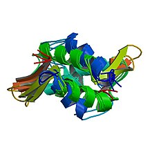
Histone H3.1 is a protein that in humans is encoded by the HIST1H3B gene.

<span class="mw-page-title-main">H2AFY</span> Protein-coding gene in the species Homo sapiens

Core histone macro-H2A.1 is a protein that in humans is encoded by the H2AFY gene.

<span class="mw-page-title-main">HMGN1</span> Protein-coding gene in the species Homo sapiens

Non-histone chromosomal protein HMG-14 is a protein that in humans is encoded by the HMGN1 gene.

<span class="mw-page-title-main">HIST1H1A</span>

Histone H1.1 is a protein that in humans is encoded by the HIST1H1A gene.

<span class="mw-page-title-main">Histone H3.1</span>

Histone H3.1 is a protein in humans that is encoded by the H3C1 gene.

<span class="mw-page-title-main">HIST1H1E</span>

Histone H1.4 is a protein that in humans is encoded by the HIST1H1E gene.

<span class="mw-page-title-main">HIST1H3F</span>

Histone H3.1 is a protein that in humans is encoded by the HIST1H3F gene.

<span class="mw-page-title-main">HIST1H1D</span>

Histone H1.3 is a protein that in humans is encoded by the HIST1H1D gene.

<span class="mw-page-title-main">H1FX</span>

Histone H1x is a protein that in humans is encoded by the H1FX gene.

<span class="mw-page-title-main">H2AFJ</span> Protein-coding gene in humans

Histone H2A.J is a protein that in humans is encoded by the H2AFJ gene.

<span class="mw-page-title-main">H2AFB1</span> Protein-coding gene in the species Homo sapiens

Histone H2A-Bbd type 1 also known as H2A Barr body-deficient is a histone protein variant that in humans is encoded by the H2AFB1 gene.

<span class="mw-page-title-main">H2AFB2</span> Protein-coding gene in the species Homo sapiens

Histone H2A-Bbd type 2/3 also known as H2A Barr body-deficient is a histone protein that in humans is encoded by the H2AFB2 gene.

<span class="mw-page-title-main">Linker histone H1 variants</span> Protein family which binds DNA wrapped around the core in a nucleosome

In molecular biology, the linker histone H1 is a protein family forming a critical component of eukaryotic chromatin. H1 histones bind to the linker DNA exiting from the nucleosome core particle, while the core histones form the octamer core of the nucleosome around which the DNA is wrapped.

References

  1. Ramakrishnan V, Finch JT, Graziano V, Lee PL, Sweet RM (March 1993). "Crystal structure of globular domain of histone H5 and its implications for nucleosome binding". Nature. 362 (6417): 219–23. Bibcode:1993Natur.362..219R. doi:10.1038/362219a0. PMID   8384699. S2CID   4301198.
  2. Kasinsky HE, Lewis JD, Dacks JB, Ausió J (January 2001). "Origin of H1 linker histones". FASEB Journal. 15 (1): 34–42. doi:10.1096/fj.00-0237rev. PMID   11149891. S2CID   10089116.
  3. 1 2 3 Izzo A, Kamieniarz K, Schneider R (April 2008). "The histone H1 family: specific members, specific functions?". Biological Chemistry. 389 (4): 333–43. doi:10.1515/BC.2008.037. PMID   18208346. S2CID   1516241.
  4. Jeon, Kwang W.; Berezney, Ronald (1995). Structural and functional organization of the nuclear matrix . Boston: Academic Press. pp.  214–7. ISBN   978-0-12-364565-4.
  5. Finch JT, Klug A (June 1976). "Solenoidal model for superstructure in chromatin". Proceedings of the National Academy of Sciences of the United States of America. 73 (6): 1897–901. Bibcode:1976PNAS...73.1897F. doi: 10.1073/pnas.73.6.1897 . PMC   430414 . PMID   1064861.
  6. Thoma F, Koller T (September 1977). "Influence of histone H1 on chromatin structure" (PDF). Cell. 12 (1): 101–7. doi:10.1016/0092-8674(77)90188-X. PMID   561660. S2CID   11155120.
  7. Thoma F, Koller T, Klug A (November 1979). "Involvement of histone H1 in the organization of the nucleosome and of the salt-dependent superstructures of chromatin". The Journal of Cell Biology. 83 (2 Pt 1): 403–27. doi:10.1083/jcb.83.2.403. PMC   2111545 . PMID   387806.
  8. 1 2 van Holde K, Zlatanova J (October 1996). "What determines the folding of the chromatin fiber?". Proceedings of the National Academy of Sciences of the United States of America. 93 (20): 10548–55. Bibcode:1996PNAS...9310548V. doi: 10.1073/pnas.93.20.10548 . PMC   38190 . PMID   8855215.
  9. Routh A, Sandin S, Rhodes D (July 2008). "Nucleosome repeat length and linker histone stoichiometry determine chromatin fiber structure". Proceedings of the National Academy of Sciences of the United States of America. 105 (26): 8872–7. Bibcode:2008PNAS..105.8872R. doi: 10.1073/pnas.0802336105 . PMC   2440727 . PMID   18583476.
  10. Varshavsky AJ, Bakayev VV, Georgiev GP (February 1976). "Heterogeneity of chromatin subunits in vitro and location of histone H1". Nucleic Acids Research. 3 (2): 477–92. doi:10.1093/nar/3.2.477. PMC   342917 . PMID   1257057.
  11. Whitlock JP, Simpson RT (July 1976). "Removal of histone H1 exposes a fifty base pair DNA segment between nucleosomes". Biochemistry. 15 (15): 3307–14. doi:10.1021/bi00660a022. PMID   952859.
  12. 1 2 Allan J, Hartman PG, Crane-Robinson C, Aviles FX (December 1980). "The structure of histone H1 and its location in chromatin". Nature. 288 (5792): 675–9. Bibcode:1980Natur.288..675A. doi:10.1038/288675a0. PMID   7453800. S2CID   4262304.
  13. Staynov DZ, Crane-Robinson C (December 1988). "Footprinting of linker histones H5 and H1 on the nucleosome". The EMBO Journal. 7 (12): 3685–91. doi:10.1002/j.1460-2075.1988.tb03250.x. PMC   454941 . PMID   3208745.
  14. Bednar J, Horowitz RA, Grigoryev SA, Carruthers LM, Hansen JC, Koster AJ, Woodcock CL (November 1998). "Nucleosomes, linker DNA, and linker histone form a unique structural motif that directs the higher-order folding and compaction of chromatin". Proceedings of the National Academy of Sciences of the United States of America. 95 (24): 14173–8. Bibcode:1998PNAS...9514173B. doi: 10.1073/pnas.95.24.14173 . PMC   24346 . PMID   9826673.
  15. Dworkin-Rastl E, Kandolf H, Smith RC (February 1994). "The maternal histone H1 variant, H1M (B4 protein), is the predominant H1 histone in Xenopus pregastrula embryos". Developmental Biology. 161 (2): 425–39. doi:10.1006/dbio.1994.1042. PMID   8313993.
  16. Brown DT, Alexander BT, Sittman DB (February 1996). "Differential effect of H1 variant overexpression on cell cycle progression and gene expression". Nucleic Acids Research. 24 (3): 486–93. doi:10.1093/nar/24.3.486. PMC   145659 . PMID   8602362.
  17. Gunjan A, Alexander BT, Sittman DB, Brown DT (December 1999). "Effects of H1 histone variant overexpression on chromatin structure". The Journal of Biological Chemistry. 274 (53): 37950–6. doi: 10.1074/jbc.274.53.37950 . PMID   10608862.
  18. Maresca TJ, Freedman BS, Heald R (June 2005). "Histone H1 is essential for mitotic chromosome architecture and segregation in Xenopus laevis egg extracts". The Journal of Cell Biology. 169 (6): 859–69. doi:10.1083/jcb.200503031. PMC   2171634 . PMID   15967810.
  19. 1 2 3 Freedman BS, Heald R (June 2010). "Functional comparison of H1 histones in Xenopus reveals isoform-specific regulation by Cdk1 and RanGTP". Current Biology. 20 (11): 1048–52. doi:10.1016/j.cub.2010.04.025. PMC   2902237 . PMID   20471264.
  20. Shen X, Yu L, Weir JW, Gorovsky MA (July 1995). "Linker histones are not essential and affect chromatin condensation in vivo". Cell. 82 (1): 47–56. doi: 10.1016/0092-8674(95)90051-9 . PMID   7606784. S2CID   14427681.
  21. Jedrusik MA, Schulze E (April 2001). "A single histone H1 isoform (H1.1) is essential for chromatin silencing and germline development in Caenorhabditis elegans". Development. 128 (7): 1069–80. doi:10.1242/dev.128.7.1069. PMID   11245572.
  22. Lu X, Wontakal SN, Emelyanov AV, Morcillo P, Konev AY, Fyodorov DV, Skoultchi AI (February 2009). "Linker histone H1 is essential for Drosophila development, the establishment of pericentric heterochromatin, and a normal polytene chromosome structure". Genes & Development. 23 (4): 452–65. doi:10.1101/gad.1749309. PMC   2648648 . PMID   19196654.
  23. Misteli T, Gunjan A, Hock R, Bustin M, Brown DT (December 2000). "Dynamic binding of histone H1 to chromatin in living cells". Nature. 408 (6814): 877–81. Bibcode:2000Natur.408..877M. doi:10.1038/35048610. PMID   11130729. S2CID   4428934.
  24. Chen D, Dundr M, Wang C, Leung A, Lamond A, Misteli T, Huang S (January 2005). "Condensed mitotic chromatin is accessible to transcription factors and chromatin structural proteins". The Journal of Cell Biology. 168 (1): 41–54. doi:10.1083/jcb.200407182. PMC   2171683 . PMID   15623580.
  25. Bustin M, Catez F, Lim JH (March 2005). "The dynamics of histone H1 function in chromatin". Molecular Cell. 17 (5): 617–20. doi: 10.1016/j.molcel.2005.02.019 . PMID   15749012.
  26. Xiao B, Freedman BS, Miller KE, Heald R, Marko JF (December 2012). "Histone H1 compacts DNA under force and during chromatin assembly". Molecular Biology of the Cell. 23 (24): 4864–71. doi:10.1091/mbc.E12-07-0518. PMC   3521692 . PMID   23097493.
  27. Freedman BS, Miller KE, Heald R (September 2010). Cimini D (ed.). "Xenopus egg extracts increase dynamics of histone H1 on sperm chromatin". PLOS ONE. 5 (9): e13111. Bibcode:2010PLoSO...513111F. doi: 10.1371/journal.pone.0013111 . PMC   2947519 . PMID   20927327.
  28. Steinbach OC, Wolffe AP, Rupp RA (September 1997). "Somatic linker histones cause loss of mesodermal competence in Xenopus". Nature. 389 (6649): 395–9. Bibcode:1997Natur.389..395S. doi:10.1038/38755. PMID   9311783. S2CID   4390287.
  29. De S, Brown DT, Lu ZH, Leno GH, Wellman SE, Sittman DB (June 2002). "Histone H1 variants differentially inhibit DNA replication through an affinity for chromatin mediated by their carboxyl-terminal domains". Gene. 292 (1–2): 173–81. doi:10.1016/S0378-1119(02)00675-3. PMID   12119111.
  30. Khochbin S (June 2001). "Histone H1 diversity: bridging regulatory signals to linker histone function". Gene. 271 (1): 1–12. doi:10.1016/S0378-1119(01)00495-4. PMID   11410360.
  31. Godde JS, Ura K (March 2008). "Cracking the enigmatic linker histone code". Journal of Biochemistry. 143 (3): 287–93. doi:10.1093/jb/mvn013. PMID   18234717.
  32. Happel N, Doenecke D (February 2009). "Histone H1 and its isoforms: contribution to chromatin structure and function". Gene. 431 (1–2): 1–12. doi:10.1016/j.gene.2008.11.003. PMID   19059319.
  33. Harshman SW, Young NL, Parthun MR, Freitas MA (November 2013). "H1 histones: current perspectives and challenges". Nucleic Acids Research. 41 (21): 9593–609. doi:10.1093/nar/gkt700. PMC   3834806 . PMID   23945933.