Horseshoe kidney

Last updated
Horseshoe kidney
Other namesRenal fusion
Specialty Nephrology

Horseshoe kidney, also known as ren arcuatus (in Latin), renal fusion or super kidney, is a congenital disorder affecting about 1 in 500 people that is more common in men, often asymptomatic, and usually diagnosed incidentally. [1] [2] In this disorder, the patient's kidneys fuse to form a horseshoe-shape during development in the womb. The fused part is the isthmus of the horseshoe kidney. The abnormal anatomy can affect kidney drainage resulting in increased frequency of kidney stones and urinary tract infections as well as increase risk of certain renal cancers. [1]

Contents

Fusion abnormalities of the kidney can be categorized into two groups: horseshoe kidney and crossed fused ectopia. The 'horseshoe kidney' is the most common renal fusion anomaly. [3]

Signs and symptoms

Axial CT image of the abdomen showing a horseshoe kidney. Hufeisenniere CT axial.jpg
Axial CT image of the abdomen showing a horseshoe kidney.

Although often asymptomatic, the most common presenting symptom of patients with a horseshoe kidney is abdominal or flank pain. However, presentation is often non-specific. [1] Approximately a third of patients with horseshoe kidneys remain asymptomatic throughout their entire life with over 50% of patients having no medical issues related to their renal fusion when followed for a 25-year period. [1] As a result, it is estimated that approximately 25% of patients with horseshoe kidneys are diagnosed incidentally with ultrasound or CT imaging. [1]

Associated conditions

Patients with a horseshoe kidney can develop sequelae related to the abnormal anatomy and present with symptoms related to them.[ citation needed ]

Individuals that have a horseshoe kidney may be at increased risk for conditions that generally fall within the following categories:

Intravenous pyelogram showing horseshoe kidney. Medical X-Ray imaging CNO03 nevit.jpg
Intravenous pyelogram showing horseshoe kidney.

Cause

There have been several proposed factors that may contribute to the development of a horseshoe kidney. Different exposures to the developing fetus such as different teratogens (e.g. thalidomide, ethanol, ACE inhibitors, cocaine, gentamicin, corticosteroids, NSAIDs, and vitamin A) have been hypothesized. [8] [2] [9] Impairment of a developing embryo's nephrogenic cell migration or abnormal migration of the kidneys due to fetal structural abnormalities is another potential factor. [8] [2] However, no definitive genetic cause has been identified. [8] [9]

Pathophysiology

Kidneys are normally located in the retroperitoneal space between the T12 and L3 vertebrae after ascending from the pelvis during development to rest underneath the adrenal glands. [1] In patients with this condition, the horseshoe kidney ascent is commonly arrested by the inferior mesenteric artery due to the central fusion of the kidneys. [10] However, this is present in only 40% of cases, and, in 20% of cases, the fused kidney remains in the pelvis. [1] Its ascension may also be restricted by its own renal artery. [11] Additionally, during normal development, the kidneys undergo a 90 degree medial rotation while ascending. However, due to the renal fusion, this rotation is impaired resulting in abnormal placement of the ureters. This in turn can lead to urinary stasis and drainage issues. [1] Furthermore, approximately 70% of kidneys in normal individuals are supplied by a single renal artery with the remaining 30% having embryonic collateral or accessory arteries. [1] With horseshoe kidneys, the majority are supplied by derivatives of the abdominal aorta or common illiac arteries depending on the final position of the kidneys. [1] [12]

Diagnosis

Horseshoe kidneys are commonly diagnosed incidentally on abdominal imaging. The diagnosis can be made with many different imaging modalities such as ultrasound, intravenous pyelogram, CT, and MRI. [1]

Common features that can be found on imaging include:

Treatment

Symphysiotomy, which involves separating the fused isthmus in order to release the kidneys, used to be a recommended treatment for this condition but has fallen out of favor due to complications and minimal benefit. [1] [15] Furthermore, kidneys can remain in their original abnormal location after the surgery. [1] [16] Instead, management focuses on treating the sequelae should the patient become symptomatic.[ citation needed ]

While treatment typically does not differ from that of patients with normal kidney anatomy, [17] kidney stones can warrant a different approach. Extracorporeal shockwave lithotripsy, a possible treatment for kidney stones, can be less effective in patients with horseshoe kidneys due to the abnormal anatomy causing difficulties with localizing the energy to the stones. Also, due to the kidney obstruction that can commonly occur with this renal fusion, clearance of the resulting stone fragments can also be impaired. [1] For this reason, prior to any treatment with shockwave lithotripsy, a UPJ obstruction must first be ruled out as it significantly impair successful treatment. [4] For stones that are less than 1.5 cm, ureteroscopy and shockwave lithotripsy can be first utilized. [4] For stones larger than 1.5 cm or when previous treatment has failed, the stones can instead be removed through a minimally invasive procedure known as percutaneous nephrolithotomy. [4]

Compared to patients with normal kidneys, patients with horseshoe kidneys who undergo treatment with percutaneous nephrolithotomy experience no difference in complications or stone clearance. [4]

Patients will also typically require imaging before any abdominal surgery as the vascular supply to the abnormal kidney can be highly variable between patients. [1] Additionally, the horseshoe kidneys can have a close association with colon which can increase risk of bowel injury. [1]

Epidemiology

There is an incidence of 1 in every 500 individuals within a normal population. [1] [2]

Males are more likely to develop a horseshoe kidney with a preponderance of 2:1. [1] [2]

Certain genetic diseases can predispose patients to developing a horseshoe kidney:

Notable cases

Related Research Articles

<span class="mw-page-title-main">Kidney</span> Organ that filters blood and produces urine

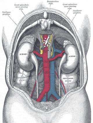
In humans, the kidneys are two reddish-brown bean-shaped blood-filtering organs that are a multilobar, multipapillary form of mammalian kidneys, usually without signs of external lobulation. They are located on the left and right in the retroperitoneal space, and in adult humans are about 12 centimetres in length. They receive blood from the paired renal arteries; blood exits into the paired renal veins. Each kidney is attached to a ureter, a tube that carries excreted urine to the bladder.

<span class="mw-page-title-main">Kidney stone disease</span> Formation of mineral stones in the urinary tract

Kidney stone disease, also known as renal calculus disease, nephrolithiasis or urolithiasis, is a crystallopathy where a solid piece of material develops in the urinary tract. Renal calculi typically form in the kidney and leave the body in the urine stream. A small calculus may pass without causing symptoms. If a stone grows to more than 5 millimeters, it can cause blockage of the ureter, resulting in sharp and severe pain in the lower back or abdomen. A calculus may also result in blood in the urine, vomiting, or painful urination. About half of people who have had a renal calculus are likely to have another within ten years.

<span class="mw-page-title-main">Prune belly syndrome</span> Medical condition

Prune belly syndrome is a rare, genetic birth defect affecting about 1 in 40,000 births. About 97% of those affected are male. Prune belly syndrome is a congenital disorder of the urinary system, characterized by a triad of symptoms. The syndrome is named for the mass of wrinkled skin that is often present on the abdomen of those with the disorder.

The excretory system is a passive biological system that removes excess, unnecessary materials from the body fluids of an organism, so as to help maintain internal chemical homeostasis and prevent damage to the body. The dual function of excretory systems is the elimination of the waste products of metabolism and to drain the body of used up and broken down components in a liquid and gaseous state. In humans and other amniotes, most of these substances leave the body as urine and to some degree exhalation, mammals also expel them through sweating.

<span class="mw-page-title-main">Ureter</span> Tubes used in the urinary system in most animals

The ureters are tubes made of smooth muscle that propel urine from the kidneys to the urinary bladder. In a human adult, the ureters are usually 20–30 cm (8–12 in) long and around 3–4 mm (0.12–0.16 in) in diameter. The ureter is lined by urothelial cells, a type of transitional epithelium, and has an additional smooth muscle layer that assists with peristalsis in its lowest third.

<span class="mw-page-title-main">Renal calyx</span> Anatomical structure in the kidneys

The renal calyces are conduits in the kidney through which urine passes. The minor calyces form a cup-shaped drain around the apex of the renal pyramids. Urine formed in the kidney passes through a renal papilla at the apex into the minor calyx; 4-5 minor calyces converge to form a major calyx through which urine passes into the renal pelvis.

<span class="mw-page-title-main">Ureterocele</span> Medical condition

A ureterocele is a congenital abnormality found in the ureter. In this condition the distal ureter balloons at its opening into the bladder, forming a sac-like pouch. It is most often associated with a duplicated collection system, where two ureters drain their respective kidney instead of one. Simple ureterocele, where the condition involves only a single ureter, represents only twenty percent of cases.

<span class="mw-page-title-main">Pyelonephritis</span> Medical condition

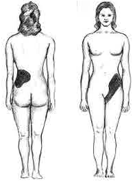
Pyelonephritis is inflammation of the kidney, typically due to a bacterial infection. Symptoms most often include fever and flank tenderness. Other symptoms may include nausea, burning with urination, and frequent urination. Complications may include pus around the kidney, sepsis, or kidney failure.

<span class="mw-page-title-main">Renal artery</span> Vessel supplying blood to kidney

The renal arteries are paired arteries that supply the kidneys with blood. Each is directed across the crus of the diaphragm, so as to form nearly a right angle.

<span class="mw-page-title-main">Hydronephrosis</span> Medical condition

Hydronephrosis describes hydrostatic dilation of the renal pelvis and calyces as a result of obstruction to urine flow downstream. Alternatively, hydroureter describes the dilation of the ureter, and hydronephroureter describes the dilation of the entire upper urinary tract.

<span class="mw-page-title-main">Renal colic</span> Medical condition

Renal colic, also known as ureteric colic, is a type of abdominal pain commonly caused by obstruction of ureter from dislodged kidney stones. The most frequent site of obstruction is the vesico-ureteric junction (VUJ), the narrowest point of the upper urinary tract. Acute obstruction and the resultant urinary stasis can distend the ureter (hydroureter) and cause a reflexive peristaltic smooth muscle spasm, which leads to a very intense visceral pain transmitted via the ureteric plexus.

<span class="mw-page-title-main">Vesicoureteral reflux</span> Medical condition

Vesicoureteral reflux (VUR), also known as vesicoureteric reflux, is a condition in which urine flows retrograde, or backward, from the bladder into one or both ureters and then to the renal calyx or kidneys. Urine normally travels in one direction from the kidneys to the bladder via the ureters, with a one-way valve at the vesicoureteral (ureteral-bladder) junction preventing backflow. The valve is formed by oblique tunneling of the distal ureter through the wall of the bladder, creating a short length of ureter (1–2 cm) that can be compressed as the bladder fills. Reflux occurs if the ureter enters the bladder without sufficient tunneling, i.e., too "end-on".

Pyelogram is a form of imaging of the renal pelvis and ureter.

<span class="mw-page-title-main">Ovarian vein syndrome</span> Medical condition

Ovarian vein syndrome is a rare condition in which a dilated ovarian vein compresses the ureter. This causes chronic or colicky abdominal pain, back pain and/or pelvic pain. The pain can worsen on lying down or between ovulation and menstruation. There can also be an increased tendency towards urinary tract infection or pyelonephritis. The right ovarian vein is most commonly involved, although the disease can be left-sided or affect both sides. It is currently classified as a form of pelvic congestion syndrome.

Urologic diseases or conditions include urinary tract infections, kidney stones, bladder control problems, and prostate problems, among others. Some urologic conditions do not affect a person for that long and some are lifetime conditions. Kidney diseases are normally investigated and treated by nephrologists, while the specialty of urology deals with problems in the other organs. Gynecologists may deal with problems of incontinence in women.

Bladder outlet obstruction occurs when urine is unable to flow from the kidneys through the ureters and out of the bladder through the urethra. Decreased flow of urine leads to swelling of the urinary tract, called hydronephrosis. This process of decreased flow of urine through the urinary tract can begin as early as during intrauterine life and it prevents normal development of fetal kidneys and fetal urine. Low levels of fetal urine leads to low amniotic fluid levels and incomplete lung maturation. Older children and adults can also experience bladder outlet obstruction; however, this process is usually reversible and isn't associated with as many poor outcomes as in infants with congenital bladder outlet obstruction.

<span class="mw-page-title-main">Fraley syndrome</span> Medical condition

Fraley syndrome is a condition where the superior infundibulum of the upper calyx of the kidney is obstructed by the crossing renal artery branch, causing distension and dilatation of the calyx and presenting clinically as haematuria and nephralgia. Furthermore, when the renal artery obstructs the proximal collecting system, filling defects can occur anywhere in the calyces, pelvis, or ureter.

<span class="mw-page-title-main">Ureteral cancer</span> Medical condition

Ureteral cancer is cancer of the ureters, muscular tubes that propel urine from the kidneys to the urinary bladder. It is also known as ureter cancer, renal pelvic cancer, and rarely ureteric cancer or uretal cancer. Cancer in this location is rare. Ureteral cancer becomes more likely in older adults, usually ages 70–80, who have previously been diagnosed with bladder cancer.

<span class="mw-page-title-main">Renal ultrasonography</span> Examination of one or both kidneys using medical ultrasound

Renal ultrasonography is the examination of one or both kidneys using medical ultrasound.

Ureteric stricture (ureteral stricture) is the pathological narrowing of the ureter which may lead to serious complications such as kidney failure.

References

  1. 1 2 3 4 5 6 7 8 9 10 11 12 13 14 15 16 17 18 19 20 21 22 23 24 25 Kirkpatrick JJ, Leslie SW (2018). Horseshoe Kidney. StatPearls Publishing. PMID   28613757 . Retrieved 2019-01-16.{{cite book}}: |work= ignored (help)
  2. 1 2 3 4 5 6 7 8 Taghavi K, Kirkpatrick J, Mirjalili SA (October 2016). "The horseshoe kidney: Surgical anatomy and embryology". Journal of Pediatric Urology. 12 (5): 275–280. doi:10.1016/j.jpurol.2016.04.033. PMID   27324557.
  3. Glodny B, Petersen J, Hofmann KJ, Schenk C, Herwig R, Trieb T, et al. (January 2009). "Kidney fusion anomalies revisited: clinical and radiological analysis of 209 cases of crossed fused ectopia and horseshoe kidney". BJU International. 103 (2): 224–35. doi:10.1111/j.1464-410X.2008.07912.x. PMID   18710445.
  4. 1 2 3 4 5 6 Wein, Alan J.; Kavoussi, Louis R.; Partin, Alan W.; Peters, Craig Andrew (2015-10-23). Campbell-Walsh Urology (Eleventh ed.). Philadelphia, PA. ISBN   9780323263740. OCLC   931870910.{{cite book}}: CS1 maint: location missing publisher (link)
  5. Jung M, Rai A, Wang L, Puttmann K, Kukreja K, Koh CJ (October 2018). "Nephrolithiasis in a 17-Year-Old Male With Seckel Syndrome and Horseshoe Kidneys: Case Report and Review of the Literature". Urology. 120: 241–243. doi:10.1016/j.urology.2018.05.023. PMID   29894776. S2CID   48353132.
  6. Pawar AS, Thongprayoon C, Cheungpasitporn W, Sakhuja A, Mao MA, Erickson SB (January 2018). "Incidence and characteristics of kidney stones in patients with horseshoe kidney: A systematic review and meta-analysis". Urology Annals. 10 (1): 87–93. doi: 10.4103/UA.UA_76_17 . PMC   5791465 . PMID   29416282.
  7. 1 2 3 4 5 Shah HU, Ojili V (2017). "Multimodality imaging spectrum of complications of horseshoe kidney". The Indian Journal of Radiology & Imaging. 27 (2): 133–140. doi: 10.4103/ijri.IJRI_298_16 . PMC   5510309 . PMID   28744072.
  8. 1 2 3 Kirkpatrick JJ, Joshua J.; Leslie SW, Stephen W. (2021). Horseshoe Kidney. StatPearls [Internet]: Treasure Island (FL): StatPearls Publishing. PMID   28613757.
  9. 1 2 Woolf AS, Winyard PJ, Hermanns MH, Welham SJ (2003). "Maldevelopment of the human kidney and lower urinary tract: an overview.". In Vize PD, Woolf AS, Bard JB (eds.). In The kidney. Academic Press. pp. 377–393. doi:10.1016/b978-012722441-1/50023-3. ISBN   9780127224411.
  10. Oktem H, Gozil R, Calguner E, Bahcelioglu M, Mutlu S, Kurkcuoglu A, et al. (2008). "Morphometric study of a horseshoe kidney". Medical Principles and Practice. 17 (1): 80–3. doi:10.1159/000109596. PMID   18059107.
  11. Suwannakhan A, Meemon K (2019-05-28). "Horseshoe kidney with extrarenal calyces and malformed renal vessels". European Journal of Anatomy. 20 (4): 355–359.
  12. 1 2 Natsis K, Piagkou M, Skotsimara A, Protogerou V, Tsitouridis I, Skandalakis P (August 2014). "Horseshoe kidney: a review of anatomy and pathology". Surgical and Radiologic Anatomy. 36 (6): 517–26. doi:10.1007/s00276-013-1229-7. PMID   24178305. S2CID   7889789.
  13. 1 2 3 4 5 Gutiérrez M (2013). "Renal anomalies of position, shape and fusion: radiographic analysis" (PDF). Revista de la Federación Ecuatoriana de Radiología. 6: 24–30.
  14. Nahm AM, Ritz E (November 1999). "Horseshoe kidney". Nephrology, Dialysis, Transplantation. 14 (11): 2740–1. doi: 10.1093/ndt/14.11.2740 . PMID   10534525.
  15. Oderda M, Calleris G, Allasia M, Dalmasso E, Falcone M, Catti M, et al. (February 2017). "Robot-assisted laparoscopic pyeloplasty in a pediatric patient with horseshoe kidney: surgical technique and review of the literature". Urologia. 84 (1): 55–60. doi:10.5301/uro.5000188. PMID   27516351. S2CID   25468419.
  16. Pitts WR, Muecke EC (June 1975). "Horseshoe kidneys: a 40-year experience". The Journal of Urology. 113 (6): 743–6. doi:10.1016/S0022-5347(17)59571-3. PMID   1152146.
  17. Al Otay A, Sarhan O, El-Tholoth HS, Alhelaly A, Al Akrash H, Al Ghanbar M, et al. (July 2018). "Different managements of horseshoe kidney stones, any difference in the outcome?". Urology Annals. 10 (3): 287–290. doi: 10.4103/UA.UA_116_17 . PMC   6060601 . PMID   30089987.
  18. "Renal Pathology" . Retrieved 2008-11-26.
  19. Cereda A, Carey JC (October 2012). "The trisomy 18 syndrome". Orphanet Journal of Rare Diseases. 7 (1): 81. doi: 10.1186/1750-1172-7-81 . PMC   3520824 . PMID   23088440.
  20. Kleta R, Brämswig JH (July 2000). "Horseshoe kidney and Turner syndrome". Nephrology, Dialysis, Transplantation. 15 (7): 1094. doi: 10.1093/ndt/15.7.1094-b . PMID   10862660.
  21. Ranke MB, Saenger P (July 2001). "Turner's syndrome". Lancet. 358 (9278): 309–14. doi:10.1016/S0140-6736(01)05487-3. PMID   11498234. S2CID   42096888.
  22. Niamien-Attai C, Bacchetta J, Ranchin B, Sanlaville D, Cochat P (October 2017). "[Renal abnormalities in Down syndrome: A review]". Archives de Pédiatrie. 24 (10): 1013–1018. doi:10.1016/j.arcped.2017.07.014. PMID   28893484.
  23. Parkinson. "Mel Gibson Interview - part one". BBC.
  24. Paget ET. "Autopsy Report: Sam Kinison" (PDF). Autopsy Files.