
Anemia or anaemia is a blood disorder in which the blood has a reduced ability to carry oxygen due to a lower than normal number of red blood cells, or a reduction in the amount of hemoglobin. The name is derived from Ancient Greek: ἀναιμία anaimia, meaning 'lack of blood', from ἀν- an-, 'not' and αἷμα haima, 'blood'. When anemia comes on slowly, the symptoms are often vague, such as tiredness, weakness, shortness of breath, headaches, and a reduced ability to exercise. When anemia is acute, symptoms may include confusion, feeling like one is going to pass out, loss of consciousness, and increased thirst. Anemia must be significant before a person becomes noticeably pale. Symptoms of anemia depend on how quickly hemoglobin decreases. Additional symptoms may occur depending on the underlying cause. Preoperative anemia can increase the risk of needing a blood transfusion following surgery. Anemia can be temporary or long term and can range from mild to severe.
Neutropenia is an abnormally low concentration of neutrophils in the blood. Neutrophils make up the majority of circulating white blood cells and serve as the primary defense against infections by destroying bacteria, bacterial fragments and immunoglobulin-bound viruses in the blood. People with neutropenia are more susceptible to bacterial infections and, without prompt medical attention, the condition may become life-threatening.
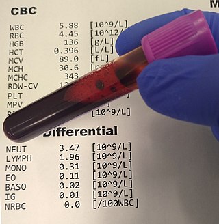
A complete blood count (CBC), also known as a full blood count (FBC), is a set of medical laboratory tests that provide information about the cells in a person's blood. The CBC indicates the counts of white blood cells, red blood cells and platelets, the concentration of hemoglobin, and the hematocrit. The red blood cell indices, which indicate the average size and hemoglobin content of red blood cells, are also reported, and a white blood cell differential, which counts the different types of white blood cells, may be included.

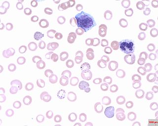
A blood smear, peripheral blood smear or blood film is a thin layer of blood smeared on a glass microscope slide and then stained in such a way as to allow the various blood cells to be examined microscopically. Blood smears are examined in the investigation of hematological (blood) disorders and are routinely employed to look for blood parasites, such as those of malaria and filariasis.

Pernicious anemia is a disease in which not enough red blood cells are produced due to a deficiency of vitamin B12. Those affected often have a gradual onset. The most common initial symptoms are feeling tired and weak. Other symptoms of anemia may include shortness of breath, lightheadedness, headaches, sore red tongue, cold hands and feet, pale or yellow skin, chest pain, and an irregular heartbeat. The digestive tract may also be disturbed giving symptoms that can include nausea and vomiting, heartburn, upset stomach and loss of appetite. Symptoms of vitamin B12 deficiency may include decreased ability to think, numbness in the hands and feet, memory problems, blurred vision, trouble walking, poor balance, muscle weakness, decreased smell and taste, poor reflexes, clumsiness, depression, and confusion. Without treatment, some of these problems may become permanent.

Megaloblastic anemia is a type of macrocytic anemia. An anemia is a red blood cell defect that can lead to an undersupply of oxygen. Megaloblastic anemia results from inhibition of DNA synthesis during red blood cell production. When DNA synthesis is impaired, the cell cycle cannot progress from the G2 growth stage to the mitosis (M) stage. This leads to continuing cell growth without division, which presents as macrocytosis. Megaloblastic anemia has a rather slow onset, especially when compared to that of other anemias. The defect in red cell DNA synthesis is most often due to hypovitaminosis, specifically vitamin B12 deficiency or folate deficiency. Loss of micronutrients may also be a cause.
An azurophilic granule is a cellular object readily stainable with a Romanowsky stain. In white blood cells and hyperchromatin, staining imparts a burgundy or merlot coloration. Neutrophils in particular are known for containing azurophils loaded with a wide variety of anti-microbial defensins that fuse with phagocytic vacuoles. Azurophils may contain myeloperoxidase, phospholipase A2, acid hydrolases, elastase, defensins, neutral serine proteases, bactericidal permeability-increasing protein, lysozyme, cathepsin G, proteinase 3, and proteoglycans.
Specific granules are secretory vesicles found exclusively in cells of the immune system called granulocytes.

Chronic myelomonocytic leukemia (CMML) is a type of leukemia, which are cancers of the blood-forming cells of the bone marrow. In adults, blood cells are formed in the bone marrow, by a process that is known as haematopoiesis. In CMML, there are increased numbers of monocytes and immature blood cells (blasts) in the peripheral blood and bone marrow, as well as abnormal looking cells (dysplasia) in at least one type of blood cell.

Toxic granulation refers to dark coarse granules found in granulocytes, particularly neutrophils, in patients with inflammatory conditions.
The term macrocytic is from Greek words meaning "large cell". A macrocytic class of anemia is an anemia in which the red blood cells (erythrocytes) are larger than their normal volume. The normal erythrocyte volume in humans is about 80 to 100 femtoliters. In metric terms the size is given in equivalent cubic micrometers. The condition of having erythrocytes which are too large, is called macrocytosis. In contrast, in microcytic anemia, the erythrocytes are smaller than normal.
Basophilic stippling, also known as punctate basophilia, is the presence of numerous basophilic granules that are dispersed through the cytoplasm of erythrocytes in a peripheral blood smear. They can be demonstrated to be RNA. They are composed of aggregates of ribosomes; degenerating mitochondria and siderosomes may be included in the aggregates. In contrast to Pappenheimer bodies, they are negative with Perls' acid ferrocyanide stain for iron. Basophilic stippling is indicative of disturbed erythropoiesis. It can also be found in some normal individuals.

Elliptocytes, also known as ovalocytes or cigar cells, are abnormally shaped red blood cells that appear oval or elongated, from slightly egg-shaped to rod or pencil forms. They have normal central pallor with the hemoglobin appearing concentrated at the ends of the elongated cells when viewed through a light microscope. The ends of the cells are blunt and not sharp like sickle cells.

In hematology, red cell agglutination or autoagglutination is a phenomenon in which red blood cells clump together, forming aggregates. It is caused by the surface of the red cells being coated with antibodies. This often occurs in cold agglutinin disease, a type of autoimmune hemolytic anemia in which people produce antibodies that bind to their red blood cells at cold temperatures and destroy them. People may develop cold agglutinins from lymphoproliferative disorders, from infection with Mycoplasma pneumoniae or Epstein–Barr virus, or idiopathically. Red cell agglutination can also occur in paroxysmal nocturnal hemoglobinuria and warm autoimmune hemolytic anemia. In cases of red cell agglutination, the direct antiglobulin test can be used to demonstrate the presence of antibodies bound to the red cells.
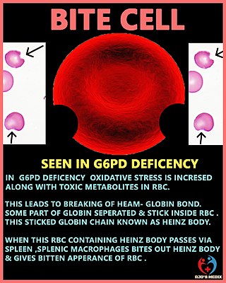
A degmacyte or bite cell is an abnormally shaped mature red blood cell with one or more semicircular portions removed from the cell margin, known as "bites". These "bites" result from the mechanical removal of denatured hemoglobin during splenic filtration as red cells attempt to migrate through endothelial slits from splenic cords into the splenic sinuses. Bite cells are known to be a result from processes of oxidative hemolysis, such as Glucose-6-phosphate dehydrogenase deficiency, in which uncontrolled oxidative stress causes hemoglobin to denature and form Heinz bodies. Bite cells can contain more than one "bite." The "bites" in degmacytes are smaller than the missing red blood cell fragments seen in schistocytes.
Armas Ralph Gustaf Gräsbeck, best known as Ralph Gräsbeck, was a Finnish physician and clinical biochemist.
Erythrocyte fragility refers to the propensity of erythrocytes to hemolyse (rupture) under stress. It can be thought of as the degree or proportion of hemolysis that occurs when a sample of red blood cells are subjected to stress. Depending on the application as well as the kind of fragility involved, the amount of stress applied and/or the significance of the resultant hemolysis may vary.

Allan Victor Hoffbrand is Emeritus Professor of Haematology at University College, London. He is distinguished for his research and as an author of internationally read textbooks of haematology. He was born in Bradford, Yorkshire in 1935. After education at Bradford Grammar School, he gained an Open Scholarship in 1953 to The Queen's College, Oxford. He gained a BA degree in Physiology and began clinical studies at The London Hospital in 1957 and qualified in medicine at University of Oxford, BM BCH in 1959.

Toxic vacuolation, also known as toxic vacuolization, is the formation of vacuoles in the cytoplasm of neutrophils in response to severe infections or inflammatory conditions.

A white blood cell differential is a medical laboratory test that provides information about the types and amounts of white blood cells in a person's blood. The test, which is usually ordered as part of a complete blood count (CBC), measures the amounts of the five normal white blood cell types – neutrophils, lymphocytes, monocytes, eosinophils and basophils – as well as abnormal cell types if they are present. These results are reported as percentages and absolute values, and compared against reference ranges to determine whether the values are normal, low, or high. Changes in the amounts of white blood cells can aid in the diagnosis of many health conditions, including viral, bacterial, and parasitic infections and blood disorders such as leukemia.
