Hereditary spherocytosis

Last updated
Hereditary spherocytosis
Other namesMinkowski–Chauffard syndrome
Hereditary Spherocytosis smear 2010-03-17.JPG
Peripheral blood smear from patient with hereditary spherocytosis
Specialty Hematology

Hereditary spherocytosis (HS) is a congenital hemolytic disorder, wherein a genetic mutation coding for a structural membrane protein phenotype leads to a spherical shaping of erythrocytic cellular morphology. As erythrocytes are sphere-shaped (spherocytosis), rather than the normal biconcave disk-shaped, their morphology interferes with these cells' abilities to be flexible during circulation throughout the entirety of the body – arteries, arterioles, capillaries, venules, veins, and organs. This difference in shape also makes the red blood cells more prone to rupture [1] under osmotic stress, mechanical stress, or both. Cells with these dysfunctional proteins are degraded in the spleen, which leads to a shortage of erythrocytes resulting in hemolytic anemia.

Contents

HS was first described in 1871, and is the most common cause of inherited hemolysis in populations of northern European descent, with an incidence of 1 in 5000 births. The clinical severity of HS varies from mild (symptom-free carrier), to moderate (anemic, jaundiced, and with splenomegaly), to severe (hemolytic crisis, in-utero hydrops fetalis), because HS is caused by genetic mutations in a multitude of structural membrane proteins and exhibits incomplete penetrance in its expression.[ citation needed ]

Early symptoms include anemia, jaundice, splenomegaly, and fatigue. [2] Acute cases can threaten to cause hypoxia secondary to anemia and acute kernicterus through high blood levels of bilirubin, particularly in newborns. Most cases can be detected soon after birth. Testing for HS is available for the children of affected adults. Occasionally, the disease will go unnoticed until the child is about 4 or 5 years of age. A person may also be a carrier of the disease and show no signs or symptoms of the disease. Late complications may result in the development of pigmented gallstones, which is secondary to the detritus of the broken-down blood cells (unconjugated or indirect bilirubin) accumulating within the gallbladder. Also, patients who are heterozygous for a hemochromatosis gene may exhibit iron overload, despite the hemochromatosis genes being recessive. [3] [4] In chronic patients, an infection or other illness can cause an increase in the destruction of red blood cells, resulting in the appearance of acute symptoms, a hemolytic crisis. On a blood smear, Howell-Jolly bodies may be seen within red blood cells. Primary treatment for patients with symptomatic HS has been total splenectomy, which eliminates the hemolytic process, allowing normal hemoglobin, reticulocyte and bilirubin levels. The resultant asplenic patient is susceptible to encapsulated bacterial infection, and prevented with vaccination. If other symptoms, such as abdominal pain persist, the removal of the gallbladder may be warranted for symptomatic cholelithiasis.[ citation needed ]

Epidemiology

Hereditary spherocytosis is the heritable hemolytic disorder, affecting 1 in 2,000 people of Northern European ancestry. [5] According to Harrison's Principles of Internal Medicine , the frequency is at least 1 in 5,000 within the United States of America. [6] While HS is most commonly (though not exclusively) found in Northern European and Japanese families, an estimated 25% of cases are due to spontaneous mutations.

Etiology

Hereditary spherocytosis is an erythrocytic disorder of that affects the following red cell membrane proteins in a congenital fashion:

Hereditary spherocytosis can be an autosomal recessive or autosomal dominant trait. [9] The autosomal recessive inheritance pattern accounts for close to 25% of the clinical cases. The autosomal dominant inheritance pattern accounts for over 75% of the clinical cases. Many positive individuals will not present clinically, thus the etiologic data may be artificially skewed towards the more prominent dominant forms. These dominant forms tend to leave a family history that yields generational splenectomies and black gallstones cholelithiasis. Lastly, an estimated 25% of cases are due to spontaneous mutations.[ citation needed ]

Pathophysiology

Causative genetic mutations and phenotypic expressions

Hereditary spherocytosis is caused by a variety of molecular defects in the genes that code for the red blood cell proteins spectrin (alpha and beta), ankyrin, [7] band 3 protein, protein 4.2, [8] and other red blood cell membrane proteins: [6]

Hereditary Spherocytosis TypeGenotypic EtiologyPhenotypic Expression
OMIM*GeneLocusErythrocyte membrane protein Affected
HS-1 182900 ANK1 8p11.2Ankyrin
HS-2 182870 SPTB 14q22-q23Spectrin (Beta)*
HS-3 270970 SPTA 1q21Spectrin (Alpha-1)*
HS-4 612653 SLC4A1 17q21-q22Band-3 Protein
HS-5 612690 EPB42 15q15Protein-4.2

*Online Mendelian Inheritance in Man (OMIM). The Alpha-1 refers the alpha-1 subunit of the spectrin protein. The Beta refers the beta subunit of the spectrin protein.

These genetic mutations are acted upon and executed by erythrocyte progenitor cells within the bone marrow, where red blood cells are normally produced in the non-pathological state (see Extramedullary hematopoiesis for pathological production outside of the bone marrow).

Pathophysiology of mutated erythrocytic membrane proteins

These proteins are necessary to maintain the normal shape of a red blood cell, which is a biconcave disk. The integrating protein that is most commonly defective is spectrin which is responsible for incorporation and binding of spectrin to the greater actin cytoskeleton. This dysfunction of cytoskeletal instabilities ensue, and leave the plasma membrane of the cell less supported and/or weakened.[ citation needed ]

Erythrocyte Membrane ProteinProtein Function CompromisedProcess EffectPathogenesis of HSLikelihoodMechanical Effect

(In Order)

Spectrin (Alpha-1)Alpha-1 subunit actin associationFailure in plasma membrane tethering to actin cytoskeleton.
Caption: The deficiency of alpha-1 subunits in spectrin protein network (top). The resulting failed or weakened tethering of spectrin interior to the plasma membrane to the greater actin cytoskeleton (bottom). Spectrin Alpha Deficiency.jpg
Caption: The deficiency of alpha-1 subunits in spectrin protein network (top). The resulting failed or weakened tethering of spectrin interior to the plasma membrane to the greater actin cytoskeleton (bottom).
Caption: The deficiency of alpha-1 subunits in spectrin protein network (top). The resulting failed or weakened tethering of Spectrin interior to the plasma membrane to the greater actin cytoskeleton (bottom).
Most Common
  1. Erythrocytic plasma membrane loss.
  2. Formation of spherocytes.
  3. Decreased surface area.
  4. Decreased plasma membrane compliance.
  5. Fragility
  6. Hemolysis
Micrograph of a spherocyte (center). Micrograph of a spherocyte.jpg
Micrograph of a spherocyte (center).
Spectrin (Beta) Hydrophobic interactions and electrostatic attraction to ankyrin and actinFailure in plasma membrane tethering to actin cytoskeleton.
Caption: The deficiency of Beta subunits in spectrin protein network (top). The resulting failed or weakened tethering of spectrin interior to the plasma membrane to the greater actin cytoskeleton (bottom). Spectrin Beta Deficiency.jpg
Caption: The deficiency of Beta subunits in spectrin protein network (top). The resulting failed or weakened tethering of spectrin interior to the plasma membrane to the greater actin cytoskeleton (bottom).
Caption: The deficiency of Beta subunits in spectrin protein network (top). The resulting failed orweakened tethering of Spectrin interior to the plasma membrane to the greater actin cytoskeleton (bottom).
Ankyrin Hydrophobic interactions and electrostatic attraction to Beta subunitFailure to mediate anchorage of integral plasma membrane proteins to spectrin.
Caption: The deficiency of ankyrin that normally associates with plasma membrane proteins (top). The resulting failed or weakened anchorage of spectin to interior plasma membrane, and subsequently weakened association between the greater actin cytoskeleton and the plasma membrane (bottom). Ankyrin Deficiency.jpg
Caption: The deficiency of ankyrin that normally associates with plasma membrane proteins (top). The resulting failed or weakened anchorage of spectin to interior plasma membrane, and subsequently weakened association between the greater actin cytoskeleton and the plasma membrane (bottom).
Caption: The deficiency of ankyrin that normally associates with plasma membrane proteins (top). The resulting failed or weakened anchorage of spectrin to interior plasma membrane, and subsequently weakened association between the greater actin cytoskeleton and the plasma membrane (bottom).
Common
Band-3 Failure to mediate the exchange of chloride (Cl) with bicarbonate (HCO3) across plasma membranes.Deficiency causes reduced lipid-stabilization at the plasma membrane.
Caption: The deficiency of band-3 protein (top). The resulting destabilized plasma membrane without integral band-3 PROTEIN for association with protein-4.2 and subsequently ANKYRIN (bottom). Band-3 Protein Deficiency.jpg
Caption: The deficiency of band-3 protein (top). The resulting destabilized plasma membrane without integral band-3 PROTEIN for association with protein-4.2 and subsequently ANKYRIN (bottom).
Caption: The deficiency of band-3 protein (top). The resulting destabilized plasma membrane without integral band-3 protein for association with protein-4.2 and subsequently ankyrin (bottom).
Less Common
Protein-4.2 Failure of ATP binding that regulates the association of band-3 with ankyrin.Reduced density of plasma cell membrane skeleton, destabilizing the plasma lipid bilayer, thus releasing band-3 protein-containing microvesicles.
Caption: The deficiency of protein-4.2 (top). The resulting inability of the greater actin cytoskeleton to associate with the plasma membrane via spectrin and ankyrin, and the subsequent loss of band-3 protein within microvesicles (bottom). Protein-4.2 Deficiency.jpg
Caption: The deficiency of protein-4.2 (top). The resulting inability of the greater actin cytoskeleton to associate with the plasma membrane via spectrin and ankyrin, and the subsequent loss of band-3 protein within microvesicles (bottom).
Caption: The deficiency of protein-4.2 (top). The resulting inability of the greater actin cytoskeleton to associate with the plasma membrane via spectrin and ankyrin, and the subsequent loss of Band-3 Protein within microvesicles (bottom).
Least Common

Cellular implications

A secondary defect in hereditary spherocytosis is a deficiency of membrane surface area. The decrease in surface area leads to less efficient gas exchange of the erythrocyte at the alveoli of the lungs and throughout circulation. Decreased surface area may be produced by two different mechanisms:

  1. Defects of spectrin, ankyrin (most commonly), or PROTEIN 4.2 lead to reduced structural integrity of the plasma membrane, destabilizing the overlying lipid bilayer, and releasing band 3-containing microvesicles. Band-3 is important for gas exchange (as seen above).
  2. Defects of band 3 lead to band 3 deficiency and loss of its lipid-stabilizing effect within the plasma membrane lipid bilayer. This results in the release of band 3-free microvesicles.

Both pathways result in compromised plasma membrane integrity, decreased surface area, and formation of spherocytes with decreased mechanical compliance during circulation.[ citation needed ]

Cardiovascular and organ sequelae

Spherocytes have less plasma membrane compliance and fluidity, and this has implications throughout the entirety of circulation within the body, i.e. arteries, arterioles, capillaries, venules, veins, and organs. The lack of compliance and fluidity lead to a higher viscosity value for blood (thickened blood), which can have implications for both larger and smaller diameter vasculature. However, the most pronounced issues with the lack of compliance and fluidity declare themselves in the failure of the erythrocyte to deform itself when transiting arterioles, capillary beds, and venules. These vessels are smaller, and can become congested or blocked altogether. Studies have demonstrated that HS is related to deep vein thrombosis (DVT) and arterial cardiovascular disease for this reason.

The spleen typically acts as a filter for blood, and targets pathogens and other damaged cells within circulation. Removing blood contaminants promotes entire-body homeostasis. The following facts about the spleen's role in normal body functioning are crucial to understanding the implications of HS on the overall health of the individual:

In relation to the three facts above, the spleen misguidedly filters spherocytes – regardless of the age or functional status of the cell. The spleen is not dysfunctional itself when selecting spherocytes for destruction, as this is the regular function of the spleen both at an anatomic structure level, and at the molecular cellular level. At the anatomic level, the passage from the cords of Billroth into the sinusoids may be seen as a "bottleneck", where red blood cells need to be flexible in order to pass through. In HS, the erythrocytes fail to pass through fenestrations, and this is where resident splenic macrophages sample, or "bite", part of the stuck spherocytes' plasma membranes. The macrophages recognize that the spherocyte is not normal, and the macrophage is "programmed" to destroy irregularities filtered at the spleen from the blood. The resident splenic macrophages therefore phagocytose the spherocytes, causing extravascular hemolysis. [10] This leads to both splenomegaly and anemia. Should this process continue unchecked chronically, inappropriate regulation of erythropoeisis leads to extramedullary hematopoiesis.

Clinical presentation

HS patients present in a vast array of presentations, from being asymptomatic to the extreme situations of splenic rupture, hemolytic crisis, or in-utero demise.

The most common presentation will demonstrate jaundice (due to increased unconjugated bilirubin), anemia (with secondary pallor), and a palpable spleen sometimes with concomitant tenderness (due to splenic congestion and splenomegaly). Its worth noting that a subsection of HS patients will also have incidental black pigmented gallstones (made of calcium bilirubinate consequence of the extravascular hemolysis), and some of these patients will develop cholelithiasis or the potential complex sequelae of this condition, i.e. cholecystitis, choledocholithiasis, etc.

Diagnostics

Laboratory testing

Available lab testing that may aid in the diagnosis of HS is as follows:

Common laboratory findings

The common findings of lab testing in setting of a patient with hereditary spherocytosis:

In chronic cases, patients who have taken iron supplementation, have heterozygous hemochromatosis, or received numerous blood transfusions, iron overload may cause additional health issues. Measuring iron stores is sometimes considered part of the diagnostic approach to hereditary spherocytosis in older patients presenting with heart muscle damage of unknown etiology or liver disease without apparent cause.

Imaging

Ultrasound is often used to evaluate the dimensions of the spleen, and also the gallbladder in preparation for functionally curative splenectomy with or without cholecystectomy.

Treatment

Although research is ongoing, currently there is no genetic-level cure for the myriad of mutations that cause the various presentations of hereditary spherocytosis. [6] Common current management focuses on interventions prevent the body from inappropriately destroying the functional spherocytes produced by erythrocyte progenitor cells within the bone marrow.

Typical treatment options include:

All operative and interventional treatments require the immunization of HS patients against the influenza virus, SARS-CoV-2, and encapsulated bacteria such as Streptococcus pneumoniae and meningococcus. Antibiotics are no longer recommended for maintenance use, even in post-splenectomy HS patients. Since the spleen is important for protecting against encapsulated organisms, sepsis caused by encapsulated organisms is a possible complication of splenectomy. [2]

Additional elective treatments offered:

Experimental treatment:

Symptomatic treatments:

Complications

Common complications

Research

Modern ongoing research interests:

See also

Related Research Articles

<span class="mw-page-title-main">Red blood cell</span> Oxygen-delivering blood cell and the most common type of blood cell

Red blood cells (RBCs), scientific name erythrocytes (from Greek erythros 'red' and kytos 'hollow vessel', with -cyte translated as 'cell' in modern usage), also referred to as red cells, red blood corpuscles (in humans or other animals not having nucleus in red blood cells) or haematids, are the most common type of blood cell and the vertebrate's principal means of delivering oxygen (O2) to the body tissues—via blood flow through the circulatory system. Erythrocytes take up oxygen in the lungs, or in fish the gills, and release it into tissues while squeezing through the body's capillaries.

<span class="mw-page-title-main">Hemolysis</span> Rupturing of red blood cells and release of their contents

Hemolysis or haemolysis, also known by several other names, is the rupturing (lysis) of red blood cells (erythrocytes) and the release of their contents (cytoplasm) into surrounding fluid. Hemolysis may occur in vivo or in vitro.

<span class="mw-page-title-main">Spherocytosis</span> Medical condition

Spherocytosis is the presence of spherocytes in the blood, i.e. erythrocytes that are sphere-shaped rather than bi-concave disk shaped as normal. Spherocytes are found in all hemolytic anemias to some degree. Hereditary spherocytosis and autoimmune hemolytic anemia are characterized by having only spherocytes.

<span class="mw-page-title-main">Splenectomy</span> Surgical removal of the spleen

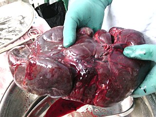
A splenectomy is the surgical procedure that partially or completely removes the spleen. The spleen is an important organ in regard to immunological function due to its ability to efficiently destroy encapsulated bacteria. Therefore, removal of the spleen runs the risk of overwhelming post-splenectomy infection, a medical emergency and rapidly fatal disease caused by the inability of the body's immune system to properly fight infection following splenectomy or asplenia.

<span class="mw-page-title-main">Paroxysmal nocturnal hemoglobinuria</span> Medical condition

Paroxysmal nocturnal hemoglobinuria (PNH) is a rare, acquired, life-threatening disease of the blood characterized by destruction of red blood cells by the complement system, a part of the body's innate immune system. This destructive process occurs due to deficiency of the red blood cell surface protein DAF, which normally inhibits such immune reactions. Since the complement cascade attacks the red blood cells within the blood vessels of the circulatory system, the red blood cell destruction (hemolysis) is considered an intravascular hemolytic anemia. There is ongoing research into other key features of the disease, such as the high incidence of venous blood clot formation. Research suggests that PNH thrombosis is caused by both the absence of GPI-anchored complement regulatory proteins on PNH platelets and the excessive consumption of nitric oxide (NO).

<span class="mw-page-title-main">Hemolytic anemia</span> Medical condition

Hemolytic anemia or haemolytic anaemia is a form of anemia due to hemolysis, the abnormal breakdown of red blood cells (RBCs), either in the blood vessels or elsewhere in the human body (extravascular). This most commonly occurs within the spleen, but also can occur in the reticuloendothelial system or mechanically. Hemolytic anemia accounts for 5% of all existing anemias. It has numerous possible consequences, ranging from general symptoms to life-threatening systemic effects. The general classification of hemolytic anemia is either intrinsic or extrinsic. Treatment depends on the type and cause of the hemolytic anemia.

An autosplenectomy is a negative outcome of disease and occurs when a disease damages the spleen to such an extent that it becomes shrunken and non-functional. The spleen is an important immunological organ that acts as a filter for red blood cells, triggers phagocytosis of invaders, and mounts an immunological response when necessary. Lack of a spleen, called asplenia, can occur by autosplenectomy or the surgical counterpart, splenectomy. Asplenia can increase susceptibility to infection. Autosplenectomy can occur in cases of sickle-cell disease where the misshapen cells block blood flow to the spleen, causing scarring and eventual atrophy of the organ. Autosplenectomy is a rare condition that is linked to certain diseases but is not a common occurrence. It is also seen in systemic lupus erythematosus (SLE).

<span class="mw-page-title-main">Howell–Jolly body</span> Cluster of DNA in red blood cells

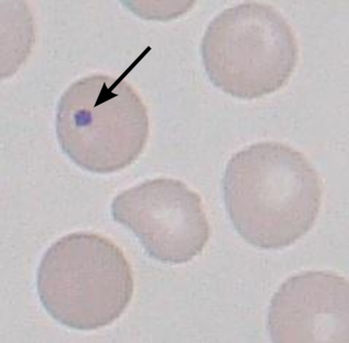
A Howell–Jolly body is a cytopathological finding of basophilic nuclear remnants in circulating erythrocytes. During maturation in the bone marrow, late erythroblasts normally expel their nuclei; but, in some cases, a small portion of DNA remains. The presence of Howell–Jolly bodies usually signifies a damaged or absent spleen, because a healthy spleen would normally filter such erythrocytes.

Warm antibody autoimmune hemolytic anemia (WAIHA) is the most common form of autoimmune haemolytic anemia. About half of the cases are of unknown cause, with the other half attributable to a predisposing condition or medications being taken. Contrary to cold autoimmune hemolytic anemia which happens in cold temperature (28–31 °C), WAIHA happens at body temperature.

<span class="mw-page-title-main">Codocyte</span> Type of red blood cell

Codocytes, also known as target cells, are red blood cells that have the appearance of a shooting target with a bullseye. In optical microscopy these cells appear to have a dark center surrounded by a white ring, followed by dark outer (peripheral) second ring containing a band of hemoglobin. However, in electron microscopy they appear very thin and bell shaped. Because of their thinness they are referred to as leptocytes. On routine smear morphology, some people like to make a distinction between leptocytes and codocytes- suggesting that in leptocytes the central spot is not completely detached from the peripheral ring, i.e. the pallor is in a C shape rather than a full ring.

<span class="mw-page-title-main">Hereditary elliptocytosis</span> Medical condition

Hereditary elliptocytosis, also known as ovalocytosis, is an inherited blood disorder in which an abnormally large number of the person's red blood cells are elliptical rather than the typical biconcave disc shape. Such morphologically distinctive erythrocytes are sometimes referred to as elliptocytes or ovalocytes. It is one of many red-cell membrane defects. In its severe forms, this disorder predisposes to haemolytic anaemia. Although pathological in humans, elliptocytosis is normal in camelids.

<span class="mw-page-title-main">Hereditary stomatocytosis</span> Medical condition

Hereditary stomatocytosis describes a number of inherited, mostly autosomal dominant human conditions which affect the red blood cell and create the appearance of a slit-like area of central pallor (stomatocyte) among erythrocytes on peripheral blood smear. The erythrocytes' cell membranes may abnormally 'leak' sodium and/or potassium ions, causing abnormalities in cell volume. Hereditary stomatocytosis should be distinguished from acquired causes of stomatocytosis, including dilantin toxicity and alcoholism, as well as artifact from the process of preparing peripheral blood smears.

<span class="mw-page-title-main">Hereditary pyropoikilocytosis</span> Medical condition

Hereditary pyropoikilocytosis (HPP) is an autosomal recessive form of hemolytic anemia characterized by an abnormal sensitivity of red blood cells to heat and erythrocyte morphology similar to that seen in thermal burns or from prolonged exposure of a healthy patient's blood sample to high ambient temperatures. Patients with HPP tend to experience severe hemolysis and anemia in infancy that gradually improves, evolving toward typical elliptocytosis later in life. However, the hemolysis can lead to rapid sequestration and destruction of red cells. Splenectomy is curative when this occurs.

Autoimmune hemolytic anemia (AIHA) occurs when antibodies directed against the person's own red blood cells (RBCs) cause them to burst (lyse), leading to an insufficient number of oxygen-carrying red blood cells in the circulation. The lifetime of the RBCs is reduced from the normal 100–120 days to just a few days in serious cases. The intracellular components of the RBCs are released into the circulating blood and into tissues, leading to some of the characteristic symptoms of this condition. The antibodies are usually directed against high-incidence antigens, therefore they also commonly act on allogenic RBCs. AIHA is a relatively rare condition, with an incidence of 5–10 cases per 1 million persons per year in the warm-antibody type and 0.45 to 1.9 cases per 1 million persons per year in the cold antibody type. Autoimmune hemolysis might be a precursor of later onset systemic lupus erythematosus.

Normocytic anemia is a type of anemia and is a common issue that occurs for men and women typically over 85 years old. Its prevalence increases with age, reaching 44 percent in men older than 85 years. The most common type of normocytic anemia is anemia of chronic disease.

<span class="mw-page-title-main">6-phosphogluconate dehydrogenase deficiency</span> Medical condition

6-Phosphogluconate dehydrogenase deficiency, or partial deficiency, is an autosomal hereditary disease characterized by abnormally low levels of 6-phosphogluconate dehydrogenase (6PGD), a metabolic enzyme involved in the Pentose phosphate pathway. It is very important in the metabolism of red blood cells (erythrocytes). 6PDG deficiency affects less than 1% of the population, and studies suggest that there may be race variant involved in many of the reported cases. Although it is similar, 6PDG deficiency is not linked to glucose-6-phosphate dehydrogenase (G6PD) deficiency, as they are located on different chromosomes. However, a few people have had both of these metabolic diseases.

Congenital hemolytic anemia (CHA) is a diverse group of rare hereditary conditions marked by decreased life expectancy and premature removal of erythrocytes from blood flow. Defects in erythrocyte membrane proteins and red cell enzyme metabolism, as well as changes at the level of erythrocyte precursors, lead to impaired bone marrow erythropoiesis. CAH is distinguished by variable anemia, chronic extravascular hemolysis, decreased erythrocyte life span, splenomegaly, jaundice, biliary lithiasis, and iron overload. Immune-mediated mechanisms may play a role in the pathogenesis of these uncommon diseases, despite the paucity of data regarding the immune system's involvement in CHAs.

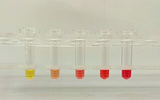
Erythrocyte fragility refers to the propensity of erythrocytes to hemolyse (rupture) under stress. It can be thought of as the degree or proportion of hemolysis that occurs when a sample of red blood cells are subjected to stress. Depending on the application as well as the kind of fragility involved, the amount of stress applied and/or the significance of the resultant hemolysis may vary.

Intravascular hemolysis describes hemolysis that happens mainly inside the vasculature. As a result, the contents of the red blood cell are released into the general circulation, leading to hemoglobinemia and increasing the risk of ensuing hyperbilirubinemia.

Hemolytic jaundice, also known as prehepatic jaundice, is a type of jaundice arising from hemolysis or excessive destruction of red blood cells, when the byproduct bilirubin is not excreted by the hepatic cells quickly enough. Unless the patient is concurrently affected by hepatic dysfunctions or is experiencing hepatocellular damage, the liver does not contribute to this type of jaundice.

References

  1. Cotran, Ramzi S.; Kumar, Vinay; Fausto, Nelson; Nelso Fausto; Robbins, Stanley L.; Abbas, Abul K. (2005). Robbins and Cotran pathologic basis of disease. St. Louis, Mo: Elsevier Saunders. p. 625. ISBN   0-7216-0187-1.
  2. 1 2 3 4 Bolton-Maggs, P. H. B.; Stevens, R. F.; Dodd, N. J.; Lamont, G.; Tittensor, P.; King, M. -J.; General Haematology Task Force of the British Committee for Standards in Haematology (2004). "Guidelines for the diagnosis and management of hereditary spherocytosis". British Journal of Haematology. 126 (4): 455–474. doi:10.1111/j.1365-2141.2004.05052.x. PMID   15287938. S2CID   5870305.
  3. J L Rasmussen; D A Odelson; F L Macrina (1987-08-01). "Complete nucleotide sequence of insertion element IS4351 from Bacteroides fragilis. - UKPMC Article - UK PubMed Central". UKPMC Article. Archived from the original on 2012-12-23. Retrieved 2012-07-03.
  4. Paula Bolton-Maggs (September 2011). "Guidelines for the Diagnosis and Management of Hereditary Spherocytosis" (PDF). The British Committee for Standards in Haematology. Archived from the original (PDF) on 12 May 2012. Retrieved 2 July 2012.
  5. "Hereditary spherocytosis: MedlinePlus Genetics".
  6. 1 2 3 4 5 Anthony S. Fauci; Eugene Braunwald; Dennis L. Kasper; Stephen L. Hauser; Dan L. Longo; J. Larry Jameson; Joseph Loscalzo (2008). Harrison's principles of internal medicine (17th ed.). New York: McGraw-Hill Medical. pp. Chapter 106. ISBN   978-0071466332.
  7. 1 2 3 Gallagher PG, Forget BG (December 1998). "Hematologically important mutations: spectrin and ankyrin variants in hereditary spherocytosis". Blood Cells Mol. Dis. 24 (4): 539–43. doi: 10.1006/bcmd.1998.0217 . PMID   9887280.
  8. 1 2 3 Perrotta S, Gallagher PG, Mohandas N (October 2008). "Hereditary spherocytosis". Lancet. 372 (9647): 1411–26. doi:10.1016/S0140-6736(08)61588-3. PMID   18940465. S2CID   10926437.
  9. Eber S, Lux SE (April 2004). "Hereditary spherocytosis--defects in proteins that connect the membrane skeleton to the lipid bilayer". Semin. Hematol. 41 (2): 118–41. doi:10.1053/j.seminhematol.2004.01.002. PMID   15071790.
  10. Mitchell, Richard Sheppard; Kumar, Vinay; Abbas, Abul K.; Fausto, Nelson (2007). "12". Robbins Basic Pathology. Philadelphia: Saunders. p. 425. ISBN   978-1-4160-2973-1. 8th edition.
  11. Won DI, Suh JS (March 2009). "Flow cytometric detection of erythrocyte osmotic fragility". Cytometry Part B. 76 (2): 135–41. doi:10.1002/cyto.b.20448. PMID   18727072. S2CID   7339411.
  12. Goljan. Rapid Review Pathology. 2010. Page 213.
  13. Buesing, K. L.; Tracy, E. T.; Kiernan, C.; Pastor, A. C.; Cassidy, L. D.; Scott, J. P.; Ware, R. E.; Davidoff, A. M.; Rescorla, F. J.; Langer, J. C.; Rice, H. E.; Oldham, K. T. (2011). "Partial splenectomy for hereditary spherocytosis: A multi-institutional review". Journal of Pediatric Surgery. 46 (1): 178–183. doi:10.1016/j.jpedsurg.2010.09.090. PMID   21238662.