Iron overload

Last updated
Iron overload
Other namesHaemochromatosis or Hemochromatosis
Hemosiderosis high mag.jpg
Micrograph of liver biopsy showing iron deposits due to haemosiderosis. Iron stain.
Specialty Hematology, gastroenterology/hepatology

Iron overload (also haemochromatosis (British English) or hemochromatosis (American English)) is the abnormal and increased accumulation of total iron in the body causing organ damage. [1] Oxidative stress is the primary mediator of organ damage, as abnormally elevated intracellular iron levels increase free radical formation (via the Fenton reaction). Iron overload is often primary (i.e., hereditary haemochromatosis) but may also be secondary to repeated blood transfusions (i.e., transfusional iron overload). [2] Iron deposition most commonly occurs in the liver, pancreas, skin, heart, and joints. People with iron overload, therefore, classically present with the triad of liver cirrhosis, secondary diabetes mellitus, and bronze skin. [3] However, due to the earlier detection nowadays, symptoms are often limited to general chronic malaise, arthralgia, and hepatomegaly. [3]

Contents

Signs and symptoms

Organs most commonly affected by hemochromatosis include the liver, heart, and endocrine glands. [4]

Hemochromatosis may present with the following clinical syndromes:

Hemochromatosis leading to secondary diabetes (through iron deposition in the insulin secreting beta cells of the pancreas), when combined with a bronzing or darkening of the skin, is sometimes known as "bronze diabetes". [10]

Causes

The term hemochromatosis was initially used to refer to what is now more specifically called hemochromatosis type 1 (or HFE-related hereditary hemochromatosis). Currently, hemochromatosis (without further specification) is mostly defined as iron overload with a hereditary or primary cause, [11] [12] or originating from a metabolic disorder. [13] However, the term is currently also used more broadly to refer to any form of iron overload, thus requiring specification of the cause, for example, hereditary hemochromatosis.

Primary hemochromatosis and hemosiderosis

Hereditary hemochromatosis

Hereditary hemochromatosis is an autosomal recessive disorder with estimated prevalence in the population of 1 in 200 among patients with European ancestry, with lower incidence in other ethnic groups. [14] Mutations of the HFE gene (hemostatic iron regulator) located on chromosome 6 (responsible for iron regulatory protein hepcidin production) are responsible for most cases of hereditary hemochromatosis; 95% of cases of hereditary hemochromatosis involve a mutation of this HFE gene. [1] [7] Non-HFE hereditary hemochromatosis involves mutations in genes coding for the iron regulatory proteins hemojuvelin, transferrin receptor-2 and ferroportin. [7]

Hereditary hemochromatosis is characterized by an accelerated rate of intestinal iron absorption and progressive iron deposition in various tissues. This typically begins to be expressed in the third to fifth decades of life, but may occur in children. The most common presentation is hepatic cirrhosis in combination with hypopituitarism, cardiomyopathy, diabetes, arthritis, or hyperpigmentation. Because of the severe sequelae of this disorder if left untreated, and recognizing that treatment is relatively simple, early diagnosis before symptoms or signs appear is important. [15] [16]

Hemosiderosis

In general, the term hemosiderosis is used to indicate the pathological effect of iron accumulation in any given organ, which mainly occurs in the form of the iron-storage complex hemosiderin. [17] [18] Sometimes, the simpler term siderosis is used instead.

Other definitions distinguishing hemochromatosis or hemosiderosis that are occasionally used include:

  • Hemosiderosis is hemochromatosis caused by excessive blood transfusions, that is, hemosiderosis is a form of secondary hemochromatosis. [19] [20]
  • Hemosiderosis is hemosiderin deposition within cells, while hemochromatosis is hemosiderin within cells and interstitium. [21]
  • Hemosiderosis is iron overload that does not cause tissue damage, [22] while hemochromatosis does. [23]
  • Hemosiderosis is arbitrarily differentiated from hemochromatosis by the reversible nature of the iron accumulation in the reticuloendothelial system. [24]

The causes of hemochromatosis broken down into two subcategories: primary cases (hereditary or genetically determined) and less frequent secondary cases (acquired during life). [25] People of Northern European descent, including Celtic (Irish, Scottish, Welsh, Cornish, Breton etc.), English, and Scandinavian origin [26] have a particularly high incidence, with about 10% being carriers of the principal genetic variant, the C282Y mutation on the HFE gene, and 1% having the condition. [27]

Non-classical hereditary hemochromatosis

The overwhelming majority depend on mutations of the HFE gene discovered in 1996, but since then others have been discovered and sometimes are grouped together as "non-classical hereditary hemochromatosis", [28] "non-HFE related hereditary hemochromatosis", [29] or "non-HFE hemochromatosis". [30]

Description OMIM Mutation
Hemochromatosis type 1: "classical" hemochromatosis 235200 HFE
Hemochromatosis type 2A: juvenile hemochromatosis 602390 Haemojuvelin (HJV, also known as RGMc and HFE2)
Hemochromatosis type 2B: juvenile hemochromatosis 606464 hepcidin antimicrobial peptide ( HAMP ) or HFE2B
Hemochromatosis type 3 604250 transferrin receptor-2 (TFR2 or HFE3)
Hemochromatosis type 4 / African iron overload 604653 ferroportin (SLC11A3/SLC40A1)
Neonatal hemochromatosis 231100 (unknown)
Acaeruloplasminaemia (very rare) 604290 caeruloplasmin
Congenital atransferrinaemia (very rare) 209300 transferrin
GRACILE syndrome (very rare) 603358 BCS1L

Most types of hereditary hemochromatosis have autosomal recessive inheritance, while type 4 has autosomal dominant inheritance. [31]

Secondary hemochromatosis

Pathophysiology

Defects in iron metabolism, specifically involving the iron regulatory protein hepcidin are thought to play an integral role in the pathogenesis of hereditary hemochromatosis. [7]

Normally, hepcidin acts to reduce iron levels in the body by inhibiting intestinal iron absorption and inhibiting iron mobilization from stores in the bone marrow and liver. [7] Iron is absorbed from the intestines (mostly in the duodenum) and transported across intestinal enterocytes or mobilized out of storage in liver hepatocytes or from macrophages in the bone marrow by the transmembrane ferroportin transporter. [7] In response to elevated plasma iron levels, hepcidin inhibits the ferroportin transporter leading to decreased iron mobilization from stores and decreased intestinal iron absorption, thus functioning as a negative iron regulatory protein. [7]

In hereditary hemochromatosis, mutations in the proteins involved in hepcidin production including HFE (hemostatic iron regulator), hemojuvelin and transferrin receptor 2 lead to a loss or decrease in hepcidin production, which subsequently leads to the loss of the inhibitory signal regulating iron absorption and mobilization and thus leads to iron overload. [7] In very rare instances, mutations in ferroportin result in ferroportin resistance to hepcidin's negative regulatory effects, and continued intestinal iron absorption and mobilization despite inhibitory signaling from hepcidin. [7] Approximately 95% of cases of hereditary hemochromatosis are due to mutations in the HFE gene. [7]

The resulting iron overload causes iron to deposit in various sites throughout the body, especially the liver and joints, which coupled with oxidative stress leads to organ damage or joint damage and the pathological findings seen in hemochromatosis. [7]

Diagnosis

Selective iron deposition (blue) in pancreatic islet beta cells (red) Left column showing selective iron deposition (blue) in pancreatic islet beta cells(red) from transfusional hemochromatosis autopsy cases versus that of controls (right lower one).jpg
Selective iron deposition (blue) in pancreatic islet beta cells (red)

There are several methods available for diagnosing and monitoring iron overload.

Blood test

Blood tests are usually the initial test if there is a clinical suspicion of iron overload. Serum ferritin testing is a low-cost, readily available, and minimally invasive method for assessing body iron stores. However ferritin levels may be elevated due to a variety of other causes including obesity, infection, inflammation (as an acute phase protein), chronic alcohol intake, liver disease, kidney disease, and cancer. [7] [32] [33] In males and postmenopausal females, normal range of serum ferritin is between 12 and 300 ng/mL (670 pmol/L) . [34] [35] [36] In premenopausal females, normal range of serum ferritin is between 12 and 150 [34] or 200 [35] ng/mL (330 or 440 pmol/L). [36] In those with hemochromatosis, the serum ferritin level correlates with the degree of iron overload. [7] Ferritin levels are usually monitored serially in those with hemochromatosis to assess response to treatment. [7]

Elevations in serum levels of the iron transporter protein transferrin saturation as well as increased red blood cell mean corpuscular volume and mean corpuscular hemoglobin concentration usually precede ferritin elevations in hemochromatosis. [7] Transferrin saturation of greater than 45% combined with an elevated ferritin level is highly sensitive in diagnosing HFE hemochromatosis. [7] Total iron binding capacity may be low in hemochromatosis, but can also be normal. [37]

Genetics

General screening for hemochromatosis is not recommended, however first-degree relatives of those affected should be screened. [7] [38] [39] [40]

Once iron overload has been established, HFE gene mutation genetic testing for hereditary causes of iron overload is indicated. [39] [15] The presence of HFE gene mutations in addition to iron overload confirms the clinical diagnosis of hereditary hemochromatosis. [39] The alleles evaluated by HFE gene analysis are evident in ~80% of patients with hemochromatosis; a negative report for HFE gene does not rule out hemochromatosis.[ citation needed ]

Biopsy

Histopathology of the liver, showing Kupffer cells with significant hemosiderin deposition (shown next to a hepatocyte with lipofuscin pigment, which is a common normal finding). H&E stain. Kupffer cell with hemosiderin and hepatocyte with lipofuscin.jpg
Histopathology of the liver, showing Kupffer cells with significant hemosiderin deposition (shown next to a hepatocyte with lipofuscin pigment, which is a common normal finding). H&E stain.
Prussian blue iron staining, highlighting the hemosiderin pigment as blue. This finding indicates mesenchymal iron overload (within Kupffer cells and/or portal macrophages) rather than parenchymal iron overload (within hepatocytes). Kupffer cell with hemosiderin and hepatocyte with lipofuscin, iron stain.jpg
Prussian blue iron staining, highlighting the hemosiderin pigment as blue. This finding indicates mesenchymal iron overload (within Kupffer cells and/or portal macrophages) rather than parenchymal iron overload (within hepatocytes).

Liver biopsy is the removal of small sample in order to be studied and can determine the cause of inflammation or cirrhosis. In someone with negative HFE gene testing, elevated iron status for no other obvious reason, and family history of liver disease, additional evaluation of liver iron concentration is indicated. In this case, diagnosis of hemochromatosis is based on biochemical analysis and histologic examination of a liver biopsy. Assessment of the hepatic iron index (HII) is considered the "gold standard" for diagnosis of hemochromatosis.[ citation needed ]

Imaging

Magnetic resonance imaging (MRI) is used as a noninvasive method to estimate iron deposition levels in the liver and heart, which may aid in determining a response to treatment or prognosis. [7] Liver elastography has limited utility in detecting liver fibrosis in hemochromatosis. [7]

Treatment

Phlebotomy

Phlebotomy, bloodletting or venesection is the mainstay of treatment in iron overload, consisting of regularly scheduled blood draws to remove red blood cells (and iron) from the body. [7] Upon initial diagnosis of iron overload, the phlebotomies may be performed weekly or twice weekly, until iron levels are normalized. Once the serum ferritin and transferrin saturation are within the normal range, maintenance phlebotomies may be needed in some (depending upon the rate of reabsorption of iron), scheduled at varying frequencies to keep iron stores within normal range. [39] A phlebotomy session typically draws between 450 and 500 mL of blood. [42] Routine phlebotomy may reverse liver fibrosis and alleviate some symptoms of hemochromatosis, but chronic arthritis is usually not responsive to treatment. [7] In those with hemochromatosis; the blood drawn during phlebotomy is safe to be donated. [43] [39]

Phlebotomy is associated with improved survival if it is initiated before the onset of cirrhosis or diabetes. [39]

Diet

The human diet contains iron in two forms: heme iron and non-heme iron. Heme iron is usually found in red meat, whereas non-heme iron is found in plant based sources. Heme iron is the most easily absorbed form of iron. In those with hemochromatosis undergoing phlebotomy for treatment; restriction of dietary iron is not required. [39] [40] [7] However, those who do restrict dietary iron usually require less blood needing to be phlebotomized (about 0.5–1.5 liters of blood less per year). [44] Vitamin C and iron supplementation should be avoided as vitamin C accelerates intestinal absorption of iron and mobilization of body iron stores. [39] [40] Raw seafood should be avoided because of increased risk of infections from iron loving pathogens such as Vibrio vulnificus . [7] [45] Alcohol consumption should be avoided due to the risk of compounded liver damage with iron overload. [7]

Medication

Medications are used for those unable to tolerate routine blood draws, there are chelating agents available for use. [46] The drug deferoxamine binds with iron in the bloodstream and enhances its elimination in urine and faeces. Typical treatment for chronic iron overload requires subcutaneous injection over a period of 8–12 hours daily.[ citation needed ] Two newer iron-chelating drugs that are licensed for use in patients receiving regular blood transfusions to treat thalassaemia (and, thus, who develop iron overload as a result) are deferasirox and deferiprone. [47] [48]

Chelating polymers

A minimally invasive approach to hereditary hemochromatosis treatment is the maintenance therapy with polymeric chelators. [49] [50] [51] These polymers or particles have a negligible or null systemic biological availability and they are designed to form stable complexes with Fe2+ and Fe3+ in the GIT and thus limiting their uptake and long-term accumulation. Although this method has only a limited efficacy, unlike small-molecular chelators, the approach has virtually no side effects in sub-chronic studies. [51] Interestingly, the simultaneous chelation of Fe2+ and Fe3+ increases the treatment efficacy. [51]

Prognosis

In general, provided there has been no liver damage, patients should expect a normal life expectancy if adequately treated by venesection. If the serum ferritin is greater than 1,000 μg/L at diagnosis there is a risk of liver damage and cirrhosis which may eventually shorten their life. [52] The presence of cirrhosis increases the risk of hepatocellular carcinoma. [53] Other risk factors for liver damage in hemochromatosis include alcohol use, diabetes, liver iron levels greater than 2,000 μmol/gram and increased aspartate transaminase levels. [7]

The risk of death and liver fibrosis are elevated in males with HFE type hemochromatosis but not in females; this is thought to be due to a protective effect of menstruation and pregnancy seen in females as well as possible hormone-related differences in iron absorption. [7]

Epidemiology

HHC is most common in certain European populations (such as those of Irish or Scandinavian descent) and occurs in 0.6% of some unspecified population. [38] Men have a 24-fold increased rate of iron-overload disease compared with women. [38]

Stone Age

Diet and the environment are thought to have had large influence on the mutation of genes related to iron overload. Starting during the Mesolithic era, communities of people lived in an environment that was fairly sunny, warm and had the dry climates of the Middle East. Most humans who lived at that time were foragers and their diets consisted largely of wild plants, fish, and game. Archaeologists studying dental plaque have found evidence of tubers, nuts, plantains, grasses and other foods rich in iron. Over many generations, the human body became well-adapted to a high level of iron content in the diet. [54]

Neolithic

In the Neolithic era, significant changes are thought to have occurred in both the environment and diet. Some communities of foragers migrated north, leading to changes in lifestyle and environment, with a decrease in temperatures and a change in the landscape which the foragers then needed to adapt to. As people began to develop and advance their tools, they learned new ways of producing food, and farming also slowly developed. These changes would have led to serious stress on the body and a decrease in the consumption of iron-rich foods. This transition is a key factor in the mutation of genes, especially those that regulated dietary iron absorption. Iron, which makes up 70% of red blood cell composition, is a critical micronutrient for effective thermoregulation in the body. [55] Iron deficiency will lead to a drop in the core temperature. In the chilly and damp environments of Northern Europe, supplementary iron from food was necessary to keep temperatures regulated, however, without sufficient iron intake the human body would have started to store iron at higher rates than normal. In theory, the pressures caused by migrating north would have selected for a gene mutation that promoted greater absorption and storage of iron. [56]

Viking hypothesis

Studies and surveys conducted to determine the frequencies of hemochromatosis help explain how the mutation migrated around the globe. In theory, the disease initially evolved from travelers migrating from the north. Surveys show a particular distribution pattern with large clusters and frequencies of gene mutations along the western European coastline. [57] This led the development of the "Viking Hypothesis". [58] Cluster locations and mapped patterns of this mutation correlate closely to the locations of Viking settlements in Europe established c.700 AD to c.1100 AD. The Vikings originally came from Norway, Sweden and Denmark. Viking ships made their way along the coastline of Europe in search of trade, riches, and land. Genetic studies suggest that the extremely high frequency patterns in some European countries are the result of migrations of Vikings and later Normans, indicating a genetic link between hereditary hemochromatosis and Viking ancestry. [59]

Modern times

In 1865, Armand Trousseau (a French internist) was one of the first to describe many of the symptoms of a diabetic patient with cirrhosis of the liver and bronzed skin color. The term hemochromatosis was first used by German pathologist Friedrich Daniel von Recklinghausen in 1889 when he described an accumulation of iron in body tissues. [60]

Identification of genetic factors

Although it was known most of the 20th century that most cases of hemochromatosis were inherited, they were incorrectly assumed to depend on a single gene. [61]

In 1935 J.H. Sheldon, a British physician, described the link to iron metabolism for the first time as well as demonstrating its hereditary nature. [60]

In 1996 Felder and colleagues identified the hemochromatosis gene, HFE gene. Felder found that the HFE gene has two main mutations, causing amino acid substitutions C282Y and H63D, which were the main cause of hereditary hemochromatosis. [60] [62] The next year the CDC and the National Human Genome Research Institute sponsored an examination of hemochromatosis following the discovery of the HFE gene, which helped lead to the population screenings and estimates that are still being used today. [63]

See also

Related Research Articles

<span class="mw-page-title-main">Hereditary haemochromatosis</span> Medical condition

Hereditary haemochromatosis type 1 is a genetic disorder characterized by excessive intestinal absorption of dietary iron, resulting in a pathological increase in total body iron stores. Humans, like most animals, have no mechanism to regulate excess iron, simply losing a limited amount through various means like sweating or menstruating.

<span class="mw-page-title-main">Ferritin</span> Iron-carrying protein

Ferritin is a universal intracellular protein that stores iron and releases it in a controlled fashion. The protein is produced by almost all living organisms, including archaea, bacteria, algae, higher plants, and animals. It is the primary intracellular iron-storage protein in both prokaryotes and eukaryotes, keeping iron in a soluble and non-toxic form. In humans, it acts as a buffer against iron deficiency and iron overload.

<span class="mw-page-title-main">Transferrin</span> Mammalian protein found in Homo sapiens

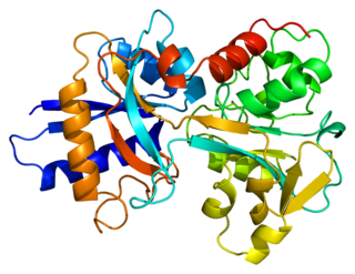
Transferrins are glycoproteins found in vertebrates which bind and consequently mediate the transport of iron (Fe) through blood plasma. They are produced in the liver and contain binding sites for two Fe3+ ions. Human transferrin is encoded by the TF gene and produced as a 76 kDa glycoprotein.

<span class="mw-page-title-main">Total iron-binding capacity</span> Medical blood test to measure transferrin

Total iron-binding capacity (TIBC) or sometimes transferrin iron-binding capacity is a medical laboratory test that measures the blood's capacity to bind iron with transferrin. Transferrin can bind two atoms of ferric iron (Fe3+) with high affinity. It means that transferrin has the capacity to transport approximately from 1.40 to 1.49 mg of iron per gram of transferrin present in the blood.

Transferrin saturation (TS), measured as a percentage, is a medical laboratory value. It is the value of serum iron divided by the total iron-binding capacity of the available transferrin, the main protein that binds iron in the blood, this value tells a clinician how much serum iron is bound. For instance, a value of 15% means that 15% of iron-binding sites of transferrin are being occupied by iron. The three results are usually reported together. A low transferrin saturation is a common indicator of iron deficiency anemia whereas a high transferrin saturation may indicate iron overload or hemochromatosis. Transferrin saturation is also called transferrin saturation index (TSI) or transferrin saturation percentage (TS%)

<span class="mw-page-title-main">Human iron metabolism</span> Iron metabolism in the body

Human iron metabolism is the set of chemical reactions that maintain human homeostasis of iron at the systemic and cellular level. Iron is both necessary to the body and potentially toxic. Controlling iron levels in the body is a critically important part of many aspects of human health and disease. Hematologists have been especially interested in systemic iron metabolism, because iron is essential for red blood cells, where most of the human body's iron is contained. Understanding iron metabolism is also important for understanding diseases of iron overload, such as hereditary hemochromatosis, and iron deficiency, such as iron-deficiency anemia.

<span class="mw-page-title-main">Hepcidin</span> Protein-coding gene in the species Homo sapiens

Hepcidin is a protein that in humans is encoded by the HAMP gene. Hepcidin is a key regulator of the entry of iron into the circulation in mammals.

<span class="mw-page-title-main">Ferroportin</span> Protein

Ferroportin-1, also known as solute carrier family 40 member 1 (SLC40A1) or iron-regulated transporter 1 (IREG1), is a protein that in humans is encoded by the SLC40A1 gene. Ferroportin is a transmembrane protein that transports iron from the inside of a cell to the outside of the cell. Ferroportin is the only known iron exporter.

<span class="mw-page-title-main">Aceruloplasminemia</span> Medical condition

Aceruloplasminemia is a rare autosomal recessive disorder in which the liver can not synthesize the protein ceruloplasmin properly, which is needed to transport copper around the blood. Copper deficiency in the brain results in neurological problems that generally appear in adulthood and worsen over time. .

<span class="mw-page-title-main">African iron overload</span> Iron overload disorder caused by consumption of home-brewed beer

African iron overload is an iron overload disorder first observed among people of African descent in Southern Africa and Central Africa. It is now recognized to actually be two disorders with different causes, possibly compounding each other:

<span class="mw-page-title-main">HFE (gene)</span> Mammalian protein found in Homo sapiens

Human homeostatic iron regulator protein, also known as the HFE protein, is a transmembrane protein that in humans is encoded by the HFE gene. The HFE gene is located on short arm of chromosome 6 at location 6p22.2

<span class="mw-page-title-main">Atransferrinemia</span> Medical condition

Atransferrinemia is an autosomal recessive metabolic disorder in which there is an absence of transferrin, a plasma protein that transports iron through the blood. Atransferrinemia is characterized by anemia and hemosiderosis in the heart and liver. The iron damage to the heart can lead to heart failure. The anemia is typically microcytic and hypochromic. Atransferrinemia was first described in 1961 and is extremely rare, with only ten documented cases worldwide.

Transfusional hemosiderosis is the accumulation of iron in the body due to frequent blood transfusions. Iron accumulates in the liver and heart, but also endocrine organs. Frequent blood transfusions may be given to many patients, such as those with thalassemia, sickle cell disease, leukemia, aplastic anemia, or myelodysplastic syndrome, among others. It is diagnosed with a blood transferrin test and a liver biopsy. It is treated with venipuncture, erythrocytapheresis, and iron chelation therapy.

<span class="mw-page-title-main">Iron in biology</span> Use of Iron by organisms

Iron is an important biological element. It is used in both the ubiquitous iron-sulfur proteins and in vertebrates it is used in hemoglobin which is essential for blood and oxygen transport.

<span class="mw-page-title-main">Transferrin receptor 2</span> Mammalian protein found in Homo sapiens

Transferrin receptor 2 (TfR2) is a protein that in humans is encoded by the TFR2 gene. This protein is involved in the uptake of transferrin-bound iron into cells by endocytosis, although its role is minor compared to transferrin receptor 1.

Juvenile hemochromatosis, also known as hemochromatosis type 2, is a rare form of hereditary hemochromatosis, which emerges in young individuals, typically between 15 and 30 years of age, but occasionally later. It is characterized by an inability to control how much iron is absorbed by the body, in turn leading to iron overload, where excess iron accumulates in many areas of the body and causes damage to the places it accumulates.

<span class="mw-page-title-main">Hemosiderosis</span> Iron metabolism disease

Hemosiderosis is a form of iron overload disorder resulting in the accumulation of hemosiderin.

Haemochromatosis type 3 is a type of iron overload disorder associated with deficiencies in transferrin receptor 2. It exhibits an autosomal recessive inheritance pattern. The first confirmed case was diagnosed in 1865 by French doctor Trousseau. Later in 1889, the German doctor von Recklinghausen indicated that the liver contains iron, and due to bleeding being considered to be the cause, he called the pigment "Haemochromatosis." In 1935, English doctor Sheldon's groundbreaking book titled, Haemochromatosis, reviewed 311 patient case reports and presented the idea that haemochromatosis was a congenital metabolic disorder. Hereditary haemochromatosis is a congenital disorder which affects the regulation of iron metabolism thus causing increased gut absorption of iron and a gradual build-up of pathologic iron deposits in the liver and other internal organs, joint capsules and the skin. The iron overload could potentially cause serious disease from the age of 40–50 years. In the final stages of the disease, the major symptoms include liver cirrhosis, diabetes and bronze-colored skin. There are four types of hereditary hemochromatosis which are classified depending on the age of onset and other factors such as genetic cause and mode of inheritance.

Hemochromatosis type 4 is a hereditary iron overload disorder that affects ferroportin, an iron transport protein needed to export iron from cells into circulation. Although the disease is rare, it is found throughout the world and affects people from various ethnic groups. While the majority of individuals with type 4 hemochromatosis have a relatively mild form of the disease, some affected individuals have a more severe form. As the disease progresses, iron may accumulate in the tissues of affected individuals over time, potentially resulting in organ damage.

<span class="mw-page-title-main">HFE H63D gene mutation</span>

The HFE H63D is a single-nucleotide polymorphism in the HFE gene, which results in the substitution of a histidine for an aspartic acid at amino acid position 63 of the HFE protein (p.His63Asp). HFE participates in the regulation of iron absorption.

References

  1. 1 2 Hsu CC, Senussi NH, Fertrin KY, Kowdley KV (June 2022). "Iron overload disorders". Hepatol Commun. 6 (8): 1842–54. doi: 10.1002/hep4.2012 . PMC   9315134 . PMID   35699322.
  2. Hider RC, Kong X (2013). "Iron: Effect of Overload and Deficiency". In Astrid Sigel, Helmut Sigel and Roland K. O. Sigel (ed.). Interrelations between Essential Metal Ions and Human Diseases. Metal Ions in Life Sciences. Vol. 13. Springer. pp. 229–94. doi:10.1007/978-94-007-7500-8_8. ISBN   978-94-007-7499-5. PMID   24470094.
  3. 1 2 Daniłowicz-Szymanowicz L, Świątczak M, Sikorska K, Starzyński RR, Raczak A, Lipiński P (2021-07-16). "Pathogenesis, Diagnosis, and Clinical Implications of Hereditary Hemochromatosis-The Cardiological Point of View". Diagnostics (Basel, Switzerland). 11 (7): 1279. doi: 10.3390/diagnostics11071279 . ISSN   2075-4418. PMC   8304945 . PMID   34359361.
  4. Andrews NC (1999). "Disorders of Iron Metabolism". New England Journal of Medicine. 341 (26): 1986–95. doi:10.1056/NEJM199912233412607. PMID   10607817.
  5. 1 2 3 John Murtagh (2007). General Practice. McGraw Hill Australia. ISBN   978-0-07-470436-3.[ page needed ]
  6. 1 2 Lu JP (1994). "Selective iron deposition in pancreatic islet B cells of transfusional iron-overloaded autopsy cases". Pathol Int. 44 (3): 194–99. doi:10.1111/j.1440-1827.1994.tb02592.x. PMID   802561. S2CID   25357672.
  7. 1 2 3 4 5 6 7 8 9 10 11 12 13 14 15 16 17 18 19 20 21 22 23 24 25 26 27 Olynyk JK, Ramm GA (8 December 2022). "Hemochromatosis". New England Journal of Medicine. 387 (23): 2159–70. doi:10.1056/NEJMra2119758. PMID   36477033. S2CID   254432917.
  8. 1 2 Bruce R Bacon, Stanley L Schrier. "Patient information: Hemochromatosis (hereditary iron overload) (Beyond the Basics)". UpToDate . Retrieved 2016-07-14. Literature review current through: Jun 2016. | This topic last updated: Apr 14, 2015.
  9. Brissot P, Pietrangelo A, Adams PC, de Graaff B, McLaren CE, Loréal O (5 April 2018). "Haemochromatosis". Nature Reviews. Disease Primers. 4: 18016. doi:10.1038/nrdp.2018.16. PMC   7775623 . PMID   29620054.
  10. "Haemochromatosis and diabetes". Diabetes UK.
  11. thefreedictionary.com > hemochromatosis, citing:
    • The American Heritage Medical Dictionary, 2004 by Houghton Mifflin Company
    • McGraw-Hill Concise Dictionary of Modern Medicine. 2002
  12. Merriam-Webster's Medical Dictionary > hemochromatosis Retrieved on December 11, 2009
  13. thefreedictionary.com, citing:
    • Dorland's Medical Dictionary for Health Consumers, 2007
    • Mosby's Medical Dictionary, 8th edition. 2009
    • Jonas: Mosby's Dictionary of Complementary and Alternative Medicine. 2005.
  14. "Hemochromatosis". Archived from the original on 2007-03-18. Retrieved 2012-10-05.
  15. 1 2 Pietrangelo A (2010). "Hereditary Hemochromatosis: Pathogenesis, Diagnosis, and Treatment". Gastroenterology. 139 (2): 393–408. doi: 10.1053/j.gastro.2010.06.013 . PMID   20542038.
  16. Brandhagen DJ, Fairbanks VF, Batts KP, Thibodeau SN (1999). "Update on hereditary hemochromatosis and the HFE gene". Mayo Clinic Proceedings. 74 (9): 917–21. doi: 10.4065/74.9.917 . PMID   10488796.
  17. Merriam-Webster's Medical Dictionary > hemosideroses Retrieved on December 11, 2009
  18. thefreedictionary.com > hemosiderosis, citing:
    • The American Heritage Medical Dictionary, 2004 by Houghton Mifflin Company
    • Mosby's Medical Dictionary, 8th edition.
  19. eMedicine Specialties > Radiology > Gastrointestinal > Hemochromatosis Author: Sandor Joffe, MD. Updated: May 8, 2009
  20. thefreedictionary.com > hemosiderosis, citing:
    • Gale Encyclopedia of Medicine. Copyright 2008
  21. Notecards on radiology gamuts, diseases, anatomy Archived 2010-07-21 at the Wayback Machine 2002, Charles E. Kahn, Jr., MD. Medical College of Wisconsin
  22. thefreedictionary.com > hemosiderosis, citing:
    • Dorland's Medical Dictionary for Health Consumers, 2007
    • Mosby's Dental Dictionary, 2nd ed.
    • Saunders Comprehensive Veterinary Dictionary, 3rd ed. 2007
  23. The HealthScout Network > Health Encyclopedia > Diseases and Conditions > Hemochromatosis Archived 2010-02-09 at the Wayback Machine Retrieved on December 11, 2009
  24. thefreedictionary.com > hemosiderosis, citing:
    • McGraw-Hill Concise Dictionary of Modern Medicine. 2002
  25. Pietrangelo A (2003). "Haemochromatosis". Gut. 52 (90002): ii23–30. doi:10.1136/gut.52.suppl_2.ii23. PMC   1867747 . PMID   12651879.
  26. The Atlantic: "The Iron in Our Blood That Keeps and Kills Us" by Bradley Wertheim January 10, 2013
  27. "Hemachromatosis". Encyclopædia Britannica.com. Retrieved 17 April 2017.
  28. Mendes AI, Ferro A, Martins R, Picanço I, Gomes S, Cerqueira R, Correia M, Nunes AR, Esteves J, Fleming R, Faustino P (2008). "Non-classical hereditary hemochromatosis in Portugal: novel mutations identified in iron metabolism-related genes" (PDF). Annals of Hematology. 88 (3): 229–34. doi:10.1007/s00277-008-0572-y. PMID   18762941. S2CID   23206256.
  29. Maddrey, Willis C., Schiff, Eugene R., Sorrell, Michael F. (2007). Schiff's diseases of the liver. Hagerstwon, MD: Lippincott Williams & Wilkins. p. 1048. ISBN   978-0-7817-6040-9.
  30. Pietrangelo A (2005). "Non-HFE Hemochromatosis". Seminars in Liver Disease. 25 (4): 450–60. doi:10.1055/s-2005-923316. PMID   16315138. S2CID   260320984.
  31. Franchini M (2006). "Hereditary iron overload: Update on pathophysiology, diagnosis, and treatment". American Journal of Hematology. 81 (3): 202–09. doi: 10.1002/ajh.20493 . PMID   16493621.
  32. Waalen J, Felitti VJ, Gelbart T, Beutler E (1 April 2008). "Screening for hemochromatosis by measuring ferritin levels: a more effective approach". Blood. 111 (7): 3373–76. doi:10.1182/blood-2007-07-102673. PMC   2275006 . PMID   18025154.
  33. Koperdanova M, Cullis JO (3 August 2015). "Interpreting raised serum ferritin levels". BMJ. 351: h3692. doi:10.1136/bmj.h3692. PMID   26239322. S2CID   44923011.
  34. 1 2 Ferritin by: Mark Levin, MD, Hematologist and Oncologist, Newark, NJ. Review provided by VeriMed Healthcare Network
  35. 1 2 Andrea Duchini. "Hemochromatosis Workup". Medscape . Retrieved 2016-07-14. Updated: Jan 02, 2016
  36. 1 2 Molar concentration is derived from mass value using molar mass of 450,000 g•mol−1 for ferritin
  37. labtestsonline.org TIBC & UIBC, Transferrin Last reviewed on October 28, 2009.
  38. 1 2 3 Crownover BK, Covey, CJ (Feb 1, 2013). "Hereditary hemochromatosis". American Family Physician. 87 (3): 183–90. PMID   23418762.
  39. 1 2 3 4 5 6 7 8 Bacon BR, Adams PC, Kowdley KV, Powell LW, Tavill AS (July 2011). "Diagnosis and management of hemochromatosis: 2011 Practice Guideline by the American Association for the Study of Liver Diseases". Hepatology. 54 (1): 328–43. doi:10.1002/hep.24330. PMC   3149125 . PMID   21452290. S2CID   9311604.
  40. 1 2 3 Kowdley KV, Brown KE, Ahn J, Sundaram V (August 2019). "ACG Clinical Guideline: Hereditary Hemochromatosis". American Journal of Gastroenterology. 114 (8): 1202–18. doi: 10.14309/ajg.0000000000000315 . PMID   31335359. S2CID   198192589.
  41. Image by Mikael Häggström, MD. Source for mesenchymal versus parenchymal iron overload Deugnier Y, Turlin B (2007). "Pathology of hepatic iron overload". World J Gastroenterol. 13 (35): 4755–60. doi: 10.3748/wjg.v13.i35.4755 . PMC   4611197 . PMID   17729397.
  42. Barton JC (1 December 1998). "Management of Hemochromatosis". Annals of Internal Medicine. 129 (11_Part_2): 932–39. doi:10.7326/0003-4819-129-11_Part_2-199812011-00003. PMID   9867745. S2CID   53087679.
  43. NIH blood bank. "Hemochromatosis Donor Program".
  44. Moretti D, Van Doorn GM, Swinkels DW, Melse-Boonstra A (2013). "Relevance of dietary iron intake and bioavailability in the management of HFE hemochromatosis: A systematic review". The American Journal of Clinical Nutrition. 98 (2): 468–79. doi: 10.3945/ajcn.112.048264 . PMID   23803887.
  45. Bullen JJ (1 August 1991). "Hemochromatosis, Iron, and Septicemia Caused by Vibrio vulnificus". Archives of Internal Medicine. 151 (8): 1606–09. doi:10.1001/archinte.1991.00400080096018. PMID   1872665.
  46. Miller MJ (1989-11-01). "Syntheses and therapeutic potential of hydroxamic acid based siderophores and analogs". Chemical Reviews. 89 (7): 1563–79. doi:10.1021/cr00097a011.
  47. Choudhry VP, Naithani R (2007). "Current status of iron overload and chelation with deferasirox". Indian J Pediatr. 74 (8): 759–64. doi:10.1007/s12098-007-0134-7. PMID   17785900. S2CID   19930076.
  48. Hoffbrand AV (20 March 2003). "Role of deferiprone in chelation therapy for transfusional iron overload". Blood. 102 (1): 17–24. doi: 10.1182/blood-2002-06-1867 . PMID   12637334.
  49. Polomoscanik SC, Cannon CP, Neenan TX, Holmes-Farley SR, Mandeville WH, Dhal PK (2005). "Hydroxamic Acid-Containing Hydrogels for Nonabsorbed Iron Chelation Therapy: Synthesis, Characterization, and Biological Evaluation". Biomacromolecules. 6 (6): 2946–53. doi:10.1021/bm050036p. ISSN   1525-7797. PMID   16283713.
  50. Qian J, Sullivan BP, Peterson SJ, Berkland C (2017). "Nonabsorbable Iron Binding Polymers Prevent Dietary Iron Absorption for the Treatment of Iron Overload". ACS Macro Letters. 6 (4): 350–53. doi:10.1021/acsmacrolett.6b00945. ISSN   2161-1653. PMID   35610854.
  51. 1 2 3 Groborz O, Poláková L, Kolouchová K, Švec P, Loukotová L, Miriyala VM, Francová P, Kučka J, Krijt J, Páral P, Báječný M, Heizer T, Pohl R, Dunlop D, Czernek J, Šefc L, Beneš J, Štěpánek P, Hobza P, Hrubý M (2020). "Chelating Polymers for Hereditary Hemochromatosis Treatment". Macromolecular Bioscience. 20 (12): 2000254. doi:10.1002/mabi.202000254. ISSN   1616-5187. PMID   32954629. S2CID   221827050.
  52. Allen KJ, Gurrin LC, Constantine CC, Osborne NJ, Delatycki MB, Nicoll AJ, McLaren CE, Bahlo M, Nisselle AE, Vulpe CD, Anderson GJ, Southey MC, Giles GG, English DR, Hopper JL, Olynyk JK, Powell LW, Gertig DM (17 January 2008). "Iron-overload-related disease in HFE hereditary hemochromatosis" (PDF). The New England Journal of Medicine. 358 (3): 221–30. doi:10.1056/NEJMoa073286. PMID   18199861.
  53. Kowdley KV (November 2004). "Iron, hemochromatosis, and hepatocellular carcinoma". Gastroenterology. 127 (5 Suppl 1): S79–86. doi:10.1016/j.gastro.2004.09.019. PMID   15508107.
  54. "The Evolution of Diet". National Geographic. Retrieved 2018-04-11.
  55. Rosenzweig PH, Volpe SL (March 1999). "Iron, thermoregulation, and metabolic rate". Critical Reviews in Food Science and Nutrition. 39 (2): 131–148. doi:10.1080/10408399908500491. ISSN   1040-8398. PMID   10198751.
  56. Heath KM, Axton JH, McCullough JM, Harris N (May 2016). "The evolutionary adaptation of the C282Y mutation to culture and climate during the European Neolithic". American Journal of Physical Anthropology. 160 (1): 86–101. doi:10.1002/ajpa.22937. ISSN   0002-9483. PMC   5066702 . PMID   26799452.
  57. "Clinical Penetrance of HFE Hereditary Hemochromatosis, Serum Ferritin Levels, and Screening Implications: Can We Iron This Out?". www.hematology.org. 2008-05-01. Archived from the original on 2018-06-15. Retrieved 2018-04-11.
  58. Symonette CJ, Adams PC (June 2011). "Do all hemochromatosis patients have the same origin? A pilot study of mitochondrial DNA and Y-DNA". Canadian Journal of Gastroenterology. 25 (6): 324–326. doi: 10.1155/2011/463810 . ISSN   0835-7900. PMC   3142605 . PMID   21766093.
  59. "Videos: Hereditary Hemochromatosis | Canadian Hemochromatosis Society". www.toomuchiron.ca. Retrieved 2018-04-11.
  60. 1 2 3 Fitzsimons EJ, Cullis JO, Thomas DW, Tsochatzis E, Griffiths WJ, the British Society for Haematology (May 2018). "Diagnosis and therapy of genetic haemochromatosis (review and 2017 update)". British Journal of Haematology. 181 (3): 293–303. doi: 10.1111/bjh.15164 . PMID   29663319.
  61. Cam Patterson, Marschall S. Runge (2006). Principles of molecular medicine. Totowa, NJ: Humana Press. p. 567. ISBN   978-1-58829-202-5.
  62. Feder J, Gnirke A, Thomas W, Tsuchihashi Z, Ruddy D, Basava A, Dormishian F, Domingo R, Ellis M (August 1996). "A novel MHC class I–like gene is mutated in patients with hereditary haemochromatosis". Nature Genetics. 13 (4): 399–408. doi:10.1038/ng0896-399. PMID   8696333. S2CID   26239768.
  63. Burke W, Thomson E, Khoury MJ, McDonnell SM, Press N, Adams PC, Barton JC, Beutler E, Brittenham G (1998-07-08). "Hereditary Hemochromatosis: Gene Discovery and Its Implications for Population-Based Screening". JAMA. 280 (2): 172–78. doi:10.1001/jama.280.2.172. ISSN   0098-7484. PMID   9669792.