Transferrin

Last updated
TF
Protein TF PDB 1a8e.png
Available structures
PDB Ortholog search: PDBe RCSB
Identifiers
Aliases TF , PRO1557, PRO2086, TFQTL1, HEL-S-71p, transferrin
External IDs OMIM: 190000 MGI: 98821 HomoloGene: 68153 GeneCards: TF
Orthologs
SpeciesHumanMouse
Entrez
Ensembl
UniProt
RefSeq (mRNA)

NM_001063
NM_001354704
NM_001354703

NM_133977

RefSeq (protein)

NP_001054
NP_001341633
NP_001341632

NP_598738

Location (UCSC) Chr 3: 133.75 – 133.8 Mb Chr 9: 103.08 – 103.11 Mb
PubMed search [3] [4]
Wikidata
View/Edit Human View/Edit Mouse
Transferrin
Identifiers
SymbolTransferrin
Pfam PF00405
InterPro IPR001156
PROSITE PDOC00182
SCOP2 1lcf / SCOPe / SUPFAM
Available protein structures:
Pfam   structures / ECOD  
PDB RCSB PDB; PDBe; PDBj
PDBsum structure summary

Transferrins are glycoproteins found in vertebrates which bind and consequently mediate the transport of iron (Fe) through blood plasma. [5] They are produced in the liver and contain binding sites for two Fe3+ ions. [6] Human transferrin is encoded by the TF gene and produced as a 76 kDa glycoprotein. [7] [8]

Transferrin glycoproteins bind iron tightly, but reversibly. Although iron bound to transferrin is less than 0.1% (4 mg) of total body iron, it forms the most vital iron pool with the highest rate of turnover (25 mg/24 h). Transferrin has a molecular weight of around 80 kDa and contains two specific high-affinity Fe(III) binding sites. The affinity of transferrin for Fe(III) is extremely high (association constant is 1020 M−1 at pH 7.4) [9] but decreases progressively with decreasing pH below neutrality. Transferrins are not limited to only binding to iron but also to different metal ions. [10] These glycoproteins are located in various bodily fluids of vertebrates. [11] [12] Some invertebrates have proteins that act like transferrin found in the hemolymph. [11] [13]

When not bound to iron, transferrin is known as "apotransferrin" (see also apoprotein).

Occurrence and function

Transferrins are glycoproteins that are often found in biological fluids of vertebrates. When a transferrin protein loaded with iron encounters a transferrin receptor on the surface of a cell, e.g., erythroid precursors in the bone marrow, it binds to it and is transported into the cell in a vesicle by receptor-mediated endocytosis. [14] The pH of the vesicle is reduced by hydrogen ion pumps (H+
ATPases
) to about 5.5, causing transferrin to release its iron ions. [11] Iron release rate is dependent on several factors including pH levels, interactions between lobes, temperature, salt, and chelator. [14] The receptor with its ligand bound transferrin is then transported through the endocytic cycle back to the cell surface, ready for another round of iron uptake. Each transferrin molecule has the ability to carry two iron ions in the ferric form (Fe3+
). [13]

Humans and other mammals

The liver is the main site of transferrin synthesis but other tissues and organs, including the brain, also produce transferrin. A major source of transferrin secretion in the brain is the choroid plexus in the ventricular system. [15] The main role of transferrin is to deliver iron from absorption centers in the duodenum and white blood cell macrophages to all tissues. Transferrin plays a key role in areas where erythropoiesis and active cell division occur. [16] The receptor helps maintain iron homeostasis in the cells by controlling iron concentrations. [16]

The gene coding for transferrin in humans is located in chromosome band 3q21. [7]

Medical professionals may check serum transferrin level in iron deficiency and in iron overload disorders such as hemochromatosis.

Other species

Drosophila melanogaster has three transferrin genes and is highly divergent from all other model clades, Ciona intestinalis one, Danio rerio has three highly divergent from each other, as do Takifugu rubripes and Xenopus tropicalis and Gallus gallus , while Monodelphis domestica has two divergent orthologs, and Mus musculus has two relatively close and one more distant ortholog. Relatedness and orthology/paralogy data are also available for Dictyostelium discoideum , Arabidopsis thaliana , and Pseudomonas aeruginosa . [17]

Structure

In humans, transferrin consists of a polypeptide chain containing 679 amino acids and two carbohydrate chains. The protein is composed of alpha helices and beta sheets that form two domains. [18] The N- and C- terminal sequences are represented by globular lobes and between the two lobes is an iron-binding site. [12]

The amino acids which bind the iron ion to the transferrin are identical for both lobes; two tyrosines, one histidine, and one aspartic acid. For the iron ion to bind, an anion is required, preferably carbonate (CO2−
3
). [18] [13]

Transferrin also has a transferrin iron-bound receptor; it is a disulfide-linked homodimer. [16] In humans, each monomer consists of 760 amino acids. It enables ligand bonding to the transferrin, as each monomer can bind to one or two atoms of iron. Each monomer consists of three domains: the protease, the helical, and the apical domains. The shape of a transferrin receptor resembles a butterfly based on the intersection of three clearly shaped domains. [18] Two main transferrin receptors found in humans denoted as transferrin receptor 1 (TfR1) and transferrin receptor 2 (TfR2). Although both are similar in structure, TfR1 can only bind specifically to human TF where TfR2 also has the capability to interact with bovine TF. [8]

Immune system

Transferrin is also associated with the innate immune system. It is found in the mucosa and binds iron, thus creating an environment low in free iron that impedes bacterial survival in a process called iron withholding. The level of transferrin decreases in inflammation. [21]

Role in disease

An increased plasma transferrin level is often seen in patients with iron deficiency anemia, during pregnancy, and with the use of oral contraceptives, reflecting an increase in transferrin protein expression. When plasma transferrin levels rise, there is a reciprocal decrease in percent transferrin iron saturation, and a corresponding increase in total iron binding capacity in iron deficient states [22]

A decreased plasma transferrin level can occur in iron overload diseases and protein malnutrition. An absence of transferrin results from a rare genetic disorder known as atransferrinemia, a condition characterized by anemia and hemosiderosis in the heart and liver that leads to heart failure and many other complications as well as to H63D syndrome.

Studies reveal that a transferrin saturation (serum iron concentration ÷ total iron binding capacity) over 60 percent in men and over 50 percent in women identified the presence of an abnormality in iron metabolism (Hereditary hemochromatosis, heterozygotes and homozygotes) with approximately 95 percent accuracy. This finding helps in the early diagnosis of Hereditary hemochromatosis, especially while serum ferritin still remains low. The retained iron in Hereditary hemochromatosis is primarily deposited in parenchymal cells, with reticuloendothelial cell accumulation occurring very late in the disease. This is in contrast to transfusional iron overload in which iron deposition occurs first in the reticuloendothelial cells and then in parenchymal cells. This explains why ferritin levels remain relative low in Hereditary hemochromatosis, while transferrin saturation is high. [23] [24]

Transferrin and its receptor have been shown to diminish tumour cells when the receptor is used to attract antibodies. [16]

Transferrin and nanomedicine

Many drugs are hindered when providing treatment when crossing the blood-brain barrier yielding poor uptake into areas of the brain. Transferrin glycoproteins are able to bypass the blood-brain barrier via receptor-mediated transport for specific transferrin receptors found in the brain capillary endothelial cells. [25] Due to this functionality, it is theorized that nanoparticles acting as drug carriers bound to transferrin glycoproteins can penetrate the blood-brain barrier allowing these substances to reach the diseased cells in the brain. [26] Advances with transferrin conjugated nanoparticles can lead to non-invasive drug distribution in the brain with potential therapeutic consequences of central nervous system (CNS) targeted diseases (e.g. Alzheimer's or Parkinson's disease). [27]

Other effects

Carbohydrate deficient transferrin increases in the blood with heavy ethanol consumption and can be monitored through laboratory testing. [28]

Transferrin is an acute phase protein and is seen to decrease in inflammation, cancers, and certain diseases (in contrast to other acute phase proteins, e.g., C-reactive protein, which increase in case of acute inflammation). [29]

Pathology

Atransferrinemia is associated with a deficiency in transferrin.

In nephrotic syndrome, urinary loss of transferrin, along with other serum proteins such as thyroxine-binding globulin, gammaglobulin, and anti-thrombin III, can manifest as iron-resistant microcytic anemia.

Reference ranges

An example reference range for transferrin is 204–360 mg/dL. [30] Laboratory test results should always be interpreted using the reference range provided by the laboratory that performed the test[ citation needed ].

Reference ranges for blood tests, comparing blood content of transferrin and other iron-related compounds (shown in brown and orange) with other constituents Blood values sorted by mass and molar concentration.png
Reference ranges for blood tests, comparing blood content of transferrin and other iron-related compounds (shown in brown and orange) with other constituents

A high transferrin level may indicate an iron deficiency anemia. Levels of serum iron and total iron binding capacity (TIBC) are used in conjunction with transferrin to specify any abnormality. See interpretation of TIBC. Low transferrin likely indicates malnutrition.

Interactions

Transferrin has been shown to interact with insulin-like growth factor 2 [31] and IGFBP3. [32] Transcriptional regulation of transferrin is upregulated by retinoic acid. [33]

Members of the family include blood serotransferrin (or siderophilin, usually simply called transferrin); lactotransferrin (lactoferrin); milk transferrin; egg white ovotransferrin (conalbumin); and membrane-associated melanotransferrin. [34]

See also

Related Research Articles

<span class="mw-page-title-main">Ferritin</span> Iron-carrying protein

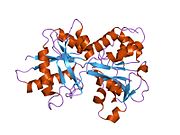
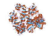
Ferritin is a universal intracellular protein that stores iron and releases it in a controlled fashion. The protein is produced by almost all living organisms, including archaea, bacteria, algae, higher plants, and animals. It is the primary intracellular iron-storage protein in both prokaryotes and eukaryotes, keeping iron in a soluble and non-toxic form. In humans, it acts as a buffer against iron deficiency and iron overload.

<span class="mw-page-title-main">Lactoferrin</span> Mammalian protein found in Homo sapiens

Lactoferrin (LF), also known as lactotransferrin (LTF), is a multifunctional protein of the transferrin family. Lactoferrin is a globular glycoprotein with a molecular mass of about 80 kDa that is widely represented in various secretory fluids, such as milk, saliva, tears, and nasal secretions. Lactoferrin is also present in secondary granules of PMNs and is secreted by some acinar cells. Lactoferrin can be purified from milk or produced recombinantly. Human colostrum has the highest concentration, followed by human milk, then cow milk (150 mg/L).

<span class="mw-page-title-main">Total iron-binding capacity</span> Medical blood test to measure transferrin

Total iron-binding capacity (TIBC) or sometimes transferrin iron-binding capacity is a medical laboratory test that measures the blood's capacity to bind iron with transferrin. Transferrin can bind two atoms of ferric iron (Fe3+) with high affinity. It means that transferrin has the capacity to transport approximately from 1.40 to 1.49 mg of iron per gram of transferrin present in the blood.

Transferrin saturation (TS), measured as a percentage, is a medical laboratory value. It is the value of serum iron divided by the total iron-binding capacity of the available transferrin, the main protein that binds iron in the blood, this value tells a clinician how much serum iron is bound. For instance, a value of 15% means that 15% of iron-binding sites of transferrin are being occupied by iron. The three results are usually reported together. A low transferrin saturation is a common indicator of iron deficiency anemia whereas a high transferrin saturation may indicate iron overload or hemochromatosis. Transferrin saturation is also called transferrin saturation index (TSI) or transferrin saturation percentage (TS%)

<span class="mw-page-title-main">Human iron metabolism</span> Iron metabolism in the body

Human iron metabolism is the set of chemical reactions that maintain human homeostasis of iron at the systemic and cellular level. Iron is both necessary to the body and potentially toxic. Controlling iron levels in the body is a critically important part of many aspects of human health and disease. Hematologists have been especially interested in systemic iron metabolism, because iron is essential for red blood cells, where most of the human body's iron is contained. Understanding iron metabolism is also important for understanding diseases of iron overload, such as hereditary hemochromatosis, and iron deficiency, such as iron-deficiency anemia.

<span class="mw-page-title-main">Hepcidin</span> Protein-coding gene in the species Homo sapiens

Hepcidin is a protein that in humans is encoded by the HAMP gene. Hepcidin is a key regulator of the entry of iron into the circulation in mammals.

<span class="mw-page-title-main">HFE (gene)</span> Mammalian protein found in Homo sapiens

Human homeostatic iron regulator protein, also known as the HFE protein, is a transmembrane protein that in humans is encoded by the HFE gene. The HFE gene is located on short arm of chromosome 6 at location 6p22.2

Iron-binding proteins are carrier proteins and metalloproteins that are important in iron metabolism and the immune response. Iron is required for life.

<span class="mw-page-title-main">Atransferrinemia</span> Medical condition

Atransferrinemia is an autosomal recessive metabolic disorder in which there is an absence of transferrin, a plasma protein that transports iron through the blood. Atransferrinemia is characterized by anemia and hemosiderosis in the heart and liver. The iron damage to the heart can lead to heart failure. The anemia is typically microcytic and hypochromic. Atransferrinemia was first described in 1961 and is extremely rare, with only ten documented cases worldwide.

<span class="mw-page-title-main">Transferrin receptor</span> Family of transport proteins

Transferrin receptor (TfR) is a carrier protein for transferrin. It is needed for the import of iron into cells and is regulated in response to intracellular iron concentration. It imports iron by internalizing the transferrin-iron complex through receptor-mediated endocytosis. The existence of a receptor for transferrin iron uptake has been recognized since the late 1950s. Earlier two transferrin receptors in humans, transferrin receptor 1 and transferrin receptor 2 had been characterized and until recently cellular iron uptake was believed to occur chiefly via these two well documented transferrin receptors. Both these receptors are transmembrane glycoproteins. TfR1 is a high affinity ubiquitously expressed receptor while expression of TfR2 is restricted to certain cell types and is unaffected by intracellular iron concentrations. TfR2 binds to transferrin with a 25-30 fold lower affinity than TfR1. Although TfR1 mediated iron uptake is the major pathway for iron acquisition by most cells and especially developing erythrocytes, several studies have indicated that the uptake mechanism varies depending upon the cell type. It is also reported that Tf uptake exists independent of these TfRs although the mechanisms are not well characterized. The multifunctional glycolytic enzyme glyceraldehyde 3-phosphate dehydrogenase has been shown to utilize post translational modifications to exhibit higher order moonlighting behavior wherein it switches its function as a holo or apo transferrin receptor leading to either iron delivery or iron export respectively.

<span class="mw-page-title-main">Ovotransferrin</span> Protein found in egg whites

Ovotransferrin (conalbumin) is a glycoprotein of egg white albumen. Egg white albumen is composed of multiple proteins, of which ovotransferrin is the most heat reliable. It has a molecular weight of 76,000 daltons and contains about 700 amino acids. Ovotransferrin makes up approximately 13% of egg albumen. As a member of the transferrin and metalloproteinase family, ovotransferrin has been found to possess antibacterial and antioxydant and immunomodulatory properties, arising primarily through its iron (Fe3+) binding capacity by locking away a key biochemical component necessary for micro-organismal survival. Bacteria starved of iron are rendered incapable of moving, making ovotransferrin a potent bacteriostatic.

<span class="mw-page-title-main">Iron-responsive element-binding protein</span> Protein family

The iron-responsive element-binding proteins, also known as IRE-BP, IRBP, IRP and IFR , bind to iron-responsive elements (IREs) in the regulation of human iron metabolism.

<span class="mw-page-title-main">Iron in biology</span> Use of Iron by organisms

Iron is an important biological element. It is used in both the ubiquitous iron-sulfur proteins and in vertebrates it is used in hemoglobin which is essential for blood and oxygen transport.

<span class="mw-page-title-main">Transferrin receptor 2</span> Mammalian protein found in Homo sapiens

Transferrin receptor 2 (TfR2) is a protein that in humans is encoded by the TFR2 gene. This protein is involved in the uptake of transferrin-bound iron into cells by endocytosis, although its role is minor compared to transferrin receptor 1.

<span class="mw-page-title-main">Transferrin receptor 1</span> Protein-coding gene in the species Homo sapiens

Transferrin receptor protein 1 (TfR1), also known as Cluster of Differentiation 71 (CD71), is a protein that in humans is encoded by the TFRC gene. TfR1 is required for iron import from transferrin into cells by endocytosis.

Haemochromatosis type 3 is a type of iron overload disorder associated with deficiencies in transferrin receptor 2. It exhibits an autosomal recessive inheritance pattern. The first confirmed case was diagnosed in 1865 by French doctor Trousseau. Later in 1889, the German doctor von Recklinghausen indicated that the liver contains iron, and due to bleeding being considered to be the cause, he called the pigment "Haemochromatosis." In 1935, English doctor Sheldon's groundbreaking book titled, Haemochromatosis, reviewed 311 patient case reports and presented the idea that haemochromatosis was a congenital metabolic disorder. Hereditary haemochromatosis is a congenital disorder which affects the regulation of iron metabolism thus causing increased gut absorption of iron and a gradual build-up of pathologic iron deposits in the liver and other internal organs, joint capsules and the skin. The iron overload could potentially cause serious disease from the age of 40–50 years. In the final stages of the disease, the major symptoms include liver cirrhosis, diabetes and bronze-colored skin. There are four types of hereditary hemochromatosis which are classified depending on the age of onset and other factors such as genetic cause and mode of inheritance.

Soluble transferrin receptor conventionally refers to the cleaved extracellular portion of transferrin receptor 1 that is released into serum. This receptor is a protein dimer of two identical subunits, linked together by two pairs of disulfide bonds. Its molecular mass 190,000 Dalton.

Clement Alfred Finch was an American physician specializing in hematology whose research on iron metabolism in the bloodstream at the University of Washington led to significant advancements in accurately diagnosing and treating anemia during a time period in which little was known about this aspect of the body. Finch was distinctively noted for using himself as a test subject by taking blood and bone marrow from his own bones before conducting similar tests on patients. He graduated in 1941 from the University of Rochester Medical School and a year later was married to the first of three wives. He experienced a 60-year tenure at the University of Washington, and has published many scholarly articles pertaining to iron in the bloodstream and is the author of three books entitled: Iron Metabolism (1962), Red Cell Manual (1969) and Fulfilling the Dream: A History of the University of Washington School of Medicine 1946 to 1988 (1990). Finch was elected as a Fellow of the National Academy of Sciences in 1974, and elected as a Fellow of the American Academy of Arts and Sciences in 1976.

<span class="mw-page-title-main">KP1019</span> Chemical compound

KP1019, or indazole trans-[tetrachlorobis(1H-indazole)ruthenate(III)], is one of four ruthenium anti-cancer drugs to enter into phase I clinical trials, the others being BOLD-100, NAMI-A and TLD-1433. Research into ruthenium-based drugs has provided novel alternatives for platinum-based chemotherapeutics such as Cisplatin and its derivatives. KP1019 is useful for metastatic tumors and cis-platin resistant tumors. It exhibits potent cytotoxicity against primary tumors, particularly in colorectal cancer.

Hemochromatosis type 4 is a hereditary iron overload disorder that affects ferroportin, an iron transport protein needed to export iron from cells into circulation. Although the disease is rare, it is found throughout the world and affects people from various ethnic groups. While the majority of individuals with type 4 hemochromatosis have a relatively mild form of the disease, some affected individuals have a more severe form. As the disease progresses, iron may accumulate in the tissues of affected individuals over time, potentially resulting in organ damage.

References

  1. 1 2 3 GRCh38: Ensembl release 89: ENSG00000091513 - Ensembl, May 2017
  2. 1 2 3 GRCm38: Ensembl release 89: ENSMUSG00000032554 - Ensembl, May 2017
  3. "Human PubMed Reference:". National Center for Biotechnology Information, U.S. National Library of Medicine.
  4. "Mouse PubMed Reference:". National Center for Biotechnology Information, U.S. National Library of Medicine.
  5. Crichton RR, Charloteaux-Wauters M (May 1987). "Iron transport and storage". European Journal of Biochemistry. 164 (3): 485–506. doi: 10.1111/j.1432-1033.1987.tb11155.x . PMID   3032619.
  6. Hall DR, Hadden JM, Leonard GA, Bailey S, Neu M, Winn M, Lindley PF (January 2002). "The crystal and molecular structures of diferric porcine and rabbit serum transferrins at resolutions of 2.15 and 2.60 A, respectively". Acta Crystallographica. Section D, Biological Crystallography. 58 (Pt 1): 70–80. Bibcode:2002AcCrD..58...70H. doi:10.1107/s0907444901017309. PMID   11752780.
  7. 1 2 Yang F, Lum JB, McGill JR, Moore CM, Naylor SL, van Bragt PH, et al. (May 1984). "Human transferrin: cDNA characterization and chromosomal localization". Proceedings of the National Academy of Sciences of the United States of America. 81 (9): 2752–6. Bibcode:1984PNAS...81.2752Y. doi: 10.1073/pnas.81.9.2752 . PMC   345148 . PMID   6585826.
  8. 1 2 Kawabata H (March 2019). "Transferrin and transferrin receptors update". Free Radical Biology & Medicine. 133: 46–54. doi:10.1016/j.freeradbiomed.2018.06.037. PMID   29969719. S2CID   49674402.
  9. Aisen P, Leibman A, Zweier J (March 1978). "Stoichiometric and site characteristics of the binding of iron to human transferrin". The Journal of Biological Chemistry. 253 (6): 1930–7. doi: 10.1016/S0021-9258(19)62337-9 . PMID   204636.
  10. Nicotra S, Sorio D, Filippi G, De Gioia L, Paterlini V, De Palo EF, et al. (November 2017). "Terbium chelation, a specific fluorescent tagging of human transferrin. Optimization of conditions in view of its application to the HPLC analysis of carbohydrate-deficient transferrin (CDT)". Analytical and Bioanalytical Chemistry. 409 (28): 6605–6612. doi:10.1007/s00216-017-0616-z. PMID   28971232. S2CID   13929228.
  11. 1 2 3 MacGillivray RT, Moore SA, Chen J, Anderson BF, Baker H, Luo Y, et al. (June 1998). "Two high-resolution crystal structures of the recombinant N-lobe of human transferrin reveal a structural change implicated in iron release". Biochemistry. 37 (22): 7919–28. doi:10.1021/bi980355j. PMID   9609685.
  12. 1 2 Dewan JC, Mikami B, Hirose M, Sacchettini JC (November 1993). "Structural evidence for a pH-sensitive dilysine trigger in the hen ovotransferrin N-lobe: implications for transferrin iron release". Biochemistry. 32 (45): 11963–8. doi:10.1021/bi00096a004. PMID   8218271.
  13. 1 2 3 Baker EN, Lindley PF (August 1992). "New perspectives on the structure and function of transferrins". Journal of Inorganic Biochemistry. 47 (3–4): 147–60. doi:10.1016/0162-0134(92)84061-q. PMID   1431877.
  14. 1 2 Halbrooks PJ, He QY, Briggs SK, Everse SJ, Smith VC, MacGillivray RT, Mason AB (April 2003). "Investigation of the mechanism of iron release from the C-lobe of human serum transferrin: mutational analysis of the role of a pH sensitive triad". Biochemistry. 42 (13): 3701–7. doi:10.1021/bi027071q. PMID   12667060.
  15. Moos T (November 2002). "Brain iron homeostasis". Danish Medical Bulletin. 49 (4): 279–301. PMID   12553165.
  16. 1 2 3 4 Macedo MF, de Sousa M (March 2008). "Transferrin and the transferrin receptor: of magic bullets and other concerns". Inflammation & Allergy - Drug Targets. 7 (1): 41–52. doi:10.2174/187152808784165162. PMID   18473900.
  17. Gabaldón T, Koonin EV (May 2013). "Functional and evolutionary implications of gene orthology". Nature Reviews. Genetics. Nature Portfolio. 14 (5): 360–6. doi:10.1038/nrg3456. PMC   5877793 . PMID   23552219.
  18. 1 2 3 "Transferrin Structure". St. Edward's University. 2005-07-18. Archived from the original on 2012-12-11. Retrieved 2009-04-24.
  19. PDB: 1suv ; Cheng Y, Zak O, Aisen P, Harrison SC, Walz T (Feb 2004). "Structure of the human transferrin receptor-transferrin complex". Cell. 116 (4): 565–76. doi:10.1016/S0092-8674(04)00130-8. PMID   14980223. S2CID   2981917.
  20. PDB: 2nsu ; Hafenstein S, Palermo LM, Kostyuchenko VA, Xiao C, Morais MC, Nelson CD, Bowman VD, Battisti AJ, Chipman PR, Parrish CR, Rossmann MG (Apr 2007). "Asymmetric binding of transferrin receptor to parvovirus capsids". Proceedings of the National Academy of Sciences of the United States of America. 104 (16): 6585–9. Bibcode:2007PNAS..104.6585H. doi: 10.1073/pnas.0701574104 . PMC   1871829 . PMID   17420467.
  21. Ritchie RF, Palomaki GE, Neveux LM, Navolotskaia O, Ledue TB, Craig WY (1999). "Reference distributions for the negative acute-phase serum proteins, albumin, transferrin and transthyretin: a practical, simple and clinically relevant approach in a large cohort". Journal of Clinical Laboratory Analysis. 13 (6): 273–9. doi:10.1002/(SICI)1098-2825(1999)13:6<273::AID-JCLA4>3.0.CO;2-X. PMC   6808097 . PMID   10633294.
  22. Miller JL (July 2013). "Iron deficiency anemia: a common and curable disease". Cold Spring Harbor Perspectives in Medicine. 3 (7): a011866. doi:10.1101/cshperspect.a011866. PMC   3685880 . PMID   23613366.
  23. Bacon BR, Adams PC, Kowdley KV, Powell LW, Tavill AS (July 2011). "Diagnosis and management of hemochromatosis: 2011 practice guideline by the American Association for the Study of Liver Diseases". Hepatology. Baltimore, Md. 54 (1): 328–43. doi:10.1002/hep.24330. PMC   3149125 . PMID   21452290.
  24. "Hemochromatosis". guidelinecentral.com.
  25. Ghadiri M, Vasheghani-Farahani E, Atyabi F, Kobarfard F, Mohamadyar-Toupkanlou F, Hosseinkhani H (October 2017). "Transferrin-conjugated magnetic dextran-spermine nanoparticles for targeted drug transport across blood-brain barrier". Journal of Biomedical Materials Research Part A. 105 (10): 2851–2864. doi:10.1002/jbm.a.36145. PMID   28639394.
  26. Gaspar R (February 2013). "Nanoparticles: Pushed off target with proteins". Nature Nanotechnology. 8 (2): 79–80. Bibcode:2013NatNa...8...79G. doi:10.1038/nnano.2013.11. PMID   23380930.
  27. Li S, Peng Z, Dallman J, Baker J, Othman AM, Blackwelder PL, Leblanc RM (September 2016). "Crossing the blood-brain-barrier with transferrin conjugated carbon dots: A zebrafish model study". Colloids and Surfaces. B, Biointerfaces. 145: 251–256. doi: 10.1016/j.colsurfb.2016.05.007 . PMID   27187189.
  28. Sharpe PC (November 2001). "Biochemical detection and monitoring of alcohol abuse and abstinence". Annals of Clinical Biochemistry. 38 (Pt 6): 652–64. doi:10.1258/0004563011901064. PMID   11732647. S2CID   12203099.
  29. Jain S, Gautam V, Naseem S (January 2011). "Acute-phase proteins: As diagnostic tool". Journal of Pharmacy & Bioallied Sciences. 3 (1): 118–27. doi: 10.4103/0975-7406.76489 . PMC   3053509 . PMID   21430962.
  30. "Normal Reference Range Table". Interactive Case Study Companion to Pathological Basis of Disease. The University of Texas Southwestern Medical Center at Dallas. Archived from the original on 2011-12-25. Retrieved 2008-10-25.
    Kumar V, Hagler HK (1999). Interactive Case Study Companion to Robbins Pathologic Basis of Disease (6th Edition (CD-ROM for Windows & Macintosh, Individual) ed.). W B Saunders Co. ISBN   0-7216-8462-9.
  31. Storch S, Kübler B, Höning S, Ackmann M, Zapf J, Blum W, Braulke T (December 2001). "Transferrin binds insulin-like growth factors and affects binding properties of insulin-like growth factor binding protein-3". FEBS Letters. 509 (3): 395–8. doi: 10.1016/S0014-5793(01)03204-5 . PMID   11749962. S2CID   22895295.
  32. Weinzimer SA, Gibson TB, Collett-Solberg PF, Khare A, Liu B, Cohen P (April 2001). "Transferrin is an insulin-like growth factor-binding protein-3 binding protein". The Journal of Clinical Endocrinology and Metabolism. 86 (4): 1806–13. doi: 10.1210/jcem.86.4.7380 . PMID   11297622.
  33. Hsu SL, Lin YF, Chou CK (April 1992). "Transcriptional regulation of transferrin and albumin genes by retinoic acid in human hepatoma cell line Hep3B". The Biochemical Journal. 283 ( Pt 2) (2): 611–5. doi:10.1042/bj2830611. PMC   1131079 . PMID   1315521.
  34. Chung MC (October 1984). "Structure and function of transferrin". Biochemical Education. 12 (4): 146–154. doi:10.1016/0307-4412(84)90118-3.

Further reading