Long-chain-fatty-acid—CoA ligase

Last updated
Long-chain-fatty-acid—CoA ligase
PDB 1v26 EBI.jpg
Long chain fatty acyl-CoA synthetase homodimer from Thermus thermophilus . [1]
Identifiers
EC no. 6.2.1.3
CAS no. 9013-18-7
Databases
IntEnz IntEnz view
BRENDA BRENDA entry
ExPASy NiceZyme view
KEGG KEGG entry
MetaCyc metabolic pathway
PRIAM profile
PDB structures RCSB PDB PDBe PDBsum
Gene Ontology AmiGO / QuickGO
Search
PMC articles
PubMed articles
NCBI proteins
acyl-CoA synthetase long-chain family member 1
Identifiers
Symbol ACSL1
Alt. symbolsFACL2
NCBI gene 2180
HGNC 3569
OMIM 152425
RefSeq NM_001995
UniProt P33121
Other data
EC number 6.2.1.3
Locus Chr. 4 q35
Search for
Structures Swiss-model
Domains InterPro

The long chain fatty acyl-CoA ligase (or synthetase) is an enzyme (EC 6.2.1.3) of the ligase family that activates the oxidation of complex fatty acids. [2] Long chain fatty acyl-CoA synthetase catalyzes the formation of fatty acyl-CoA by a two-step process proceeding through an adenylated intermediate. [3] The enzyme catalyzes the following reaction,

Contents

Fatty acid + CoA + ATPAcyl-CoA + AMP + PP i

It is present in all organisms from bacteria to humans. It catalyzes the pre-step reaction for β-oxidation of fatty acids or can be incorporated in phospholipids.

Function

Long chain fatty acyl-CoA synthetase, LC-FACS, plays a role in the physiological regulation of various cellular functions via the production of long chain fatty acyl-CoA esters, which reportedly have affected protein transport, enzyme activation, protein acylation, cell signaling, and transcriptional regulation. [1] The formation of fatty acyl-CoA is catalyzed in two steps: a stable intermediate of fatty acyl-AMP molecule and then the product is formed—fatty acid acyl-CoA molecule. [4]

Fatty acyl CoA synthetase catalyzes the activation of a long fatty acid chain to a fatty acyl CoA, requiring the energy of 1 ATP to AMP and pyrophosphate. This step uses 2 "ATP equivalents" because pyrophosphate is cleaved into 2 molecules of inorganic phosphate, breaking a high-energy phosphate bond.

Mechanism and active site

The mechanism for Long Chain Fatty Acyl-CoA Synthetase is a “bi uni uni bi ping-pong” mechanism. [1] The uni and bi prefixes refer to the number of substrates that enter the enzyme and the number of products that leave the enzyme; bi describes a situation where two substrates enter the enzyme at the same time. Ping-pong signifies that a product is released before another substrate can bind to the enzyme.

In step one, ATP and a long chain fatty acid enter the enzyme's active site. Within the active site the negatively charged oxygen on the fatty acid attacks the alpha phosphate on ATP, forming an ATP-long chain fatty acid intermediate. (Step 1, Figure 2) In the second step, Pyrophosphate (PPi) leaves, resulting in an AMP-long chain fatty acid molecule within the enzyme's active site. (Step 2, Figure 2) Coenzyme A now enters the enzyme and another intermediate is formed which consists of AMP-long chain fatty acid-Coenzyme A. (Step 3, Figure 2) At the end of this mechanism two products are released, AMP and acyl coa product. (Step 4, Figure 2)

Acyl CoA is formed from long chain fatty acids through an acyl substitution. In an ATP dependent reaction, the fatty acid carboxylate is converted to a thioester. The final products of this reaction are acyl-CoA, pyrophosphate (PPi) and AMP.

Figure 1. Long chain Fatty acyl-CoA synthetase asymmetric unit showing active site residues Trp 234, Tyr504, and Glu540, along with the potentially supporting Asn450 residue. Long Chain Fatty Acyl-CoA Synthetase.gif
Figure 1. Long chain Fatty acyl-CoA synthetase asymmetric unit showing active site residues Trp 234, Tyr504, and Glu540, along with the potentially supporting Asn450 residue.
Figure 2. Mechanism of long chain fatty acyl-CoA synthetase. Mechanismabcd.png
Figure 2. Mechanism of long chain fatty acyl-CoA synthetase.

Structure

There are several highly conserved areas and a 20-30% amino acid sequence similarity between the members of this superfamily. [1] The enzymes in the family consist of a large N-terminal and a small C-terminal domain, with the catalytic site positioned between the two domains. [1] Substrate binding may affect the relative positions of the C- and N-terminal domains. The C-terminal domain of LC-FACS is assumed to be in an open conformation when a substrate is absent and in a closed conformation when a substrate is bound. [1] The accessibility of the active site to solvent is reduced when the C- and N-terminal domains approach one another. [5]

The structure-function relationship between LC-FACS and the formation and processing of the acyl-AMP intermediate was still unclear. A domain swapped dimer is formed by LC-FACS, with monomer interacting at the N-terminal domains. [6] A large electrostatically positive concave is located at the back of the structure in the central valley of the homodimer. [1] Asp15 forms an intermolecular salt bridge with Arg176 in the dimer interactions. An intermolecular hydrogen bond is formed between the main chain carbonyl group of Glu16and the side chain of Arg199. At the interface, Glu175 forms an intermolecular salt bridge with Arg199. [5] [7] [8] [9] The L motif, a six-amino acid peptide linker, connects the large N-terminal domain and a small C-terminal domain of each LC-FACS monomer. [1] The N-terminal domain is composed of two subdomains: a distorted antiparallel β-barrel and two β-sheets surrounded by α-helices forming an αβαβα sandwich. [1] The small C-terminal globular domain consists of two-stranded β-sheet and a three-stranded antiparallel β-sheet flanked by three α-helices. [1]

Dimer interaction

Figure 3. Dimerization of LC-FACS. Dimerinteract.png
Figure 3. Dimerization of LC-FACS.

The dimerization of LC-FACS is stabilized through a salt bridge between Asp15 of sequence A and Arg176 of sequence B. Figure 3 shows this salt bridge between these two amino acids. The yellow line between Asp15 and Arg176 shows the salt bridge present.

ATP binding to the C-terminal domain

The conformations of the C-terminal domain of the LC-FACS structures are dependent on the presence of a ligand. [1] AMP-PNP, a nonhydrolyzable ATP analogue, bound to LC-FACS results in the closed conformation with the C- and N-terminal domains directly interacting. [1] In crystal structures, AMP-PNP is bound in a crevasse of each monomer at the interface between the N- and C-terminal domains. [1] The closed conformation of the C-terminal domain is retained with myristroyl-AMP. [1] Three residues in the C-terminal domain, Glu443, Glu475, and Lys527, interact noncovalently with L motif residues and the N-terminal domain to stabilize the closed conformation. [1] There are two types of open conformations in the C-terminal domains of the uncomplexed structure. The C- and N-terminal domains do not interact directly for both monomers of the dimer. [1] An extensive hydrogen bond network is used by the AMP moiety of the bound ATP molecule to hold the C- and N-terminal domains together. [1]

Figure 4. Active site of long chain fatty acyl-CoA synthetase with a long chain fatty acid. Trp444, Lys435 and Lys439 are the important residues Long chain fatty acid CoA ligase.png
Figure 4. Active site of long chain fatty acyl-CoA synthetase with a long chain fatty acid. Trp444, Lys435 and Lys439 are the important residues

Fatty acid-binding tunnel

Bulkier long chain fatty acids are bound by a fatty acid-binding tunnel that is located in the N-terminal domain of each monomer. [1] A large β-sheet and an α-helix cluster surround the tunnel which extends from the concave cavity in the central valley to the site of ATP-binding. [1] There are two distinct paths in the large central pathway of the tunnel in the complex structure, which includes the “ATP path” and the “center path,” separated by the indole ring of Trp234 in the G motif. [1] There is also another branch of the central pathway known as the “dead and branch.” The indole ring of Trp234 closes the fatty acid-binding tunnel in the uncomplexed structure. [1] It opens up once AMP-PNP binds through hydrogen bond formation between β-phosphate and the nitrogen on the ring of Trp234. [1] During this time, the closed conformation is adopted by the mobile C-terminal domain. There is a shift in the flexible loop of the G motif in the closed structures of LC-FACS, resulting in a wider dead end branch compared to the uncomplexed forms. [1]

The ATP binding site is connected to an ATP path that is a hydrophobic channel in the fatty acid-binding tunnel. [1] The fatty acid enters through the center path extending from the interface of the dimer along β-strand 13 to the ATP path. [1] The connection between the two paths is blocked by the indole ring of Trp234 in the absence of ATP. Water molecules fill the center path in the AMP-PNP and myristoyl-AMP complex structures and through the entrance of the center path, they connect to the bulk solvent regions. The basic residues from each monomer, Lys219, Arg296, Arg297, Arg321, Lys350, and Lys 354, cause the entrance of the center path to generate a positive electrostatic potential. [1] The dead end branch contains residues 235-243 and extends from the fatty acid-binding tunnel to α-helix h. [1] The bottom of the dead end branch consists of a hydrophilic environment from the water molecules and polar side chains. [1]

Domains

Figure 5. ConservedDomains.png
Figure 5.
Figure 6. ConservedDomains3.png
Figure 6.

The domains founds in Long chain fatty acyl CoA synthetase are shown both in the enzyme view (figure 5) and sequence view (figure 6). LC-FACS has five domains. After searching 1v26 in Entrez, the location of the 5 domains was shown and was used to create figure 5 and 6. The ribbons colors in figure 5 correspond to the colors of the figure 6.

Inhibition by long chain fatty acyl-CoAs

A long term and short term regulation controls fatty acid synthesis. [4] Long term fatty acid synthesis regulation is dependent on the rate of acetyl-CoA carboxylase (ACC) synthesis, the rate-limiting enzyme and first enzyme of the fatty acid synthesis, and fatty acid synthase (FAS), the second and major enzyme of the fatty acid synthesis. [4] [10] [11] [12] Cellular fatty acyl-CoA is involved in the short term regulation, but there is not a full understanding of the mechanisms. [13]

Free fatty acids inhibits the de novo fatty acid synthesis and appears to be dependent on the formation of long chain fatty acyl-CoAs. [14] Studies have shown that long chain fatty acyl-CoAs inhibit ACC and FAS via feedback inhibition. [15] [16] [17] [18] Long chain fatty acyl-CoA's inhibitory effect on the fatty acid synthesis may be a result of its regulation of lipogenic enzymes in a feedback manner through gene transcription suppression. [19]

Long-chain fatty-acid-CoA ligase in cells catalytically synthesizes long chain fatty acyl-CoAs. Long-chain fatty-acid-CoA ligase may be involved in an important role in the suppression of fatty acid synthesis and it has been reported that it played a part in fatty acid synthesis inhibition. [20] It was recently found that vitamin D3 upregulates FACL3, which forms long-chain fatty acid synthesis through the use of myristic acid, eicosapentaenoic acid (EPA), and arachidonic acid as substrates, in expression and activity levels. [21] FACL3 contributes to vitamin D3 growth inhibitory effect in human prostate cancer LNCaP cells. [21] A current study reports that the feedback inhibition of FAS expression by long chain fatty acyl-CoAs causes the downregulation of FAS mRNA by vitamin D3. [4] [22]

Clinical significance

Adrenoleukodystrophy (ALD) is the build up of long chain fatty acids in the brain and adrenal cortex, because of the decreased activity of long chain fatty acyl coa synthetase. [23] The oxidation of the long chain fatty acids normally occurs in the peroxisome where the long chain fatty acyl coa synthetase is found. Long chain fatty acids enter the peroxisome via a transporter protein, ALDP, which creates a gate in the membrane of the peroxisome. In ALD the gene for this peroximal membrane transporter, ALDP, is defective, preventing long chain fatty acids from entering the peroxisome. [24]

Examples

Human genes encoding long-chain-fatty-acid—CoA ligase enzymes (also known as acyl-CoA synthetase long-chain, or ACSL) include:

See also

Related Research Articles

<span class="mw-page-title-main">Beta oxidation</span> Process of fatty acid breakdown

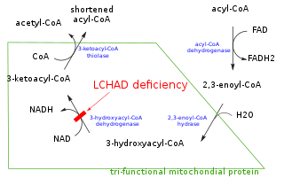
In biochemistry and metabolism, beta oxidation (also β-oxidation) is the catabolic process by which fatty acid molecules are broken down in the cytosol in prokaryotes and in the mitochondria in eukaryotes to generate acetyl-CoA, which enters the citric acid cycle, and NADH and FADH2, which are co-enzymes used in the electron transport chain. It is named as such because the beta carbon of the fatty acid undergoes oxidation to a carbonyl group. Beta-oxidation is primarily facilitated by the mitochondrial trifunctional protein, an enzyme complex associated with the inner mitochondrial membrane, although very long chain fatty acids are oxidized in peroxisomes.

<span class="mw-page-title-main">Firefly luciferase</span>

Firefly luciferase is the light-emitting enzyme responsible for the bioluminescence of fireflies and click beetles. The enzyme catalyses the oxidation of firefly luciferin, requiring oxygen and ATP. Because of the requirement of ATP, firefly luciferases have been used extensively in biotechnology.

Acyl-CoA dehydrogenases (ACADs) are a class of enzymes that function to catalyze the initial step in each cycle of fatty acid β-oxidation in the mitochondria of cells. Their action results in the introduction of a trans double-bond between C2 (α) and C3 (β) of the acyl-CoA thioester substrate. Flavin adenine dinucleotide (FAD) is a required co-factor in addition to the presence of an active site glutamate in order for the enzyme to function.

<span class="mw-page-title-main">Acyl-CoA</span>

Acyl-CoA is a group of coenzymes that metabolize fatty acids. Acyl-CoA's are susceptible to beta oxidation, forming, ultimately, acetyl-CoA. The acetyl-CoA enters the citric acid cycle, eventually forming several equivalents of ATP. In this way, fats are converted to ATP, the universal biochemical energy carrier.

Fatty acid degradation is the process in which fatty acids are broken down into their metabolites, in the end generating acetyl-CoA, the entry molecule for the citric acid cycle, the main energy supply of living organisms, including bacteria and animals. It includes three major steps:

Palmitoyl-CoA is an acyl-CoA thioester. It is an "activated" form of palmitic acid and can be transported into the mitochondrial matrix by the carnitine shuttle system, and once inside can participate in beta-oxidation. Alternatively, palmitoyl-CoA is used as a substrate in the biosynthesis of sphingosine.

Acetyl-CoA synthetase (ACS) or Acetate—CoA ligase is an enzyme involved in metabolism of acetate. It is in the ligase class of enzymes, meaning that it catalyzes the formation of a new chemical bond between two large molecules.

<span class="mw-page-title-main">Thiolase</span> Enzymes

Thiolases, also known as acetyl-coenzyme A acetyltransferases (ACAT), are enzymes which convert two units of acetyl-CoA to acetoacetyl CoA in the mevalonate pathway.

Pantothenate kinase (EC 2.7.1.33, PanK; CoaA) is the first enzyme in the Coenzyme A (CoA) biosynthetic pathway. It phosphorylates pantothenate (vitamin B5) to form 4'-phosphopantothenate at the expense of a molecule of adenosine triphosphate (ATP). It is the rate-limiting step in the biosynthesis of CoA.

In enzymology, a 4-coumarate—CoA ligase is an enzyme that catalyzes the chemical reaction

In enzymology, a 6-carboxyhexanoate—CoA ligase is an enzyme that catalyzes the chemical reaction

Butyrate—CoA ligase, also known as xenobiotic/medium-chain fatty acid-ligase (XM-ligase), is an enzyme that catalyzes the chemical reaction:

In enzymology, a long-chain-fatty-acid—[acyl-carrier-protein] ligase is an enzyme that catalyzes the chemical reaction

In enzymology, a long-chain-fatty-acid—luciferin-component ligase is an enzyme that catalyzes the chemical reaction

<span class="mw-page-title-main">Fatty-acyl-CoA synthase</span>

Fatty-acyl-CoA Synthase, or more commonly known as yeast fatty acid synthase, is an enzyme complex responsible for fatty acid biosynthesis, and is of Type I Fatty Acid Synthesis (FAS). Yeast fatty acid synthase plays a pivotal role in fatty acid synthesis. It is a 2.6 MDa barrel shaped complex and is composed of two, unique multi-functional subunits: alpha and beta. Together, the alpha and beta units are arranged in an α6β6 structure. The catalytic activities of this enzyme complex involves a coordination system of enzymatic reactions between the alpha and beta subunits. The enzyme complex therefore consists of six functional centers for fatty acid synthesis.

<span class="mw-page-title-main">ACSL1</span> Protein-coding gene in the species Homo sapiens

Long-chain-fatty-acid—CoA ligase 1 is an enzyme that in humans is encoded by the ACSL1 gene.

<span class="mw-page-title-main">Very long-chain acyl-CoA synthetase</span> Protein-coding gene in the species Homo sapiens

Very long-chain acyl-CoA synthetase is an enzyme that in humans is encoded by the SLC27A2 gene.

<span class="mw-page-title-main">ACOT11</span> Protein-coding gene in the species Homo sapiens

Acyl-coenzyme A thioesterase 11 also known as StAR-related lipid transfer protein 14 (STARD14) is an enzyme that in humans is encoded by the ACOT11 gene. This gene encodes a protein with acyl-CoA thioesterase activity towards medium (C12) and long-chain (C18) fatty acyl-CoA substrates which relies on its StAR-related lipid transfer domain. Expression of a similar murine protein in brown adipose tissue is induced by cold exposure and repressed by warmth. Expression of the mouse protein has been associated with obesity, with higher expression found in obesity-resistant mice compared with obesity-prone mice. Alternative splicing results in two transcript variants encoding different isoforms.

<span class="mw-page-title-main">SLC27A5</span> Protein-coding gene in the species Homo sapiens

Bile acyl-CoA synthetase is an enzyme that in humans is encoded by the SLC27A5 gene.

<span class="mw-page-title-main">ACOT13</span> Protein-coding gene in the species Homo sapiens

Acyl-CoA thioesterase 13 is a protein that in humans is encoded by the ACOT13 gene. This gene encodes a member of the thioesterase superfamily. In humans, the protein co-localizes with microtubules and is essential for sustained cell proliferation.

References

  1. 1 2 3 4 5 6 7 8 9 10 11 12 13 14 15 16 17 18 19 20 21 22 23 24 25 26 27 28 29 PDB: 1V26 ; Hisanaga Y, Ago H, Nakagawa N, Hamada K, Ida K, Yamamoto M, Hori T, Arii Y, Sugahara M, Kuramitsu S, Yokoyama S, Miyano M (July 2004). "Structural basis of the substrate-specific two-step catalysis of long chain fatty acyl-CoA synthetase dimer". J. Biol. Chem. 279 (30): 31717–26. doi: 10.1074/jbc.M400100200 . PMID   15145952.
  2. Soupene E, Kuypers FA (May 2008). "Mammalian long-chain acyl-CoA synthetases". Exp. Biol. Med. (Maywood). 233 (5): 507–21. doi:10.3181/0710-MR-287. PMC   3377585 . PMID   18375835.
  3. Bækdal T, Schjerling CK, Hansen JK, Knudsen J (1997). "Analysis of long-chain acyl-Coenzyme A esters". In Christie W (ed.). Advances in Lipid Methodology (Three ed.). Ayr, Scotland: Oily Press. pp. 109–131. ISBN   978-0-9514171-7-1.
  4. 1 2 3 4 Qiao S, Tuohimaa P (November 2004). "Vitamin D3 inhibits fatty acid synthase expression by stimulating the expression of long-chain fatty-acid-CoA ligase 3 in prostate cancer cells". FEBS Lett. 577 (3): 451–4. doi: 10.1016/j.febslet.2004.10.044 . PMID   15556626. S2CID   25190904.
  5. 1 2 Conti E, Stachelhaus T, Marahiel MA, Brick P (July 1997). "Structural basis for the activation of phenylalanine in the non-ribosomal biosynthesis of gramicidin S". EMBO J. 16 (14): 4174–83. doi:10.1093/emboj/16.14.4174. PMC   1170043 . PMID   9250661.
  6. Liu Y, Eisenberg D (June 2002). "3D domain swapping: as domains continue to swap". Protein Sci. 11 (6): 1285–99. doi:10.1110/ps.0201402. PMC   2373619 . PMID   12021428.
  7. Conti E, Franks NP, Brick P (March 1996). "Crystal structure of firefly luciferase throws light on a superfamily of adenylate-forming enzymes". Structure. 4 (3): 287–98. doi: 10.1016/S0969-2126(96)00033-0 . PMID   8805533.
  8. May JJ, Kessler N, Marahiel MA, Stubbs MT (September 2002). "Crystal structure of DhbE, an archetype for aryl acid activating domains of modular nonribosomal peptide synthetases". Proc. Natl. Acad. Sci. U.S.A. 99 (19): 12120–5. Bibcode:2002PNAS...9912120M. doi: 10.1073/pnas.182156699 . PMC   129408 . PMID   12221282.
  9. Gulick AM, Starai VJ, Horswill AR, Homick KM, Escalante-Semerena JC (March 2003). "The 1.75 A crystal structure of acetyl-CoA synthetase bound to adenosine-5'-propylphosphate and coenzyme A". Biochemistry. 42 (10): 2866–73. doi:10.1021/bi0271603. PMID   12627952.
  10. Burton DN, Collins JM, Kennan AL, Porter JW (August 1969). "The effects of nutritional and hormonal factors on the fatty acid synthetase level of rat liver". J. Biol. Chem. 244 (16): 4510–6. doi: 10.1016/S0021-9258(18)94347-4 . PMID   5806590.
  11. Craig MC, Dugan RE, Muesing RA, Slakey LL, Porter JW (July 1972). "Comparative effects of dietary regimens on the levels of enzymes regulating the synthesis of fatty acids and cholesterol in rat liver". Arch. Biochem. Biophys. 151 (1): 128–36. doi:10.1016/0003-9861(72)90481-X. PMID   5044513.
  12. Majerus PW, Kilburn E (November 1969). "Acetyl coenzyme A carboxylase. The roles of synthesis and degradation in regulation of enzyme levels in rat liver". J. Biol. Chem. 244 (22): 6254–62. doi: 10.1016/S0021-9258(18)63531-8 . PMID   4981792.
  13. Goodridge AG (June 1973). "Regulation of fatty acid synthesis in isolated hepatocytes. Evidence for a physiological role for long chain fatty acyl coenzyme A and citrate". J. Biol. Chem. 248 (12): 4318–26. doi: 10.1016/S0021-9258(19)43775-7 . PMID   4145797.
  14. McGee R, Spector AA (July 1975). "Fatty acid biosynthesis in Erlich cells. The mechanism of short term control by exogenous free fatty acids". J. Biol. Chem. 250 (14): 5419–25. doi: 10.1016/S0021-9258(19)41198-8 . PMID   237919.
  15. Guynn RW, Veloso D, Veech RL (November 1972). "The concentration of malonyl-coenzyme A and the control of fatty acid synthesis in vivo". J. Biol. Chem. 247 (22): 7325–31. doi: 10.1016/S0021-9258(19)44633-4 . PMID   4638549.
  16. Numa S, Ringelmann E, Lynen F (December 1965). "[On inhibition of acetyl-CoA-carboxylase by fatty acid-coenzyme A compounds]". Biochem Z (in German). 343 (3): 243–57. PMID   5875764.
  17. Goodridge AG (November 1972). "Regulation of the activity of acetyl coenzyme A carboxylase by palmitoyl coenzyme A and citrate". J. Biol. Chem. 247 (21): 6946–52. doi: 10.1016/S0021-9258(19)44677-2 . PMID   5082134.
  18. Sumper M, Träuble H (February 1973). "Membranes as acceptors for palmitoyl CoA in fatty acid biosynthesis" (PDF). FEBS Lett. 30 (1): 29–34. doi:10.1016/0014-5793(73)80612-X. PMID   11947055. S2CID   8678424.
  19. Faergeman NJ, Knudsen J (April 1997). "Role of long-chain fatty acyl-CoA esters in the regulation of metabolism and in cell signalling". Biochem. J. 323 (1): 1–12. doi:10.1042/bj3230001. PMC   1218279 . PMID   9173866.
  20. Fox SR, Hill LM, Rawsthorne S, Hills MJ (December 2000). "Inhibition of the glucose-6-phosphate transporter in oilseed rape (Brassica napus L.) plastids by acyl-CoA thioesters reduces fatty acid synthesis". Biochem. J. 352 (2): 525–32. doi:10.1042/0264-6021:3520525. PMC   1221485 . PMID   11085947.
  21. 1 2 Qiao S, Tuohimaa P (June 2004). "The role of long-chain fatty-acid-CoA ligase 3 in vitamin D3 and androgen control of prostate cancer LNCaP cell growth". Biochem. Biophys. Res. Commun. 319 (2): 358–68. doi:10.1016/j.bbrc.2004.05.014. PMID   15178414.
  22. Qiao S, Pennanen P, Nazarova N, Lou YR, Tuohimaa P (May 2003). "Inhibition of fatty acid synthase expression by 1alpha,25-dihydroxyvitamin D3 in prostate cancer cells". J. Steroid Biochem. Mol. Biol. 85 (1): 1–8. doi:10.1016/S0960-0760(03)00142-0. PMID   12798352. S2CID   54296796.
  23. "Adrenoleukodystrophy Information Page". National Institute of Neurological Disorders and Stroke (NINDS). 2009-03-18. Archived from the original on 2006-05-10. Retrieved 2010-01-16.
  24. Kemp S, Watkins P (2009-03-03). "very long-chain fatty acids and X-ALD". X-linked Adrenoleukodystrophy Database. Archived from the original on December 21, 2009. Retrieved 2010-01-16.