Mediastinal shift

Last updated
Mediastinal shift
Pneumothorax CXR.jpg
Left tension pneumothorax with a large, well-demarcated area devoid of lung markings with tracheal deviation and movement of the heart away from the affected side.

Mediastinal shift is an abnormal movement of the mediastinal structures toward one side of the chest cavity. A shift indicates a severe imbalance of pressures inside the chest. [1] Mediastinal shifts are generally caused by increased lung volume, decreased lung volume, or abnormalities in the pleural space. Additionally, masses inside the mediastinum or musculoskeletal abnormalities can also lead to abnormal mediastinal arrangement. [2] Typically, these shifts are observed on x-ray but also on computed tomography (CT) or magnetic resonance imaging (MRI). On chest x-ray, tracheal deviation, or movement of the trachea away from its midline position can be used as a sign of a shift. Other structures like the heart can also be used as reference points. [3] [4] Below are examples of pathologies that can cause a mediastinal shift and their appearance.

Contents

Pleural space abnormalities

Tension pneumothorax

Tension pneumothorax is an emergent condition in which air gets trapped in the space between the chest wall and the lung. This space is referred to as the pleural space. Because air can't escape from this space, the pocket of air grows larger and larger which results in collapse of the lung closest to the pneumothorax. Forces are transmitted to the mediastinum and effectively "push" the mediastinal structures to the opposite side of the chest. [5]


Pleural effusion

A massive left pleural effusion displacing the heart and trachea to the right Pleural effusion - Left lung (7471755836).jpg
A massive left pleural effusion displacing the heart and trachea to the right

A pleural effusion is an accumulation of fluid inside the pleural space. If this collection of fluid gets large enough, it can also push structures in the chest away from it and cause a mediastinal shift. However, a pleural effusion can also pull mediastinal structure towards itself. If this is the case, then there is an underlying condition causing collapse of the lung on that side. An example is a tumor obstructing a bronchus and causing lung collapse and pleural effusion. [3]

Hemothorax

Massive right sided pleural effusion later confirmed to be a hemothorax PMC2567296 1757-1626-1-225-2.png
Massive right sided pleural effusion later confirmed to be a hemothorax

Hemothorax, or accumulation of blood in the pleural space can result from trauma or surgical procedures in the chest. This accumulation of blood can grow large enough to compress the lung and push away other structures in the chest, thus causing a mediastinal shift. [6] On a chest x-ray, a hemothorax can appear similarly to a pleural effusion with blunting of the pleural recess and white out of normal lung zones. [7] In the setting of traumatic chest injury, rib fractures are also commonly observed on x-ray. [8]

Empyema

Empyema progression seen on left side of the chest over the course of 2 weeks. Radiology 2706 1407 empyema progression nevit.gif
Empyema progression seen on left side of the chest over the course of 2 weeks.

An empyema is a collection of pus inside the pleural cavity. It is a complication of pneumonia or thoracic injury or surgery and also requires urgent diagnosis and treatment. [9] Radiographic appearance is similar to that of a pleural effusion with costophrenic angle blunting and white out of lung zones. CT imaging is necessary to evaluate the structure of the empyema and evaluate for loculation or separation of the pus into different compartments. [10] Finally, ultrasound is becoming a more commonly used imaging technique to evaluate an empyema. Ultrasound is more readily available at the bedside, is better at detecting pleural effusion, and can be used to guide thoracentesis to remove the empyema. [11]

Masses

Axial CT image showing a large left sided mass that appears attached to the pleura. Pleural based thymoma - CT scan - Case 282 (12074545993).jpg
Axial CT image showing a large left sided mass that appears attached to the pleura.

Masses such as tumors can also cause compression and displacement of mediastinal structures. There are various mediastinal tumors and they are classified by their location in the chest. Notable examples include germ cell tumors and lymphomas. [12] Teratomas are a class of germ cell tumors that arise in the chest due to failure of germ cell migration during development. They can expand to large sizes and cause hemopytsis and pleural effusion. Radiographic features of teratomas typically include fluid and fat but also muscle, teeth, and bones inside the mass. [13]

Decreased lung volume

Atelectasis

Chest x-ray demonstrating severe atelectasis or collapse of the right lung and mediastinal shift towards the right. Atelectasia1.jpg
Chest x-ray demonstrating severe atelectasis or collapse of the right lung and mediastinal shift towards the right.

Atelectasis is partial or collapse of a lung that is reversible. There are numerous etiologies including post-operative atelectasis, surfactant deficiency, mucus plugging, and foreign body aspiration. Notably, post-operative atelectasis is thought to be caused by general anesthesia administration. Collapse of the affected lung shifts mediastinal structure towards the same side and can be observed on chest x-ray or CT. Radiographic features include increased opacification of collapsed lung and/or tracheal shift. [14]

Pulmonary hypoplasia

Chest x-ray demonstrating pulmonary agenesis. There is a mediastinal toward the left and hyperinflation of the right lung. Chest X-ray showing left pulmonary agenesis with mediastinal shift and right lung hyperinflation.png
Chest x-ray demonstrating pulmonary agenesis. There is a mediastinal toward the left and hyperinflation of the right lung.

Fetal conditions can also cause mediastinal shift during development. For example, pulmonary hypoplasia is the underevelopment of a lung due to various etiologies. These include agenesis due to gene mutation, fetal hydrothorax, and congenital diaphragmatic hernia. These conditions lead to incomplete development lung tissue or hypoplasia. This can be unilateral or bilateral and is seen on x-ray as a mediastinal shift towards the side of the underdeveloped lung. [15] [16] Additionally, mediastinal shifts can also be detected using antenatal ultrasonography. [17]

Pectus excavatum

Chest x-ray showing pectus excavatum with leftward shift of heart shadow. Trichterbrust im Roentgenbild des Thorax pa.jpg
Chest x-ray showing pectus excavatum with leftward shift of heart shadow.
CT axial view showing pectus excavatum of the chest. Pectusexcavatum.png
CT axial view showing pectus excavatum of the chest.

This condition is often referred to as "funnel chest" and is observed as depression of the anterior chest at the xiphisternum. Pectus excavatum is commonly unilateral and therefore can lead to asymmetric distribution of thoracic organs. Therefore, a mediastinal shift can be seen in severe cases. Radiographic features include a leftward deviation of the heart and deformed third to seventh ribs. Patients often present with exercise tolerance, cardiac arrythmias, and heart murmur. [18]

Post-operative changes

Chest x-ray showing an individual who had their right lung removed with fluid accumulating in the operated side. PneumonectomyXray.PNG
Chest x-ray showing an individual who had their right lung removed with fluid accumulating in the operated side.

A pneumonectomy is a surgical procedure in which an entire lung is removed. A common reason for performing this procedure is for lung cancer originating in the lung itself. [19] This leads to a mediastinal shift towards the empty side of the thorax. Notably, patients can experience postpneumoectomy syndrome due to a severe mediastinal shift. This presents as difficulty breathing due to shift of airways and rotation of the heart and great vessels. On x-ray, white out of the operated side and hyperinflation of the remaining lung is often observed. [20]


Increased lung volume

Foreign body aspiration

Chest x-ray in an infant showing aspiration of a metallic coin without signs of mediastinal shift. Foreign body aspiration.jpg
Chest x-ray in an infant showing aspiration of a metallic coin without signs of mediastinal shift.

Foreign body aspiration is a major cause death in young children due their underdeveloped swallowing coordination. Young children most commonly ingest toys, coins, or food. [21] On chest x-ray, the most frequent sign is air trapping that can lead to a mediastinal shift. Atelectasis and pneumothorax may also occur in the setting of foreign body aspiration. The diagnosis is made in conjunction with clinical symptoms and confirmed and treated with bronchoscopy. [22]

Asymmetric bullous emphysema

An axial CT image showing bullous emphysema of the lungs. There are larger air pockets on the right than left. Bullus emphasemaCT.png
An axial CT image showing bullous emphysema of the lungs. There are larger air pockets on the right than left.

Bullous emphysema is a condition seen in patients with chronic obstructive pulmonary disease (COPD). The units making up the substructre of the lung (alveoli) become permanently enlarged due to destruction of their walls. This leads to hyperinflation of the alveoli and thus the lungs. When this occurs asyemmetrically, one lung can be larger than the other. [23] A severe variant of this condition is called giant bullous emphysema. On chest x-ray, one lung will be significantly more inflated than the other, causing a mediastinal shift. Bullous emphysema's radiographic appearance on x-ray mimics a tension pneumothorax. This presents a medical challenge as these diseases are treated differently despite appearing similarly on x-ray. [24] [25]

Congenital pulmonary airway malformation

Chest x-ray of infant showing CPAM in the left lung causing a mediastinal shift towards the right. The cysts appear as bubbles in the left lung. Zystisch adenomatoide Malformation bei Neugeborenem-Roe.jpg
Chest x-ray of infant showing CPAM in the left lung causing a mediastinal shift towards the right. The cysts appear as bubbles in the left lung.

Congenital pulmonary airway malformation (CPAM), is a rare disease in which the lung airways develop abnormally in the fetus. This leads to infants having pockets of air and cystic masses in their lungs. These can expand in size and cause a mediastinal shift, especially in the higher grades of CPAM. Diagnosis is usually made on ultrasound and can supplemented with x-ray, CT, or MRI to further define the malformation. On chest x-ray, CPAM has varying appearances but may look like "bubbles" within the lung fields. [26]

Related Research Articles

<span class="mw-page-title-main">Pleurisy</span> Disease of the lungs

Pleurisy, also known as pleuritis, is inflammation of the membranes that surround the lungs and line the chest cavity (pleurae). This can result in a sharp chest pain while breathing. Occasionally the pain may be a constant dull ache. Other symptoms may include shortness of breath, cough, fever, or weight loss, depending on the underlying cause. Pleurisy can be caused by a variety of conditions, including viral or bacterial infections, autoimmune disorders, and pulmonary embolism.

<span class="mw-page-title-main">Pleural effusion</span> Accumulation of excess fluid in the pleural cavity

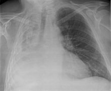
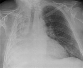
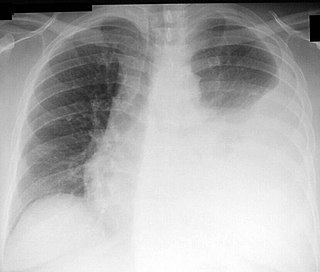
A pleural effusion is accumulation of excessive fluid in the pleural space, the potential space that surrounds each lung. Under normal conditions, pleural fluid is secreted by the parietal pleural capillaries at a rate of 0.6 millilitre per kilogram weight per hour, and is cleared by lymphatic absorption leaving behind only 5–15 millilitres of fluid, which helps to maintain a functional vacuum between the parietal and visceral pleurae. Excess fluid within the pleural space can impair inspiration by upsetting the functional vacuum and hydrostatically increasing the resistance against lung expansion, resulting in a fully or partially collapsed lung.

<span class="mw-page-title-main">Pleural empyema</span> Medical condition

Pleural empyema is a collection of pus in the pleural cavity caused by microorganisms, usually bacteria. Often it happens in the context of a pneumonia, injury, or chest surgery. It is one of the various kinds of pleural effusion. There are three stages: exudative, when there is an increase in pleural fluid with or without the presence of pus; fibrinopurulent, when fibrous septa form localized pus pockets; and the final organizing stage, when there is scarring of the pleura membranes with possible inability of the lung to expand. Simple pleural effusions occur in up to 40% of bacterial pneumonias. They are usually small and resolve with appropriate antibiotic therapy. If however an empyema develops additional intervention is required.

<span class="mw-page-title-main">Chest tube</span> Type of surgical drain

A chest tube is a surgical drain that is inserted through the chest wall and into the pleural space or the mediastinum. The insertion of the tube is sometimes a lifesaving procedure. The tube can be used to remove clinically undesired substances such as air (pneumothorax), excess fluid, blood (hemothorax), chyle (chylothorax) or pus (empyema) from the intrathoracic space. An intrapleural chest tube is also known as a Bülau drain or an intercostal catheter (ICC), and can either be a thin, flexible silicone tube, or a larger, semi-rigid, fenestrated plastic tube, which often involves a flutter valve or underwater seal.

<span class="mw-page-title-main">Atelectasis</span> Partial collapse of a lung causing reduced gas exchange

Atelectasis is the partial collapse or closure of a lung resulting in reduced or absent gas exchange. It is usually unilateral, affecting part or all of one lung. It is a condition where the alveoli are deflated down to little or no volume, as distinct from pulmonary consolidation, in which they are filled with liquid. It is often referred to informally as a collapsed lung, although more accurately it usually involves only a partial collapse, and that ambiguous term is also informally used for a fully collapsed lung caused by a pneumothorax.

<span class="mw-page-title-main">Chest radiograph</span> Projection X-ray of the chest

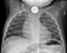
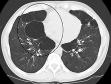
A chest radiograph, chest X-ray (CXR), or chest film is a projection radiograph of the chest used to diagnose conditions affecting the chest, its contents, and nearby structures. Chest radiographs are the most common film taken in medicine.

<span class="mw-page-title-main">Hemothorax</span> Blood accumulation in the pleural cavity

A hemothorax is an accumulation of blood within the pleural cavity. The symptoms of a hemothorax may include chest pain and difficulty breathing, while the clinical signs may include reduced breath sounds on the affected side and a rapid heart rate. Hemothoraces are usually caused by an injury, but they may occur spontaneously due to cancer invading the pleural cavity, as a result of a blood clotting disorder, as an unusual manifestation of endometriosis, in response to pneumothorax, or rarely in association with other conditions.

<span class="mw-page-title-main">Chylothorax</span> Medical condition

A chylothorax is an abnormal accumulation of chyle, a type of lipid-rich lymph, in the space surrounding the lung. The lymphatics of the digestive system normally returns lipids absorbed from the small bowel via the thoracic duct, which ascends behind the esophagus to drain into the left brachiocephalic vein. If normal thoracic duct drainage is disrupted, either due to obstruction or rupture, chyle can leak and accumulate within the negative-pressured pleural space. In people on a normal diet, this fluid collection can sometimes be identified by its turbid, milky white appearance, since chyle contains emulsified triglycerides.

<span class="mw-page-title-main">Thoracentesis</span> Removal of fluids/air from the pleural cavity of the lungs

Thoracentesis, also known as thoracocentesis, pleural tap, needle thoracostomy, or needle decompression, is an invasive medical procedure to remove fluid or air from the pleural space for diagnostic or therapeutic purposes. A cannula, or hollow needle, is carefully introduced into the thorax, generally after administration of local anesthesia. The procedure was first performed by Morrill Wyman in 1850 and then described by Henry Ingersoll Bowditch in 1852.

<span class="mw-page-title-main">Esophageal rupture</span> Medical condition

Esophageal rupture is a rupture of the esophageal wall. Iatrogenic causes account for approximately 56% of esophageal perforations, usually due to medical instrumentation such as an endoscopy or paraesophageal surgery. The 10% of esophageal perforations caused specifically by vomiting are termed Boerhaave syndrome.

<span class="mw-page-title-main">Respiratory disease</span> Disease of the respiratory system

Respiratory diseases, or lung diseases, are pathological conditions affecting the organs and tissues that make gas exchange difficult in air-breathing animals. They include conditions of the respiratory tract including the trachea, bronchi, bronchioles, alveoli, pleurae, pleural cavity, the nerves and muscles of respiration. Respiratory diseases range from mild and self-limiting, such as the common cold, influenza, and pharyngitis to life-threatening diseases such as bacterial pneumonia, pulmonary embolism, tuberculosis, acute asthma, lung cancer, and severe acute respiratory syndromes, such as COVID-19. Respiratory diseases can be classified in many different ways, including by the organ or tissue involved, by the type and pattern of associated signs and symptoms, or by the cause of the disease.

<span class="mw-page-title-main">Pericardial effusion</span> Medical condition

A pericardial effusion is an abnormal accumulation of fluid in the pericardial cavity. The pericardium is a two-part membrane surrounding the heart: the outer fibrous connective membrane and an inner two-layered serous membrane. The two layers of the serous membrane enclose the pericardial cavity between them. This pericardial space contains a small amount of pericardial fluid, normally 15-50 mL in volume. The pericardium, specifically the pericardial fluid provides lubrication, maintains the anatomic position of the heart in the chest, and also serves as a barrier to protect the heart from infection and inflammation in adjacent tissues and organs.

<span class="mw-page-title-main">Costodiaphragmatic recess</span>

The costodiaphragmatic recess, also called the costophrenic recess or phrenicocostal sinus, is the posterolateral fringe of the pleural space, a potential space around the lung inside the pleural cavity. It is located at the acutely angled junction ("reflection") between the costal and diaphragmatic parietal pleurae, and is interpreted two-dimensionally on plain X-rays as the costophrenic angle. It measures approximately 5 cm (2.0 in) vertically and extends from the eighth to the tenth rib along the mid-axillary line.

<span class="mw-page-title-main">Subcutaneous emphysema</span> Medical condition

Subcutaneous emphysema occurs when gas or air accumulates and seeps under the skin, where normally no gas should be present. Subcutaneous refers to the subcutaneous tissue, and emphysema refers to trapped air pockets. Since the air generally comes from the chest cavity, subcutaneous emphysema usually occurs around the upper torso, such as on the chest, neck, face, axillae and arms, where it is able to travel with little resistance along the loose connective tissue within the superficial fascia. Subcutaneous emphysema has a characteristic crackling-feel to the touch, a sensation that has been described as similar to touching warm Rice Krispies. This sensation of air under the skin is known as subcutaneous crepitation, a form of crepitus.

Pleural disease occurs in the pleural space, which is the thin fluid-filled area in between the two pulmonary pleurae in the human body. There are several disorders and complications that can occur within the pleural area, and the surrounding tissues in the lung.

Obstructive shock is one of the four types of shock, caused by a physical obstruction in the flow of blood. Obstruction can occur at the level of the great vessels or the heart itself. Causes include pulmonary embolism, cardiac tamponade, and tension pneumothorax. These are all life-threatening. Symptoms may include shortness of breath, weakness, or altered mental status. Low blood pressure and tachycardia are often seen in shock. Other symptoms depend on the underlying cause.

<span class="mw-page-title-main">Fibrothorax</span> Medical condition involving fibrosis of the pleural space

Fibrothorax is a medical condition characterised by severe scarring (fibrosis) and fusion of the layers of the pleural space surrounding the lungs resulting in decreased movement of the lung and ribcage. The main symptom of fibrothorax is shortness of breath. There also may be recurrent fluid collections surrounding the lungs. Fibrothorax may occur as a complication of many diseases, including infection of the pleural space known as an empyema or bleeding into the pleural space known as a haemothorax.

Tumor-like disorders of the lung pleura are a group of conditions that on initial radiological studies might be confused with malignant lesions. Radiologists must be aware of these conditions in order to avoid misdiagnosing patients. Examples of such lesions are: pleural plaques, thoracic splenosis, catamenial pneumothorax, pleural pseudotumor, diffuse pleural thickening, diffuse pulmonary lymphangiomatosis and Erdheim–Chester disease.

<span class="mw-page-title-main">Tracheal deviation</span>

Tracheal deviation is a clinical sign that results from unequal intrathoracic pressure within the chest cavity. It is most commonly associated with traumatic pneumothorax, but can be caused by a number of both acute and chronic health issues, such as pneumonectomy, atelectasis, pleural effusion, fibrothorax, or some cancers and certain lymphomas associated with the mediastinal lymph nodes.

<span class="mw-page-title-main">Lung surgery</span>

Lung surgery is a type of thoracic surgery involving the repair or removal of lung tissue, and can be used to treat a variety of conditions ranging from lung cancer to pulmonary hypertension. Common operations include anatomic and nonanatomic resections, pleurodesis and lung transplants. Though records of lung surgery date back to the Classical Age, new techniques such as VATS continue to be developed.

References

  1. Reed, James C. (2018). Chest radiology: patterns and differential diagnoses (Seventh ed.). Philadelphia, PA: Elsevier. ISBN   9780323510219. OCLC   1012134513.
  2. "Mediastinal Position". www.meddean.luc.edu. Retrieved 2024-02-21.
  3. 1 2 Khajotia, R. (2012). "Respiratory Clinics: MEDIASTINAL SHIFT: A SIGN OF SIGNIFICANT CLINICAL AND RADIOLOGICAL IMPORTANCE IN DIAGNOSIS OF MALIGNANT PLEURAL EFFUSION". Malaysian Family Physician. 7 (1): 34–36. ISSN   1985-207X. PMC   4170449 . PMID   25606244.
  4. Stoddard, Nathan; Heil, Jenna R.; Lowery, David R. (2024), "Anatomy, Thorax, Mediastinum", StatPearls, Treasure Island (FL): StatPearls Publishing, PMID   30969641 , retrieved 2024-02-21
  5. Jalota Sahota, Ruchi; Sayad, Edouard (2024), "Tension Pneumothorax", StatPearls, Treasure Island (FL): StatPearls Publishing, PMID   32644516 , retrieved 2024-02-21
  6. Taghavi, Sharven; Askari, Reza (2024), "Mediastinal Trauma", StatPearls, Treasure Island (FL): StatPearls Publishing, PMID   30252287 , retrieved 2024-03-06
  7. Salim, Shihas; Ganeshram, Prasanthi; Patel, Amish Dilip; Kumar, Anita A.; Vemuri, Divya; Jeyachandran, Vijay; Rajamanickam, Deepan; Shantha, Ghanshyam Palamaner Subash (2008-10-07). "Unilateral hemothorax in a 46 year old South Indian male due to a giant arteriovenous hemodialysis fistula: a case report". Cases Journal. 1 (1): 225. doi: 10.1186/1757-1626-1-225 . ISSN   1757-1626. PMC   2567296 . PMID   18840271.
  8. Kim, Michelle; Moore, James E. (2020). "Chest Trauma: Current Recommendations for Rib Fractures, Pneumothorax, and Other Injuries". Current Anesthesiology Reports. 10 (1): 61–68. doi:10.1007/s40140-020-00374-w. ISSN   1523-3855. PMC   7223697 . PMID   32435162.
  9. Feller-Kopman, David; Light, Richard (2018-02-22). "Pleural Disease". The New England Journal of Medicine. 378 (8): 740–751. doi:10.1056/NEJMra1403503. ISSN   1533-4406. PMID   29466146.
  10. Garvia, Veronica; Paul, Manju (2024), "Empyema", StatPearls, Treasure Island (FL): StatPearls Publishing, PMID   29083780 , retrieved 2024-03-06
  11. Ohara, Gen; Iguchi, Kesato; Satoh, Hiroaki (2018-10-19). "VATS and Intrapleural Fibrinolytic Therapy for Parapneumonic Empyema". Annals of Thoracic and Cardiovascular Surgery: Official Journal of the Association of Thoracic and Cardiovascular Surgeons of Asia. 24 (5): 263–264. doi:10.5761/atcs.lte.18-00092. ISSN   2186-1005. PMC   6198002 . PMID   29962389.
  12. Ghigna, Maria-Rosa; Montpreville, Vincent Thomas de (2021-12-31). "Mediastinal tumours and pseudo-tumours: a comprehensive review with emphasis on multidisciplinary approach". European Respiratory Review. 30 (162). doi:10.1183/16000617.0309-2020. ISSN   0905-9180. PMC   9488622 . PMID   34615701.
  13. Jilani, Talha N.; Killeen, Robert B.; Siddiqui, Abdul H. (2024), "Mediastinal Cancer", StatPearls, Treasure Island (FL): StatPearls Publishing, PMID   30020603 , retrieved 2024-03-06
  14. Grott, Kelly; Chauhan, Shaylika; Dunlap, Julie D. (2024), "Atelectasis", StatPearls, Treasure Island (FL): StatPearls Publishing, PMID   31424900 , retrieved 2024-03-07
  15. Cao, Li; Du, Yan; Wang, Ling (2017). "Fetal pleural effusion and Down syndrome". Intractable & Rare Diseases Research. 6 (3): 158–162. doi:10.5582/irdr.2017.01028. ISSN   2186-3644. PMC   5608924 . PMID   28944136.
  16. Tisekar, Owais R.; Ak, Ajith Kumar (2024), "Hypoplastic Lung Disease", StatPearls, Treasure Island (FL): StatPearls Publishing, PMID   32965810 , retrieved 2024-03-07
  17. Colombani, M.; Rubesova, E.; Potier, A.; Quarello, E.; Barth, R. A.; Devred, P.; Petit, P.; Gorincour, G. (2011). "[Management of fetal mediastinal shift: a practical approach]". Journal de Radiologie. 92 (2): 118–124. doi:10.1016/j.jradio.2010.12.002. ISSN   1773-0384. PMID   21352743.
  18. Sharma, Girish; Carter, Yvonne M. (2024), "Pectus Excavatum", StatPearls, Treasure Island (FL): StatPearls Publishing, PMID   28613668 , retrieved 2024-03-07
  19. Beshara, Michael; Bora, Vaibhav (2024), "Pneumonectomy", StatPearls, Treasure Island (FL): StatPearls Publishing, PMID   32310429 , retrieved 2024-03-10
  20. de Groot, Patricia M.; Truong, Mylene T.; Godoy, Myrna C. B. (2018). "Postoperative Imaging and Complications in Resection of Lung Cancer". Seminars in Ultrasound, CT, and MR. 39 (3): 289–296. doi:10.1053/j.sult.2018.02.008. ISSN   1558-5034. PMID   29807639. S2CID   44142767.
  21. Rose, David; Dubensky, Laurence (2024), "Airway Foreign Bodies", StatPearls, Treasure Island (FL): StatPearls Publishing, PMID   30969578 , retrieved 2024-03-20
  22. Cramer, Natan; Jabbour, Noel; Tavarez, Melissa M.; Taylor, Roger S. (2024), "Foreign Body Aspiration", StatPearls, Treasure Island (FL): StatPearls Publishing, PMID   30285375 , retrieved 2024-03-20
  23. Siddiqui, Najam A.; Mansour, Mohamed K.; Nookala, Vinod (2024), "Bullous Emphysema", StatPearls, Treasure Island (FL): StatPearls Publishing, PMID   30725928 , retrieved 2024-03-20
  24. Ascano, Maria Patricia; Kramer, Nicholas; Le, Khoa; Ascano, Maria Patricia; Kramer, Nicholas; Le, Khoa (2024-03-11). "Differentiating Giant Bullous Emphysema From Tension Pneumothorax: A Case Report". Cureus. 16 (3). doi: 10.7759/cureus.55988 . ISSN   2168-8184. PMC   11007189 .
  25. Samanta, Rudra P.; Agarwal, Srikant; Sengupta, Susmita; Samanta, Rudra P.; Agarwal, Srikant; Sengupta, Susmita (2022-11-07). "Giant Bullous Emphysema Mimicking Spontaneous Pneumothorax". Cureus. 14 (11): e31182. doi: 10.7759/cureus.31182 . ISSN   2168-8184. PMC   9727579 . PMID   36505170.
  26. Mehta, Pooja A.; Sharma, Girish (2024), "Congenital Pulmonary Airway Malformation", StatPearls, Treasure Island (FL): StatPearls Publishing, PMID   31869128 , retrieved 2024-03-20