MicroDNA

Last updated
CpG-islands characteristic in microDNA compared to a single C-G bp. CpG vs C-G bp.svg
CpG-islands characteristic in microDNA compared to a single C-G bp.

MicroDNA is the most abundant subtype of Extrachromosomal Circular DNA (eccDNA) in humans, typically ranging from 200-400 base pairs in length and enriched in non-repetitive genomic sequences with a high density of exons. [2] [3] [4] Additionally, microDNA has been found to come from regions with CpG-islands which are commonly found within the 5' and 3' UTRs. [3] [4] [5] Being produced from regions of active transcription, it is hypothesized that microDNA may be formed as a by-product of transcriptional DNA damage repair. [5] MicroDNA is also thought to arise from other DNA repair pathways, mainly due to the parental sequences of microDNA having 2- to 15 bp direct repeats at the ends, resulting in replication slippage repair. [3] While only recently discovered, the role microDNA plays in and out of the cell is still not completely understood. [5] However, microDNA is currently thought to affect cellular homeostasis through transcription factor binding and have been used as a cancer biomarker. [5] [6] [7]

Contents

Discovery

MicroDNA was discovered through protocols similar to that of eccDNA extraction. [5] Specifically, eccDNA clones were generated through multiple displacement amplification and sequenced with Sanger sequencing, leading to microDNA's discovery. [5] Now with high-throughput sequencing being a more common practice, the complete genomic sequence of mammalian eccDNA has been obtained through the sequencing of the rolling amplification products of eccDNA. [5] Computational methods were then used to identify junctional sequences in the DNA. [4] The peaks found at lengths of 180 and 380 bp were discovered as microDNA and characterized by their CpG-islands and flanking 2- to 15 bp direct repeats. [4]

Since its discovery, microDNA has been identified in all tissue types and various samples, including mouse tissues and human cancer cell lines. [5] [6] However, different species have unique genomic sites that specifically produce microDNA. [5] Because there are common genomic spots that produce microDNA in multiple cell and tissue types within a given species, there is evidence that they may not be produced solely as a DNA synthesis by-product. [5] However, studies have revealed separate clustering of microDNA extracted from cell-lines of different tissues, suggesting that formation may be linked to cell-lineage and unique transcriptional environments found in different cell types. [4] [5]

Biogenesis

Typical R-loop formation where the single-stranded DNA can become microDNA. R-loop promoting factors.jpg
Typical R-loop formation where the single-stranded DNA can become microDNA.

While the formation of microDNA is still uncertain, it has been linked to transcriptional activity and multiple DNA repair pathways. [3] [5] As microDNA is produced from areas of high transcription activity/exon density, it could be formed from DNA repair during transcription. [5] Interestingly, triple-stranded DNA:RNA hybrids formed during transcription, termed R-loops, tend to form at CpG-islands within the 5' and 3' UTRs, similar to microDNA. [3] [5] R-loops are correlated with DNA damage and genetic instability which is suggestive that microDNA may form from the single-stranded DNA (ssDNA) loop during the DNA damage response for R-loops. [3] [5] [6]

In DNA replication of short direct repeats (as found in the flanking regions of microDNA gene sources), it is possible for DNA loops to form, on the parent or product strand, through replication slippage. [3] [5] To repair this, the mismatch repair (MMR) pathway can remove the loop and upon ligation of the repeating ends, single-strand microDNA can be produced. [3] The ss microDNA is then converted to double-stranded DNA; this process is still unknown. [5] It is important to note that if the loop is formed on the newly replicated strand, there is no consequential deletion in the genome while microdeletions can form from excisions in the template strand. [3] [5] To understand the role MMR may have in microDNA biogenesis, analysis of microDNA abundance was performed in DT40 cells upon removal of MSH3, an essential protein in MMR. [3] [5] The resulting microDNA from the DT40 MSH3-/- cell line had a higher enrichment of CpG-islands compared to the wild-type as well as an over 80% reduction of double-stranded microDNA. [3] [5] Thus, it is hypothesized that the MMR pathway is essential for microDNA production from non-CpG islands in the genome while CpG enriched microDNA are formed by a different repair pathway. [3]

Transmission electron microscope image of isolated microDNA from DT40 cells. DT40 microDNA.tif
Transmission electron microscope image of isolated microDNA from DT40 cells.

Again, because of the microhomology on the template genome, if there is a DNA break or a pause in replication (replication fork stalling), the newly synthesized DNA can circularize into ss microDNA. [3] [5] This means when the template DNA is repaired after the creation of the microDNA, there is no deletion. [3]

MicroDNA created through the MMR pathway and replication fork stalling is a result of errors in DNA replication, however, there is evidence of microDNA being present in non-dividing cells as well. [5] This means that some microDNA is produced through repair pathways that also occur in quiescent cells, such as from 5' ends of LINE1 elements that are known to transpose. [3] [5] To move around the genome, DNA transposons require transposase to remove the transposon from its original site and catalyze its insertion elsewhere in the genome. [3] Thus, the transposon is created by two double-stranded DNA breaks, also creating a microdeletion in the DNA. [3] This dsDNA fragment can be circularized through microhomology-mediated circularization, creating a ds microDNA. [3]

Implications

Transcription Factor Binding

Being 200-400 bp long, microDNA is too small to encode proteins, however, they may be important for molecular sponging. [4] [5] Transcription factors often bind to promoter or regulatory sequences at the 5' end of DNA to initiate transcription. [5] These transcription factors can also bind to their respective recognition sites on microDNA because the microDNA often originates from the 5' UTRs of its parental gene, therefore, acting as a sponge for transcription factors. [4] [5] This means microDNA can indirectly control gene expression and transcription homeostasis. [4] [5]

Cancer Applications

In general, nucleic acid molecules that are found in the bloodstream, termed circulating or cell-free, are a relatively new disease biomarker being investigated, including for diagnosis and progression of cancer. [7] These molecules, such as cell-free DNA (cfDNA), are released into the blood upon cell death and in cases of cancer, can be identified based on the known mutations in oncogenes. [7]

Recent studies have extended the use of cell-free nucleic acids as cancer biomarkers to microDNA. [7] The cfmicroDNA was obtained from human and mouse serum and because of their similarities to cell-derived microDNA, as described above, it was concluded that cfmicroDNA is produced in the cell. [7] Similarly, when comparing lung tissue pre- and post-tumor removal, there was no found difference in circulating microDNA key characteristics other than an unexpected trend of longer circulating microDNA sequences in cancer patients pre-tumor removal. [7] The length of cfmicroDNA was found to be shorter post-surgery. [7]

Cell-free DNA is quickly cleared from the blood, making it a difficult cancer biomarker. [7] However, because circular DNA is not susceptible to DNA breakage by RNAse and exonuclease, it is more stable than linear DNA. [5] [7] In combination with the observed lengthening of cfmicroDNA in cancer patient serum, this makes circulating microDNA a good cancer biomarker for both diagnosis and progression after treatment. [7]

See also

Related Research Articles

Transcription (biology) Process of copying a segment of DNA into RNA

Transcription is the process of copying a segment of DNA into RNA. The segments of DNA transcribed into RNA molecules that can encode proteins are said to produce messenger RNA (mRNA). Other segments of DNA are copied into RNA molecules called non-coding RNAs (ncRNAs). Averaged over multiple cell types in a given tissue, the quantity of mRNA is more than 10 times the quantity of ncRNA. The general preponderance of mRNA in cells is valid even though less than 2% of the human genome can be transcribed into mRNA, while at least 80% of mammalian genomic DNA can be actively transcribed, with the majority of this 80% considered to be ncRNA.

CpG site Region of often-methylated DNA with a cytosine followed by a guanine

The CpG sites or CG sites are regions of DNA where a cytosine nucleotide is followed by a guanine nucleotide in the linear sequence of bases along its 5' → 3' direction. CpG sites occur with high frequency in genomic regions called CpG islands.

Regulation of gene expression

Regulation of gene expression, or gene regulation, includes a wide range of mechanisms that are used by cells to increase or decrease the production of specific gene products. Sophisticated programs of gene expression are widely observed in biology, for example to trigger developmental pathways, respond to environmental stimuli, or adapt to new food sources. Virtually any step of gene expression can be modulated, from transcriptional initiation, to RNA processing, and to the post-translational modification of a protein. Often, one gene regulator controls another, and so on, in a gene regulatory network.

DNA repair Cellular mechanism

DNA repair is a collection of processes by which a cell identifies and corrects damage to the DNA molecules that encode its genome. In human cells, both normal metabolic activities and environmental factors such as radiation can cause DNA damage, resulting in tens of thousands of individual molecular lesions per cell per day. Many of these lesions cause structural damage to the DNA molecule and can alter or eliminate the cell's ability to transcribe the gene that the affected DNA encodes. Other lesions induce potentially harmful mutations in the cell's genome, which affect the survival of its daughter cells after it undergoes mitosis. As a consequence, the DNA repair process is constantly active as it responds to damage in the DNA structure. When normal repair processes fail, and when cellular apoptosis does not occur, irreparable DNA damage may occur, including double-strand breaks and DNA crosslinkages. This can eventually lead to malignant tumors, or cancer as per the two hit hypothesis.

DNA methylation Biological process

DNA methylation is a biological process by which methyl groups are added to the DNA molecule. Methylation can change the activity of a DNA segment without changing the sequence. When located in a gene promoter, DNA methylation typically acts to repress gene transcription. In mammals, DNA methylation is essential for normal development and is associated with a number of key processes including genomic imprinting, X-chromosome inactivation, repression of transposable elements, aging, and carcinogenesis.

Malignant transformation is the process by which cells acquire the properties of cancer. This may occur as a primary process in normal tissue, or secondarily as malignant degeneration of a previously existing benign tumor.

Nucleotide excision repair DNA repair mechanism

Nucleotide excision repair is a DNA repair mechanism. DNA damage occurs constantly because of chemicals, radiation and other mutagens. Three excision repair pathways exist to repair single stranded DNA damage: Nucleotide excision repair (NER), base excision repair (BER), and DNA mismatch repair (MMR). While the BER pathway can recognize specific non-bulky lesions in DNA, it can correct only damaged bases that are removed by specific glycosylases. Similarly, the MMR pathway only targets mismatched Watson-Crick base pairs.

DNA mismatch repair

DNA mismatch repair (MMR) is a system for recognizing and repairing erroneous insertion, deletion, and mis-incorporation of bases that can arise during DNA replication and recombination, as well as repairing some forms of DNA damage.

Extrachromosomal DNA is any DNA that is found off the chromosomes, either inside or outside the nucleus of a cell. Most DNA in an individual genome is found in chromosomes contained in the nucleus. Multiple forms of extrachromosomal DNA exist, and, while some of these serve important biological functions, they can also play a role in diseases, such as ecDNA in cancer.

Microsatellite instability

Microsatellite instability (MSI) is the condition of genetic hypermutability that results from impaired DNA mismatch repair (MMR). The presence of MSI represents phenotypic evidence that MMR is not functioning normally.

Oncogenomics Sub-field of genomics

Oncogenomics is a sub-field of genomics that characterizes cancer-associated genes. It focuses on genomic, epigenomic and transcript alterations in cancer.

Flap structure-specific endonuclease 1

Flap endonuclease 1 is an enzyme that in humans is encoded by the FEN1 gene.

RAD52

RAD52 homolog , also known as RAD52, is a protein which in humans is encoded by the RAD52 gene.

Complementarity (molecular biology)

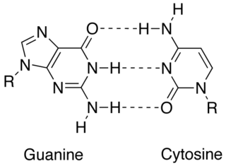
In molecular biology, complementarity describes a relationship between two structures each following the lock-and-key principle. In nature complementarity is the base principle of DNA replication and transcription as it is a property shared between two DNA or RNA sequences, such that when they are aligned antiparallel to each other, the nucleotide bases at each position in the sequences will be complementary, much like looking in the mirror and seeing the reverse of things. This complementary base pairing allows cells to copy information from one generation to another and even find and repair damage to the information stored in the sequences.

Genome instability refers to a high frequency of mutations within the genome of a cellular lineage. These mutations can include changes in nucleic acid sequences, chromosomal rearrangements or aneuploidy. Genome instability does occur in bacteria. In multicellular organisms genome instability is central to carcinogenesis, and in humans it is also a factor in some neurodegenerative diseases such as amyotrophic lateral sclerosis or the neuromuscular disease myotonic dystrophy.

R-loop Three-stranded nucleic acid structure, composed of a DNA:RNA hybrid and the associated non-template single-stranded DNA.

An R-loop is a three-stranded nucleic acid structure, composed of a DNA:RNA hybrid and the associated non-template single-stranded DNA. R-loops may be formed in a variety of circumstances, and may be tolerated or cleared by cellular components. The term "R-loop" was given to reflect the similarity of these structures to D-loops; the "R" in this case represents the involvement of an RNA moiety.

Generally, in progression to cancer, hundreds of genes are silenced or activated. Although silencing of some genes in cancers occurs by mutation, a large proportion of carcinogenic gene silencing is a result of altered DNA methylation. DNA methylation causing silencing in cancer typically occurs at multiple CpG sites in the CpG islands that are present in the promoters of protein coding genes.

Extrachromosomal circular DNA (eccDNA) is a type of double-stranded circular DNA structure that was first discovered in 1964 by Alix Bassel and Yasuo Hotta. In contrast to previously identified circular DNA structures, eccDNA are circular DNA found in the eukaryotic nuclei of human, plant, and animal cells. eccDNA is derived from chromosomal DNA, can range in size from 50 base pairs to several mega-base pairs in length, and can encode regulatory elements and full-length genes. eccDNA has been observed in various eukaryotic species and it is proposed to be a byproduct of programmed DNA recombination events, such as V(D)J recombination.

LIANTI

Linear Amplification via Transposon Insertion (LIANTI) is a linear whole genome amplification (WGA) method. To analyze or sequence very small amount of DNA, i.e. genomic DNA from a single cell, the picograms of DNA is subject to WGA to amplify at least thousands of times into nanogram scale, before DNA analysis or sequencing can be carried out. Previous WGA methods use exponential/nonlinear amplification schemes, leading to bias accumulation and error propagation. LIANTI achieved linear amplification of the whole genome for the first time, enabling more uniform and accurate amplification.

Anindya Dutta is an Indian-born American biochemist and cancer researcher, a Chair of the Department of Genetics at the University of Alabama at Birmingham School of Medicine since 2021, who has served as Chair of the Department of Biochemistry and Molecular Genetics at the University of Virginia School of Medicine in 2011–2021. Dutta's research has focused on the mammalian cell cycle with an emphasis on DNA replication and repair and on noncoding RNAs. He is particularly interested in how de-regulation of these processes promote cancer progression. For his accomplishments he has been elected a Fellow of the American Association for the Advancement of Science, received the Ranbaxy Award in Biomedical Sciences, the Outstanding Investigator Award from the American Society for Investigative Pathology, the Distinguished Scientist Award from the University of Virginia and the Mark Brothers Award from the Indiana University School of Medicine.

References

  1. "File:CpG vs C-G bp.svg - Wikipedia". commons.wikimedia.org. 31 January 2016. Retrieved 2021-11-16.
  2. Shibata Y, Kumar P, Layer R, Willcox S, Gagan JR, Griffith JD, Dutta A (April 2012). "Extrachromosomal microDNAs and chromosomal microdeletions in normal tissues". Science. 336 (6077): 82–86. Bibcode:2012Sci...336...82S. doi:10.1126/science.1213307. PMC   3703515 . PMID   22403181.
  3. 1 2 3 4 5 6 7 8 9 10 11 12 13 14 15 16 17 18 Dillon LW, Kumar P, Shibata Y, Wang YH, Willcox S, Griffith JD, et al. (June 2015). "Production of Extrachromosomal MicroDNAs Is Linked to Mismatch Repair Pathways and Transcriptional Activity". Cell Reports. 11 (11): 1749–1759. doi:10.1016/j.celrep.2015.05.020. PMC   4481157 . PMID   26051933.
  4. 1 2 3 4 5 6 7 8 Paulsen T, Kumar P, Koseoglu MM, Dutta A (April 2018). "Discoveries of Extrachromosomal Circles of DNA in Normal and Tumor Cells". Trends in Genetics. 34 (4): 270–278. doi:10.1016/j.tig.2017.12.010. PMC   5881399 . PMID   29329720.
  5. 1 2 3 4 5 6 7 8 9 10 11 12 13 14 15 16 17 18 19 20 21 22 23 24 25 26 27 28 Reon, Brian J.; Dutta, Anindya (2016-04-01). "Biological Processes Discovered by High-Throughput Sequencing". The American Journal of Pathology. 186 (4): 722–732. doi:10.1016/j.ajpath.2015.10.033. ISSN   0002-9440. PMC   5807928 . PMID   26828742.
  6. 1 2 3 Kumar P, Dillon LW, Shibata Y, Jazaeri AA, Jones DR, Dutta A (September 2017). "Normal and Cancerous Tissues Release Extrachromosomal Circular DNA (eccDNA) into the Circulation". Molecular Cancer Research. 15 (9): 1197–1205. doi:10.1158/1541-7786.MCR-17-0095. PMC   5581709 . PMID   28550083.
  7. 1 2 3 4 5 6 7 8 9 10 Kumar, Pankaj; Dillon, Laura W.; Shibata, Yoshiyuki; Jazaeri, Amir A.; Jones, David R.; Dutta, Anindya (2017-09-01). "Normal and Cancerous Tissues Release Extrachromosomal Circular DNA (eccDNA) into the Circulation". Molecular Cancer Research. 15 (9): 1197–1205. doi:10.1158/1541-7786.MCR-17-0095. ISSN   1541-7786. PMC   5581709 . PMID   28550083.
  8. "File:R-loop promoting factors.jpg - Wikipedia". commons.wikimedia.org. 24 January 2021. Retrieved 2021-11-16.
  9. "File:DT40 microDNA.tif - Wikipedia". commons.wikimedia.org. 20 March 2015. Retrieved 2021-11-16.