N-sulfoglucosamine sulfohydrolase

Last updated
N-sulfoglucosamine sulfohydrolase
C183-4MHX Enzyme.png
Cartoon depiction of N-sulfoglucosamine sulfohydrolase.
Identifiers
EC no. 3.10.1.1
CAS no. 37289-41-1
Databases
IntEnz IntEnz view
BRENDA BRENDA entry
ExPASy NiceZyme view
KEGG KEGG entry
MetaCyc metabolic pathway
PRIAM profile
PDB structures RCSB PDB PDBe PDBsum
Gene Ontology AmiGO / QuickGO
Search
PMC articles
PubMed articles
NCBI proteins

In enzymology, a N-sulfoglucosamine sulfohydrolase (EC 3.10.1.1), otherwise known as SGSH, is an enzyme that catalyzes the chemical reaction

Contents

N-sulfo-D-glucosamine + H2O D-glucosamine + sulfate

Thus, the two substrates of this enzyme are N-sulfo-D-glucosamine and H2O, whereas its two products are D-glucosamine and sulfate.

This enzyme belongs to the family of hydrolases, specifically those acting on sulfur-nitrogen bonds. The systematic name of this enzyme class is N-sulfo-D-glucosamine sulfohydrolase. Other names in common use include sulfoglucosamine sulfamidase, heparin sulfamidase, 2-desoxy-D-glucoside-2-sulphamate sulphohydrolase (sulphamate, and sulphohydrolase). This enzyme participates in glycosaminoglycan degradation and glycan structures - degradation.

Structure

N-glusoamine sulfohydrolase is a homodimer of two identical monomeric subunits that associate non-covalently. The crystal structure, solved using molecular replacement, consists of two domains: a large N-terminal domain (domain 1) and a smaller C-terminal domain (domain 2) that are both centered about a β-sheet. The core of Domain 1 is formed by eight β-strands surrounded by nine ⍺-helices while the core of Domain 2 is formed by a four-stranded antiparallel β-sheet surrounded by 4 ⍺-helices, accompanied by a C-terminal extension of a small two-stranded β-sheet. There are two disulfide bonds that serve to stabilize a large loop segment in Domain 1 and connect the C-terminal extension to a nearby loop in Domain 2.

The active site is located through a short tunnel at the bottom of the surface cleft of the enzyme close to the end of the first β-strand in Domain 1 of the dimer subunits. A calcium ion, Ca2+, is coordinated in an octahedral arrangement by oxygen atoms of nearby side chains of residues Asp31, Asp32, Asp273, Asn274, and phosphorylated formylglycine, PFGly70. The PFGly70, which is post-translationally converted from a cysteine and is key in the catalytic activity of the enzyme, is stabilized by the side chains of residues Arg74, Lys123, His125, His181, Asp273, and Arg282 and the coordinate calcium ion. [1] [2]

Active site of N-sulfoglucosamine sulfohydrolase enzyme. Generated from 4MHX. C183-4MHX Active Site.png
Active site of N-sulfoglucosamine sulfohydrolase enzyme. Generated from 4MHX.

Mechanism

N-sulfoglucosamine sulfohydrolase mechanism for the removal of sulfate from heparin or heparan sulfate substrate. N-sulfoglucosamine sulfohydrolase Generalized Mechanism.jpg
N-sulfoglucosamine sulfohydrolase mechanism for the removal of sulfate from heparin or heparan sulfate substrate.

One of the proposed mechanisms for the N-sulfoglucosamine sulfohydrolase, depicted in the figure above, was inspired by a mechanism determined for a sulfatase enzymes, which similarly cleave sulfate ester groups from their substrates. [4] The N-sulfoglucosamine sulfohydrolase mechanism involves four key residues found in the enzyme active site: formylglycine (FGly70), two histidines (His125 and His181), and aspartic acid (Asp273). The formylglycine residue (FGly70) is first hydrated to form a geminal diol whose hydroxyl group subsequently coordinates with a Calcium(II) ion in the active site. A neighboring aspartic acid residue (Asp273) then acts as a base to deprotonate the coordinated hydroxyl group in the geminal diol, which acts as a nucleophile to the sulfate group on the substrate and leads to the cleavage of the substrate nitrogen-sulfur bond. A proximal histidine residue (His181) is proposed to donate a proton as a replacement for the sulfate group that was involved in the nitrogen-sulfur bond. At this point, the formylglycine (FGly70) has the sulfate group attached adjacent to a hydroxyl group, which is believed to be deprotonated by a second histidine group (His125) so that the negatively-charged oxygen atom can participate in the removal of the sulfate group from the residue. This final step liberates the enzyme from the sulfate group. [2]

This mechanism, however, is still being studied for N-sulfoglucosamine sulfohydrolase, meaning other mechanisms are still plausible for the reaction. [2] For instance, a second mechanism proposal involves the aldehyde form of the formylglycine residue (FGly70) serving as an electrophile as it is attacked by one of the oxygen atoms on the sulfate group to transfer the sulfate from the substrate to the enzyme. [4] The order of attack for the sulfate transfer step in this proposed mechanism is therefore inverted to that of the proposed mechanism above.

Overall, the N-sulfoglucosamine sulfohydrolase mechanisms proposed involve the cleavage of the substrate nitrogen-sulfur bond - transferring the sulfate group from the substrate to the enzyme - and freeing the enzyme from its bond to the transferred sulfate group. [4] [2]

Function

As previously noted, N-sulfoglucosamine sulfohydrolase plays a crucial role in the degradation of glycosaminoglycans (GAGs), including heparin and heparan sulfate. [5] [1] Since these GAGs, particularly haparan sulfates, are integral to several biochemical processes, such as signaling pathways, any disruption in N-sulfoglucosamine sulfohydrolase activity could lead to serious diseases as noted below. [5]

Disease relevance

Sanfillipo Syndrome or Mucopolysaccharidosis III, MPS III, is a lysosomal storage disease resulting from a deficiency in one of five lysosomal enzymes: N-glusoamine sulfohydrolase (Type A), a-N-acetylglucoaminidase (Type B), acetyl CoA a-glusoaminide acetyltransferase (Type C), a-N-acetylglusoamine 6-sulfatase (Type D), and N-glusoamine 3-O-sulfatase (Type E) caused by a dysfunction of one of the genes encoding the enzyme. [6] [5] [7] These enzymes are responsible for the degradation of heparin sulfate which, in MPS III, accumulates in the lysosomes and outside of the cell, as the primary storage material. [8] In Mucopolysaccharidosis type IIIA, where there are genetic changes in the SGSH gene, there are initial signs of neurodegeneration, developmental delays, and behavioral problems with a wide phenotypic variability. [9] This is the most common form of Mucopolysaccharidosis III with a prevalence of 1 in every 100,000 individuals. [10]

Related Research Articles

<span class="mw-page-title-main">Glycosaminoglycan</span> Polysaccharides found in animal tissue

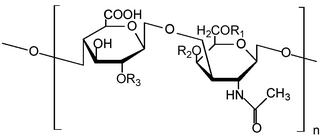
Glycosaminoglycans (GAGs) or mucopolysaccharides are long, linear polysaccharides consisting of repeating disaccharide units. The repeating two-sugar unit consists of a uronic sugar and an amino sugar, except in the case of the sulfated glycosaminoglycan keratan, where, in place of the uronic sugar there is a galactose unit. GAGs are found in vertebrates, invertebrates and bacteria. Because GAGs are highly polar molecules and attract water; the body uses them as lubricants or shock absorbers.

β-Glucuronidase Class of enzymes

β-Glucuronidases are members of the glycosidase family of enzymes that catalyze breakdown of complex carbohydrates. Human β-glucuronidase is a type of glucuronidase that catalyzes hydrolysis of β-D-glucuronic acid residues from the non-reducing end of mucopolysaccharides such as heparan sulfate. Human β-glucuronidase is located in the lysosome. In the gut, brush border β-glucuronidase converts conjugated bilirubin to the unconjugated form for reabsorption. β-Glucuronidase is also present in breast milk, which contributes to neonatal jaundice. The protein is encoded by the GUSB gene in humans and by the uidA gene in bacteria.

<span class="mw-page-title-main">Sulfatase</span> Class of enzymes

Sulfatases EC 3.1.6.- are enzymes of the esterase class that catalyze the hydrolysis of sulfate esters. These may be found on a range of substrates, including steroids, carbohydrates and proteins. Sulfate esters may be formed from various alcohols and amines. In the latter case the resultant N-sulfates can also be termed sulfamates.

Sulfation is the chemical reaction that entails the addition of SO3 group. In principle, many sulfations would involve reactions of sulfur trioxide (SO3). In practice, most sulfations are effected less directly. Regardless of the mechanism, the installation of a sulfate-like group on a substrate leads to substantial changes.

<span class="mw-page-title-main">Iduronate-2-sulfatase</span> Class of enzymes

Iduronate 2-sulfatase is a sulfatase enzyme associated with Hunter syndrome. It catalyses hydrolysis of the 2-sulfate groups of the L-iduronate 2-sulfate units of dermatan sulfate, heparan sulfate and heparin.

<span class="mw-page-title-main">Galactosamine-6 sulfatase</span> Protein-coding gene in the species Homo sapiens

N-acetylgalactosamine-6-sulfatase is an enzyme that, in humans, is encoded by the GALNS gene.

<span class="mw-page-title-main">N-acetylglucosamine-6-sulfatase</span> Protein-coding gene in the species Homo sapiens

N-acetylglucosamine-6-sulfatase (EC 3.1.6.14, glucosamine (N-acetyl)-6-sulfatase, systematic name N-acetyl-D-glucosamine-6-sulfate 6-sulfohydrolase) is an enzyme that in humans is encoded by the GNS gene. It is deficient in Sanfilippo Syndrome type IIId. It catalyses the hydrolysis of the 6-sulfate groups of the N-acetyl-D-glucosamine 6-sulfate units of heparan sulfate and keratan sulfate

<span class="mw-page-title-main">Heparosan-N-sulfate-glucuronate 5-epimerase</span>

Heparosan-N-sulfate-glucuronate 5-epimerase is an enzyme with systematic name poly( -beta-D-glucuronosyl- -N-sulfo-alpha-D-glucosaminyl) glucurono-5-epimerase. This enzyme catalyses the following chemical reaction

In enzymology, a phosphoadenylylsulfatase (EC 3.6.2.2) is an enzyme that catalyzes the chemical reaction

The enzyme chondro-4-sulfatase (EC 3.1.6.9) catalyzes the reaction

The enzyme disulfoglucosamine-6-sulfatase (EC 3.1.6.1) catalyzes the reaction

In enzymology, a glucuronate-2-sulfatase is an enzyme that catalyzes the chemical reaction of cleaving off the 2-sulfate groups of the 2-O-sulfo-D-glucuronate residues of chondroitin sulfate, heparin and heparitin sulfate.

The enzyme N-acetylgalactosamine-6-sulfatase catalyzes the chemical reaction of cleaving off the 6-sulfate groups of the N-acetyl-D-galactosamine 6-sulfate units of the macromolecule chondroitin sulfate and, similarly, of the D-galactose 6-sulfate units of the macromolecule keratan sulfate.

The enzyme N-sulfoglucosamine-3-sulfatase catalyzes cleaving off the 3-sulfate groups of the N-sulfo-D-glucosamine 3-O-sulfate units of heparin.

<span class="mw-page-title-main">SGSH</span> Protein-coding gene in the species Homo sapiens

N-sulphoglucosamine sulphohydrolase is an enzyme that in humans is encoded by the SGSH gene.

<span class="mw-page-title-main">SULF1</span> Protein-coding gene in the species Homo sapiens

Sulfatase 1, also known as SULF1, is an enzyme which in humans is encoded by the SULF1 gene.

<span class="mw-page-title-main">Multiple sulfatase deficiency</span> Medical condition

Multiple sulfatase deficiency (MSD), also known as Austin disease, or mucosulfatidosis, is a very rare autosomal recessive lysosomal storage disease caused by a deficiency in multiple sulfatase enzymes, or in formylglycine-generating enzyme, which activates sulfatases. It is similar to mucopolysaccharidosis.

<span class="mw-page-title-main">Formylglycine-generating enzyme</span>

Formylglycine-generating enzyme (FGE), located at 3p26.1 in humans, is the name for an enzyme present in the endoplasmic reticulum that catalyzes the conversion of cysteine to formylglycine (fGly). There are two main classes of FGE, aerobic and anaerobic. FGE activates sulfatases, which are essential for the degradation of sulfate esters. The catalytic activity of sulfatases is dependent upon a formylglycine residue in the active site.

Heparanase is an enzyme with systematic name heparan sulfate N-sulfo-D-glucosamine endoglucanase. This enzyme catalyses the following chemical reaction

<span class="mw-page-title-main">NAGLU</span> Protein-coding gene in the species Homo sapiens

N-acetylglucosaminidase, alpha is a protein that in humans is encoded by the NAGLU gene.

References

  1. 1 2 Dierks T, Lecca MR, Schlotterhose P, Schmidt B, von Figura K (April 1999). "Sequence determinants directing conversion of cysteine to formylglycine in eukaryotic sulfatases". The EMBO Journal. 18 (8): 2084–2091. doi:10.1093/emboj/18.8.2084. PMC   1171293 . PMID   10205163.
  2. 1 2 3 4 5 6 Sidhu NS, Schreiber K, Pröpper K, Becker S, Usón I, Sheldrick GM, et al. (May 2014). "Structure of sulfamidase provides insight into the molecular pathology of mucopolysaccharidosis IIIA". Acta Crystallographica. Section D, Biological Crystallography. 70 (Pt 5): 1321–1335. doi:10.1107/S1399004714002739. PMC   4014121 . PMID   24816101.
  3. Madej T, Lanczycki CJ, Zhang D, Thiessen PA, Geer RC, Marchler-Bauer A, Bryant SH (January 2014). "MMDB and VAST+: tracking structural similarities between macromolecular complexes". Nucleic Acids Research. 42 (Database issue): D297–D303. doi:10.1093/nar/gkt1208. PMC   3965051 . PMID   24319143.
  4. 1 2 3 Boltes I, Czapinska H, Kahnert A, von Bülow R, Dierks T, Schmidt B, et al. (June 2001). "1.3 A structure of arylsulfatase from Pseudomonas aeruginosa establishes the catalytic mechanism of sulfate ester cleavage in the sulfatase family". Structure. 9 (6): 483–491. doi: 10.1016/S0969-2126(01)00609-8 . PMID   11435113.
  5. 1 2 3 Webber DL, Choo A, Hewson LJ, Trim PJ, Snel MF, Hopwood JJ, et al. (May 2018). "Neuronal-specific impairment of heparan sulfate degradation in Drosophila reveals pathogenic mechanisms for Mucopolysaccharidosis type IIIA". Experimental Neurology. 303: 38–47. doi:10.1016/j.expneurol.2018.01.020. PMID   29408731. S2CID   3545023.
  6. Gorbunova VN, Buchinskaia NV (2021-12-13). "Lysosomal storage diseases. Mucopolysaccharidosis type III, sanfilippo syndrome". Pediatrician (St. Petersburg). 12 (4): 69–81. doi: 10.17816/ped12469-81 . ISSN   2587-6252. S2CID   245186102.
  7. Fedele AO (2015-11-25). "Sanfilippo syndrome: causes, consequences, and treatments". The Application of Clinical Genetics. 8: 269–281. doi: 10.2147/TACG.S57672 . PMC   4664539 . PMID   26648750.
  8. Jakobkiewicz-Banecka J, Gabig-Ciminska M, Kloska A, Malinowska M, Piotrowska E, Banecka-Majkutewicz Z, et al. (June 2016). "Glycosaminoglycans and mucopolysaccharidosis type III". Frontiers in Bioscience. 21 (7): 1393–1409. doi:10.2741/4463. PMID   27100513.
  9. Valstar MJ, Neijs S, Bruggenwirth HT, Olmer R, Ruijter GJ, Wevers RA, et al. (December 2010). "Mucopolysaccharidosis type IIIA: clinical spectrum and genotype-phenotype correlations". Annals of Neurology. 68 (6): 876–887. doi:10.1002/ana.22092. PMID   21061399. S2CID   205342620.
  10. Wagner VF, Northrup H (1993). "Mucopolysaccharidosis Type III". In Adam MP, Everman DB, Mirzaa GM, Pagon RA (eds.). GeneReviews®. Seattle (WA): University of Washington, Seattle. PMID   31536183 . Retrieved 2023-03-03.

Further reading