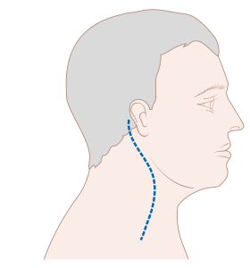
The neck is the part of the body on many vertebrates that connects the head with the torso and provides the mobility and movements of the head. The structures of the human neck are anatomically grouped into four compartments; vertebral, visceral and two vascular compartments. Within these compartments, the neck houses the cervical vertebrae and cervical part of the spinal cord, upper parts of the respiratory and digestive tracts, endocrine glands, nerves, arteries and veins. Muscles of the neck are described separately from the compartments. They bound the neck triangles.

The hyoid bone is a horseshoe-shaped bone situated in the anterior midline of the neck between the chin and the thyroid cartilage. At rest, it lies between the base of the mandible and the third cervical vertebra.
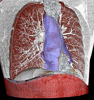
The mediastinum is the central compartment of the thoracic cavity. Surrounded by loose connective tissue, it is an undelineated region that contains a group of structures within the thorax, namely the heart and its vessels, the esophagus, the trachea, the phrenic and cardiac nerves, the thoracic duct, the thymus and the lymph nodes of the central chest.
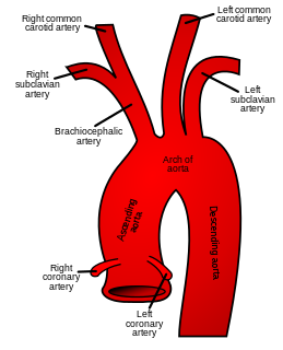
In anatomy, the left and right common carotid arteries (carotids) are arteries that supply the head and neck with oxygenated blood; they divide in the neck to form the external and internal carotid arteries.
The thoracic inlet, also known as the superior thoracic aperture, refers to the opening at the top of the thoracic cavity. It is also clinically referred to as the thoracic outlet, in the case of thoracic outlet syndrome; this refers to the superior thoracic aperture, and not to the lower, larger opening, the inferior thoracic aperture.

The sternohyoid muscle is a thin, narrow muscle attaching the hyoid bone to the sternum. It is one of the paired strap muscles of the infrahyoid muscles. It is supplied by the ansa cervicalis. It depresses the hyoid bone.

The middle pharyngeal constrictor is a fan-shaped muscle located in the neck. It is one of three pharyngeal constrictors. Similarly to the superior and inferior pharyngeal constrictor muscles, the middle pharyngeal constrictor is innervated by a branch of the vagus nerve through the pharyngeal plexus. The middle pharyngeal constrictor is smaller than the inferior pharyngeal constrictor muscle.

Anatomists use the term triangles of the neck to describe the divisions created by the major muscles in the region.

The posterior triangle is a region of the neck.

The superior thyroid artery arises from the external carotid artery just below the level of the greater cornu of the hyoid bone and ends in the thyroid gland.

The deep cervical fascia lies under cover of the platysma, and invests the muscles of the neck; it also forms sheaths for the carotid vessels, and for the structures situated in front of the vertebral column. Its attachment to the hyoid bone prevents the formation of a dewlap.

The submental triangle is a division of the anterior triangle of the neck.

The submandibular triangle corresponds to the region of the neck immediately beneath the body of the mandible.

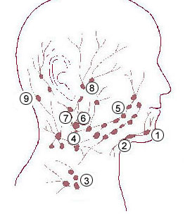
Cervical lymph nodes are lymph nodes found in the neck. Of the 800 lymph nodes in the human body, 300 are in the neck. Cervical lymph nodes are subject to a number of different pathological conditions including tumours, infection and inflammation.
The submental glands are situated between the anterior bellies of the digastric muscle and the hyoid bone.

The following outline is provided as an overview of and topical guide to human anatomy:

The parapharyngeal space, is a potential space in the head and the neck. It has clinical importance in otolaryngology due to parapharyngeal space tumours and parapharyngeal abscess developing in this area. It is also a key anatomic landmark for localizing disease processes in the surrounding spaces of the neck; the direction of its displacement indirectly reflects the site of origin for masses or infection in adjacent areas, and consequently their appropriate differential diagnosis.

The submandibular space is a fascial space of the head and neck. It is a potential space, and is paired on either side, located on the superficial surface of the mylohyoid muscle between the anterior and posterior bellies of the digastric muscle. The space corresponds to the anatomic region termed the submandibular triangle, part of the anterior triangle of the neck.

The submental space is a fascial space of the head and neck. It is a potential space located between the mylohyoid muscle superiorly, the platysma muscle inferiorly, under the chin in the midline. The space coincides with the anatomic region termed the submental triangle, part of the anterior triangle of the neck.