NiFe hydrogenase

Last updated

[NiFe] hydrogenase is a type of hydrogenase, which is an oxidative enzyme that reversibly converts molecular hydrogen in prokaryotes including Bacteria and Archaea. [1] [2] The catalytic site on the enzyme provides simple hydrogen-metabolizing microorganisms a redox mechanism by which to store and utilize energy via the reaction

Contents

This is particularly essential for the anaerobic, sulfate-reducing bacteria of the genus Desulfovibrio [3] [4] as well as pathogenic organisms Escherichia coli and Helicobacter pylori . [2] The mechanisms, maturation, and function of [NiFe] hydrogenases are actively being researched for applications to the hydrogen economy and as potential antibiotic targets.

Structure

The structure of [NiFe] hydrogenase isolated from D. vulgaris Miyazaki F consists of two subunits: the large subunit (blue) and the small subunit (magenta). The Figure was prepared using Jmol and the coordinates from PDB: 1H2A . Desulfovibrio vulgaris Miyazaki F.jpg
The structure of [NiFe] hydrogenase isolated from D. vulgaris Miyazaki F consists of two subunits: the large subunit (blue) and the small subunit (magenta). The Figure was prepared using Jmol and the coordinates from PDB: 1H2A .
The active site of [NiFe] hydrogenase in the oxidized form. L refers to non-protein ligand (1 C[?]O and 2 C[?]N). X can be an oxide, sulfur, hydroperoxide, or a hydroxide. Nickel Iron Hydrogenase Active Site.png
The active site of [NiFe] hydrogenase in the oxidized form. L refers to non-protein ligand (1 C≡O and 2 C≡N). X can be an oxide, sulfur, hydroperoxide, or a hydroxide.

The structure of [NiFe] hydrogenase was obtained from X-ray crystallography studies of five different sulfate-reducing bacteria: Desulfovibrio vulgaris Miyazaki F, [6] D. gigas, [7] D. frutosovorans, [8] [9] D. desulfuricans, [10] and Desulfomicrobium baculatum. [11] The [NiFe] hydrogenase isolated from D. vulgaris Miyazaki F is shown on the right. The larger subunit is in blue, has a molecular mass of 62.5 kDa, and houses the Ni-Fe active site. The smaller subunit is in magenta, has a molecular mass of 28.8 kDa, and contains the Fe-S clusters.

From the infrared spectra and X-ray crystallography studies, the [NiFe] hydrogenase active site was found to be (S-Cys)4Ni(μ-X)Fe(CO)(CN)2, in which the generic ligand X is either an oxide, sulfur, hydroperoxide, or a hydroxide found in an oxidized state only. [12] While the nickel atom participates in redox reactions, the iron atom is consistently in a Fe(II) oxidation state. [12] The exact geometry of the three non-protein ligands (denoted as L) coordinating to the Fe ion is not known; however, they were identified as one carbon monoxide (C≡O) molecule and two cyanide (C≡N) molecules. [13]

Fe-S clusters

Almost all hydrogenases contain at least one iron-sulfur cluster (Fe-S cluster). As previously mentioned, these Fe-S clusters connect the nickel active site of the enzyme to the surface of the protein because they serve as an electron transport chain from the Ni-Fe redox site to the electron acceptor cytochrome c3 (see cytochrome c family). [13] These electrons are produced from the heterolytic cleavage of the hydrogen molecule at the Ni-Fe active site. Crystal structures of the hydrogenase show a Fe3S4 in the center of the chain, and a Fe4S4 cluster at the molecular surface. The distance between the internal Fe4S4 cluster and the active site is approximately 12 Å. [13]

The [NiFe] and [NiFeSe] hydrogenases have remarkably similar structures, leading to the suggestion that one sulfur on a Fe-S cluster was replaced by a selenium atom, but these hydrogenases differ in catalytic reactivity and sensitivity to enzyme inhibitors. [4]

Illustration of [NiFe] hydrogenase enzyme with three Fe-S clusters in the small subunit and with Mg and Ni-Fe dimetal active site in the large subunit. Figure was prepared using Jmol and the coordinates from PDB: 1H2A . Mg ion = neon green; Ni ion = dark green; Fe ion = orange; sulfur = yellow; oxygen = red; carbon = dark gray NiFe hydrogenase active site.jpg
Illustration of [NiFe] hydrogenase enzyme with three Fe-S clusters in the small subunit and with Mg and Ni-Fe dimetal active site in the large subunit. Figure was prepared using Jmol and the coordinates from PDB: 1H2A . Mg ion = neon green; Ni ion = dark green; Fe ion = orange; sulfur = yellow; oxygen = red; carbon = dark gray

Mg ion and the proton pathways

[NiFe] hydrogenase has a Mg2+ cation bound in the C-terminus region of the larger subunit. This cation is bonded to three water molecules and three amino acids, and it stabilizes this solvent-free region. At approximately 13 Å away from the [NiFe] moiety, this cation connects the active site to a hydrogen bonding network and serves as a proton (H+) transfer pathway. [13]

The gas-access channel

Studies, in which xenon was bound to the hydrogenase, suggest a hydrophobic gas channel through which H2, CO, and O2 gases could reach the deeply buried active site within the enzyme. Crystal structure revealed several small channels at the surface, which combined into one larger channel that reached the [Ni-Fe] active site. [13]

Since hydrogenases are well known to be oxygen sensitive, the diffusion of gas to the active site depends on the size and environment of the gas-access channel, the reaction of molecular oxygen (O2) at the active site, and the recovery of the active site after oxidation. [13]

Mechanism

The exact reaction mechanism of [NiFe] hydrogenases has been a matter of great debate. In 2009, a mechanism was proposed by Higuchi and coworkers based on X-ray crystallography and spectroscopic data of Desulfovibrio vulgaris Miyazaki F. [13] During the catalytic process, the Fe ion in the active site does not change its oxidation state while the Ni metal ion participates in redox chemistry. There are two main groups of redox states that [NiFe] hydrogenases pass through during catalysis:

  1. Inactive redox states, and
  2. Active redox states.
Figure 5. Different redox states of [NiFe] hydrogenase's metal active site. The redox states in the red are the inactive redox states. The redox states in the green are the active redox states. (Adapted from ). Redox states of NiFe hydrogenase.png
Figure 5. Different redox states of [NiFe] hydrogenase's metal active site. The redox states in the red are the inactive redox states. The redox states in the green are the active redox states. (Adapted from ).

Inactive redox states

Ni-A (the “unready” state) and Ni-B (the “ready” state) are the most oxidized forms of the [NiFe] metal center and are activated via one-electron reduction with proton transfer. The rate of reductive activation of Ni-A to Ni-SU can take hours while the rate of reductive activation of Ni-B to Ni-SIr happens in seconds. [14] The reason for this disparity in activation kinetics between Ni-A and Ni-B was proposed to be a result of the difference in bridging ligands between the two different redox states. At the Ni-SIr state, a water molecule was released to form the Ni-SIa state, the first catalytic redox active state of [NiFe] hydrogenases.

Active redox states

The three most important catalytic redox active states of [NiFe] hydrogenases are Ni-SIa, Ni-C and Ni-R (which have three different variations). [13] The light-sensitive Ni-C state can be obtained via one electron reduction of Ni-SIa. The electron paramagnetic resonance spectroscopic studies of the Ni-C state, which contained a Ni3+ with S = 1/2 and a hydride bridging the two metals, Ni and Fe, showed that the heterolytic cleavage of H2 takes place in the [NiFe] hydrogenase active site.

The CO-inhibited states

Ni-SIa state can be inhibited by CO, which binds directly to Ni metal ion in a bent conformation to form Ni-SCO (see below). [15] Since Ni-C is light sensitive, illumination at 100 K results in Ni-L redox state. During this process, nickel is reduced. In the presence of CO, Ni-L forms Ni-CO state.

Figure 6. Illustration of the [NiFe] hydrogenase active site inhibited by CO. Top-down view (left). Side view (center). Chemdraw depiction of the inhibited active site (right). The figure was prepared with Jmol and coordinates from 1UBK.pdb. Ni ion = green; Fe ion = orange; sulfur = yellow; oxygen = red; carbon = dark gray; nitrogen = blue. NiFe-CO inhibited.jpg
Figure 6. Illustration of the [NiFe] hydrogenase active site inhibited by CO. Top-down view (left). Side view (center). Chemdraw depiction of the inhibited active site (right). The figure was prepared with Jmol and coordinates from 1UBK.pdb. Ni ion = green; Fe ion = orange; sulfur = yellow; oxygen = red; carbon = dark gray; nitrogen = blue.

Maturation and genetic arrangement

The maturation of [NiFe] hydrogenases requires a set of accessory proteins that synthesize the NiFe active site and modify the precursor enzyme so that it has the correct structure and location. [2] [16] [17] The maturation of the active site is of special interest because of the synthesis of cyanide (CN) and carbon monoxide (CO) metal ligands which are usually toxic to living organism. [16] This step is completed by the proteins HypC, HypD, HypE, and HypF. [17] [18] After, synthesis of the iron center, nickel is inserted using metallochaperones HypA, HypB, and SlyD. [17] [18] Once the catalytic center is completed, the hydrogenase precursor undergoes a C-terminal cleavage that prompts rearrangement of its structure and association with the small subunit. [16] [17] [18] Finally, the completed enzyme is transported to its correct position within the cell. [16] [17] [18] Hydrogenase promoter, PSH, can be studied constructing a PSH promoter-gfp fusion by using green fluorescent protein (gfp) reporter gene. [19]

Application

Since [NiFe] hydrogenase is a member of the hydrogenase family, these enzymes can catalyze both the consumption and production of hydrogen. [1] By studying [NiFe] hydrogenase, scientists can optimize a condition in which the protein will only produce hydrogen. Additionally, small enzyme mimic of [NiFe] hydrogenase can also be synthesized to act as hydrogen gas generator. The soluble [NiFe] hydrogenase from Ralstonia eutropha H16 is a promising candidate enzyme for H2-based biofuel application as it favours H2 oxidation and is relatively oxygen-tolerant. It can be produced on heterotrophic growth media [20] and purified via anion exchange and size exclusion chromatography matrices. [21]

See also

Related Research Articles

<span class="mw-page-title-main">Urease</span> Multiprotein Nickel-containing complex which hydrolyses urea

Ureases, functionally, belong to the superfamily of amidohydrolases and phosphotriesterases. Ureases are found in numerous bacteria, fungi, algae, plants, and some invertebrates, as well as in soils, as a soil enzyme. They are nickel-containing metalloenzymes of high molecular weight.

<span class="mw-page-title-main">Metalloprotein</span> Protein that contains a metal ion cofactor

Metalloprotein is a generic term for a protein that contains a metal ion cofactor. A large proportion of all proteins are part of this category. For instance, at least 1000 human proteins contain zinc-binding protein domains although there may be up to 3000 human zinc metalloproteins.

Artificial photosynthesis is a chemical process that biomimics the natural process of photosynthesis to convert sunlight, water, and carbon dioxide into carbohydrates and oxygen. The term artificial photosynthesis is commonly used to refer to any scheme for capturing and storing the energy from sunlight in the chemical bonds of a fuel. Photocatalytic water splitting converts water into hydrogen and oxygen and is a major research topic of artificial photosynthesis. Light-driven carbon dioxide reduction is another process studied that replicates natural carbon fixation.

Iron–sulfur proteins are proteins characterized by the presence of iron–sulfur clusters containing sulfide-linked di-, tri-, and tetrairon centers in variable oxidation states. Iron–sulfur clusters are found in a variety of metalloproteins, such as the ferredoxins, as well as NADH dehydrogenase, hydrogenases, coenzyme Q – cytochrome c reductase, succinate – coenzyme Q reductase and nitrogenase. Iron–sulfur clusters are best known for their role in the oxidation-reduction reactions of electron transport in mitochondria and chloroplasts. Both Complex I and Complex II of oxidative phosphorylation have multiple Fe–S clusters. They have many other functions including catalysis as illustrated by aconitase, generation of radicals as illustrated by SAM-dependent enzymes, and as sulfur donors in the biosynthesis of lipoic acid and biotin. Additionally, some Fe–S proteins regulate gene expression. Fe–S proteins are vulnerable to attack by biogenic nitric oxide, forming dinitrosyl iron complexes. In most Fe–S proteins, the terminal ligands on Fe are thiolate, but exceptions exist.

A hydrogenase is an enzyme that catalyses the reversible oxidation of molecular hydrogen (H2), as shown below:

Bioorganometallic chemistry is the study of biologically active molecules that contain carbon directly bonded to metals or metalloids. The importance of main-group and transition-metal centers has long been recognized as important to the function of enzymes and other biomolecules. However, only a small subset of naturally-occurring metal complexes and synthetically prepared pharmaceuticals are organometallic; that is, they feature a direct covalent bond between the metal(loid) and a carbon atom. The first, and for a long time, the only examples of naturally occurring bioorganometallic compounds were the cobalamin cofactors (vitamin B12) in its various forms. In the 21st century, discovery of new systems containing carbon-metal bonds in biology, bioorganometallic chemistry is rapidly emerging as a distinct subdiscipline of bioinorganic chemistry that straddles organometallic chemistry and biochemistry. Naturally occurring bioorganometallics include enzymes and sensor proteins. Also within this realm are synthetically prepared organometallic compounds that serve as new drugs and imaging agents (technetium-99m sestamibi) as well as the principles relevant to the toxicology of organometallic compounds (e.g., methylmercury). Consequently, bioorganometallic chemistry is increasingly relevant to medicine and pharmacology.

<i>Cupriavidus necator</i> Species of bacterium

Cupriavidus necator is a Gram-negative soil bacterium of the class Betaproteobacteria.

A hydrogenase mimic or bio-mimetic is an enzyme mimic of hydrogenases.

In chemistry, a (redox) non-innocent ligand is a ligand in a metal complex where the oxidation state is not clear. Typically, complexes containing non-innocent ligands are redox active at mild potentials. The concept assumes that redox reactions in metal complexes are either metal or ligand localized, which is a simplification, albeit a useful one.

In enzymology, carbon monoxide dehydrogenase (CODH) (EC 1.2.7.4) is an enzyme that catalyzes the chemical reaction

In enzymology, a cytochrome-c3 hydrogenase (EC 1.12.2.1) is an enzyme that catalyzes the chemical reaction

<span class="mw-page-title-main">Ferredoxin hydrogenase</span> Class of enzymes

In enzymology, ferredoxin hydrogenase, also referred to as [Fe-Fe]hydrogenase, H2 oxidizing hydrogenase, H2 producing hydrogenase, bidirectional hydrogenase, hydrogenase (ferredoxin), hydrogenlyase, and uptake hydrogenase, is found in Clostridium pasteurianum, Clostridium acetobutylicum,Chlamydomonas reinhardtii, and other organisms. The systematic name of this enzyme is hydrogen:ferredoxin oxidoreductase

<span class="mw-page-title-main">Dioxygenase</span>

Dioxygenases are oxidoreductase enzymes. Aerobic life, from simple single-celled bacteria species to complex eukaryotic organisms, has evolved to depend on the oxidizing power of dioxygen in various metabolic pathways. From energetic adenosine triphosphate (ATP) generation to xenobiotic degradation, the use of dioxygen as a biological oxidant is widespread and varied in the exact mechanism of its use. Enzymes employ many different schemes to use dioxygen, and this largely depends on the substrate and reaction at hand.

An enzymatic biofuel cell is a specific type of fuel cell that uses enzymes as a catalyst to oxidize its fuel, rather than precious metals. Enzymatic biofuel cells, while currently confined to research facilities, are widely prized for the promise they hold in terms of their relatively inexpensive components and fuels, as well as a potential power source for bionic implants.

<span class="mw-page-title-main">Iron–nickel clusters</span>

Iron–nickel (Fe–Ni) clusters are metal clusters consisting of iron and nickel, i.e. Fe–Ni structures displaying polyhedral frameworks held together by two or more metal–metal bonds per metal atom, where the metal atoms are located at the vertices of closed, triangulated polyhedra.

Hydrogenases are enzymes that catalyze the reversible activation of hydrogen and which occur widely in prokaryotes as well as in some eukaryotes. There are various types of hydrogenases, but all of them seem to contain at least one iron-sulphur cluster. They can be broadly divided into two groups: hydrogenases containing nickel and, in some cases, also selenium and those lacking nickel.

<span class="mw-page-title-main">Iron hydride</span> Index of articles associated with the same name

An iron hydride is a chemical system which contains iron and hydrogen in some associated form.

<span class="mw-page-title-main">Nickel superoxide dismutase</span>

Nickel superoxide dismutase (Ni-SOD) is a metalloenzyme that, like the other superoxide dismutases, protects cells from oxidative damage by catalyzing the disproportionation of the cytotoxic superoxide radical to hydrogen peroxide and molecular oxygen. Superoxide is a reactive oxygen species that is produced in large amounts during photosynthesis and aerobic cellular respiration. The equation for the disproportionation of superoxide is shown below:

<span class="mw-page-title-main">CO-methylating acetyl-CoA synthase</span>

Acetyl-CoA synthase (ACS), not to be confused with Acetyl-CoA synthetase or Acetate-CoA ligase, is a nickel-containing enzyme involved in the metabolic processes of cells. Together with Carbon monoxide dehydrogenase (CODH), it forms the bifunctional enzyme Acetyl-CoA Synthase/Carbon Monoxide Dehydrogenase (ACS/CODH) found in anaerobic organisms such as archaea and bacteria. The ACS/CODH enzyme works primarily through the Wood–Ljungdahl pathway which converts carbon dioxide to Acetyl-CoA. The recommended name for this enzyme is CO-methylating acetyl-CoA synthase.

<span class="mw-page-title-main">Wolfgang Lubitz</span> German chemist and biophysicist

Wolfgang Lubitz is a German chemist and biophysicist. He is currently a director emeritus at the Max Planck Institute for Chemical Energy Conversion. He is well known for his work on bacterial photosynthetic reaction centres, hydrogenase enzymes, and the oxygen-evolving complex using a variety of biophysical techniques. He has been recognized by a Festschrift for his contributions to electron paramagnetic resonance (EPR) and its applications to chemical and biological systems.
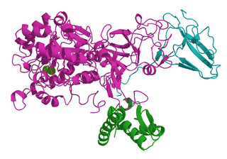
References

  1. 1 2 Jugder, Bat-Erdene; Welch, Jeffrey; Aguey-Zinsou, Kondo-Francois; Marquis, Christopher P. (2013-05-14). "Fundamentals and electrochemical applications of [Ni–Fe]-uptake hydrogenases". RSC Advances. 3 (22): 8142. doi:10.1039/c3ra22668a. ISSN   2046-2069.
  2. 1 2 3 Vignais, Paulette M.; Billoud, Bernard (October 2007). "Occurrence, Classification, and Biological Function of Hydrogenases: An Overview". Chemical Reviews. 107 (10): 4206–4272. doi:10.1021/cr050196r. PMID   17927159.
  3. Volbeda, A.; Garcin, E.; Piras, C.; de Lacey, A. L.; Fernandez, V. M.; Hatchikian, E. C.; Frey, M.; Fontecilla-Camps, J. C. (1996). "Structure of the [NiFe] Hydrogenase Active Site: Evidence for Biologically Uncommon Fe Ligands". J. Am. Chem. Soc. 118 (51): 12989–12996. doi:10.1021/ja962270g.
  4. 1 2 Eidsness, M. K.; Scott, R. A.; Prickril, B. C.; DerVartanian, D. V.; Legall, J.; Moura, I.; Moura, J. J.; Peck, H. D. (1989). "Evidence for selenocysteine coordination to the active site nickel in the [NiFeSe]hydrogenases from Desulfovibrio baculatus". Proceedings of the National Academy of Sciences. 86 (1): 147–151. doi: 10.1073/pnas.86.1.147 . PMC   286421 . PMID   2521386.
  5. 1 2 3 Jmol: an open-source Java viewer for chemical structures in 3D. http://www.jmol.org/
  6. Higuchi, Y.; Yagi, T.; Yasuoka, N. (1997). "Unusual ligand structure in Ni-Fe active center and an additional Mg site in hydrogenase revealed by high resolution X-ray structure analysis". Structure. 5 (12): 1671–1680. doi: 10.1016/s0969-2126(97)00313-4 . PMID   9438867.
  7. Volbeda, A.; Charon, M.-H.; Piras, C.; Hatchikian, E. C.; Frey, M.; Fontecilla-Camps, J. C. (1995). "Crystal structure of the nickel-iron hydrogenase from Desulfovibrio gigas". Nature. 373 (6515): 580–587. doi:10.1038/373580a0. PMID   7854413. S2CID   4335445.
  8. Volbeda, A.; Martin, L.; Cavazza, C.; Matho, M.; Faber, B. W.; Roseboom, W.; Albracht, S. P. J.; Garcin, E.; Rousset, M.; Fontecilla-Camps, J. C. (2005). "Structural differences between the ready and unready oxidized states of [NiFe] hydrogenases". Journal of Biological Inorganic Chemistry. 10 (3): 239–249. doi:10.1007/s00775-005-0632-x. PMID   15803334. S2CID   25953517.
  9. Montet, Y.; Amara, P.; Volbeda, A.; Vernede, X.; Hatchikian E. C.; Field, M. J.; Frey, M.; Fontecilla-Camps, J. C. (1997). "Gas access to the active site of Ni-Fe hydrogenases probed by X-ray crystallography and molecular dynamics". Nature Structural & Molecular Biology. 4 (7): 523–526. doi:10.1038/nsb0797-523. PMID   9228943. S2CID   19356968.
  10. Matias, P. M.; Soares, C. M.; Saraiva, L. M.; Coelho, R.; Morais, J.; Le Gall, J.; Carrondo, M. A. (2001). "[NiFe] hydrogenase from Desulfovibrio desulfuricans ATCC 27774: gene sequencing, three-dimensional structure determination and refinement at 1.8 Å and modelling studies of its interaction with the tetrahaem cytochrome c3". Journal of Biological Inorganic Chemistry. 6 (1): 63–81. doi:10.1007/s007750000167. PMID   11191224. S2CID   9661059.
  11. Garcin, E.; Vernede, X.; Hatchikian, E. C.; Volbeda, A.; Frey, M.; Fontecilla-Camps, J. C. (1999). "The crystal structure of a reduced [NiFeSe] hydrogenase provides an image of the activated catalytic center". Structure. 7 (5): 557–566. doi: 10.1016/s0969-2126(99)80072-0 . PMID   10378275.
  12. 1 2 Chiou, T.-W.; Liaw, W.-F. (2008). "Nickel–thiolate and iron–thiolate cyanocarbonyl complexes: Modeling the nickel and iron sites of [NiFe] hydrogenase". Comptes Rendus Chimie. 11 (8): 818–833. doi:10.1016/j.crci.2008.04.003.
  13. 1 2 3 4 5 6 7 8 9 Ogata, H.; Lubitz, W.; Higuchi, Y. (2009). "[NiFe] hydrogenases: structural and spectroscopic studies of the reaction mechanism". Dalton Trans. 37 (37): 7577–7587. doi:10.1039/b903840j. PMID   19759926.
  14. Lamele, S. E.; Albracht, S. P. J.; Armstrong, F. A. (2004). "Electrochemical Potential-Step Investigations of the Aerobic Interconversions of [NiFe]-Hydrogenase from Allochromatium vinosum: Insights into the Puzzling Difference between Unready and Ready Oxidized Inactive States". Journal of the American Chemical Society. 126 (45): 14899–14909. doi:10.1021/ja047939v. PMID   15535717.
  15. Ogata, H.; Mizoguchi, Y.; Mizuno, N.; Miki, K.; Adachi, S.-i.; Yasuoka, N.; Yagi, T.; Yamauchi, O.; Hirota, S.; Higuchi, Y. (2002). "Structural Studies of the Carbon Monoxide Complex of [NiFe]hydrogenase from Desulfovibrio vulgaris Miyazaki F: Suggestion for the Initial Activation Site for Dihydrogen". Journal of the American Chemical Society. 124 (39): 11628–11635. doi:10.1021/ja012645k. PMID   12296727.
  16. 1 2 3 4 Lubitz, Wolfgang; Ogata, Hideaki; Rüdiger, Olaf; Reijerse, Edward (23 April 2014). "Hydrogenases". Chemical Reviews. 114 (8): 4081–4148. doi:10.1021/cr4005814. PMID   24655035.
  17. 1 2 3 4 5 Lacasse, Michael J.; Zamble, Deborah B. (29 March 2016). "[NiFe]-Hydrogenase Maturation". Biochemistry. 55 (12): 1689–1701. doi:10.1021/acs.biochem.5b01328. PMID   26919691.
  18. 1 2 3 4 Peters, John W.; Schut, Gerrit J.; Boyd, Eric S.; Mulder, David W.; Shepard, Eric M.; Broderick, Joan B.; King, Paul W.; Adams, Michael W.W. (June 2015). "[FeFe]- and [NiFe]-hydrogenase diversity, mechanism, and maturation". Biochimica et Biophysica Acta (BBA) - Molecular Cell Research. 1853 (6): 1350–1369. doi: 10.1016/j.bbamcr.2014.11.021 . PMID   25461840.
  19. Jugder, Bat-Erdene; Welch, Jeffrey; Braidy, Nady; Marquis, Christopher P. (2016-07-26). "Construction and use of aCupriavidus necatorH16 soluble hydrogenase promoter (PSH) fusion togfp(green fluorescent protein)". PeerJ. 4: e2269. doi:10.7717/peerj.2269. ISSN   2167-8359. PMC   4974937 . PMID   27547572.
  20. Jugder, Bat-Erdene; Chen, Zhiliang; Ping, Darren Tan Tek; Lebhar, Helene; Welch, Jeffrey; Marquis, Christopher P. (2015-03-25). "An analysis of the changes in soluble hydrogenase and global gene expression in Cupriavidus necator ( Ralstonia eutropha ) H16 grown in heterotrophic diauxic batch culture". Microbial Cell Factories. 14 (1): 42. doi:10.1186/s12934-015-0226-4. ISSN   1475-2859. PMC   4377017 . PMID   25880663.
  21. Jugder, Bat-Erdene; Lebhar, Helene; Aguey-Zinsou, Kondo-Francois; Marquis, Christopher P. (2016-01-01). "Production and purification of a soluble hydrogenase from Ralstonia eutropha H16 for potential hydrogen fuel cell applications". MethodsX. 3: 242–250. doi:10.1016/j.mex.2016.03.005. PMC   4816682 . PMID   27077052.