Nucleic acid double helix

Last updated
Two complementary regions of nucleic acid molecules will bind and form a double helical structure held together by base pairs. DNA orbit animated static thumb.png
Two complementary regions of nucleic acid molecules will bind and form a double helical structure held together by base pairs.

In molecular biology, the term double helix [1] refers to the structure formed by double-stranded molecules of nucleic acids such as DNA. The double helical structure of a nucleic acid complex arises as a consequence of its secondary structure, and is a fundamental component in determining its tertiary structure. The structure was discovered by Rosalind Franklin and her student Raymond Gosling, [2] but the term "double helix" entered popular culture with the publication in 1968 of The Double Helix: A Personal Account of the Discovery of the Structure of DNA by James Watson.

Contents

The DNA double helix biopolymer of nucleic acid is held together by nucleotides which base pair together. [3] In B-DNA, the most common double helical structure found in nature, the double helix is right-handed with about 10–10.5 base pairs per turn. [4] The double helix structure of DNA contains a major groove and minor groove. In B-DNA the major groove is wider than the minor groove. [3] Given the difference in widths of the major groove and minor groove, many proteins which bind to B-DNA do so through the wider major groove. [5]

History

The double-helix model of DNA structure was first published in the journal Nature by James Watson and Francis Crick in 1953, [6] (X,Y,Z coordinates in 1954 [7] ) based on the work of Rosalind Franklin and her student Raymond Gosling, who took the crucial X-ray diffraction image of DNA labeled as "Photo 51", [8] [9] and Maurice Wilkins, Alexander Stokes, and Herbert Wilson, [10] and base-pairing chemical and biochemical information by Erwin Chargaff. [11] [12] [13] [14] [15] [16] Before this, Linus Pauling—who had already accurately characterised the conformation of protein secondary structure motifs—and his collaborator Robert Corey had posited, erroneously, that DNA would adopt a triple-stranded conformation. [17]

The realization that the structure of DNA is that of a double-helix elucidated the mechanism of base pairing by which genetic information is stored and copied in living organisms and is widely considered one of the most important scientific discoveries of the 20th century. Crick, Wilkins, and Watson each received one-third of the 1962 Nobel Prize in Physiology or Medicine for their contributions to the discovery. [18]

Nucleic acid hybridization

Hybridization is the process of complementary base pairs binding to form a double helix. Melting is the process by which the interactions between the strands of the double helix are broken, separating the two nucleic acid strands. These bonds are weak, easily separated by gentle heating, enzymes, or mechanical force. Melting occurs preferentially at certain points in the nucleic acid. [19] T and A rich regions are more easily melted than C and G rich regions. Some base steps (pairs) are also susceptible to DNA melting, such as T A and T G. [20] These mechanical features are reflected by the use of sequences such as TATA at the start of many genes to assist RNA polymerase in melting the DNA for transcription.

Strand separation by gentle heating, as used in polymerase chain reaction (PCR), is simple, providing the molecules have fewer than about 10,000 base pairs (10 kilobase pairs, or 10 kbp). The intertwining of the DNA strands makes long segments difficult to separate. [21] The cell avoids this problem by allowing its DNA-melting enzymes (helicases) to work concurrently with topoisomerases, which can chemically cleave the phosphate backbone of one of the strands so that it can swivel around the other. [22] Helicases unwind the strands to facilitate the advance of sequence-reading enzymes such as DNA polymerase. [23]

Base pair geometry

Base pair geometries CCF10292011 00000.jpg
Base pair geometries

The geometry of a base, or base pair step can be characterized by 6 coordinates: shift, slide, rise, tilt, roll, and twist. These values precisely define the location and orientation in space of every base or base pair in a nucleic acid molecule relative to its predecessor along the axis of the helix. Together, they characterize the helical structure of the molecule. In regions of DNA or RNA where the normal structure is disrupted, the change in these values can be used to describe such disruption.

For each base pair, considered relative to its predecessor, there are the following base pair geometries to consider: [24] [25] [26]

Rise and twist determine the handedness and pitch of the helix. The other coordinates, by contrast, can be zero. Slide and shift are typically small in B-DNA, but are substantial in A- and Z-DNA. Roll and tilt make successive base pairs less parallel, and are typically small.

"Tilt" has often been used differently in the scientific literature, referring to the deviation of the first, inter-strand base-pair axis from perpendicularity to the helix axis. This corresponds to slide between a succession of base pairs, and in helix-based coordinates is properly termed "inclination".

Helix geometries

At least three DNA conformations are believed to be found in nature, A-DNA, B-DNA, and Z-DNA. The B form described by James Watson and Francis Crick is believed to predominate in cells. [27] It is 23.7 Å wide and extends 34 Å per 10 bp of sequence. The double helix makes one complete turn about its axis every 10.4–10.5 base pairs in solution. This frequency of twist (termed the helical pitch) depends largely on stacking forces that each base exerts on its neighbours in the chain. The absolute configuration of the bases determines the direction of the helical curve for a given conformation.

A-DNA and Z-DNA differ significantly in their geometry and dimensions to B-DNA, although still form helical structures. It was long thought that the A form only occurs in dehydrated samples of DNA in the laboratory, such as those used in crystallographic experiments, and in hybrid pairings of DNA and RNA strands, but DNA dehydration does occur in vivo, and A-DNA is now known to have biological functions. Segments of DNA that cells have methylated for regulatory purposes may adopt the Z geometry, in which the strands turn about the helical axis the opposite way to A-DNA and B-DNA. There is also evidence of protein-DNA complexes forming Z-DNA structures.

Other conformations are possible; A-DNA, B-DNA, C-DNA, E-DNA, [28] L-DNA (the enantiomeric form of D-DNA), [29] P-DNA, [30] S-DNA, Z-DNA, etc. have been described so far. [31] In fact, only the letters F, Q, U, V, and Y are now available to describe any new DNA structure that may appear in the future. [32] [33] However, most of these forms have been created synthetically and have not been observed in naturally occurring biological systems.[ citation needed ] There are also triple-stranded DNA forms and quadruplex forms such as the G-quadruplex and the i-motif.

The structures of A-, B-, and Z-DNA. Dnaconformations.png
The structures of A-, B-, and Z-DNA.
The helix axis of A-, B-, and Z-DNA. B&Z&A DNA formula.svg
The helix axis of A-, B-, and Z-DNA.
Structural features of the three major forms of DNA [34] [35] [36]
Geometry attributeA-DNAB-DNAZ-DNA
Helix senseright-handedright-handedleft-handed
Repeating unit1 bp1 bp2 bp
Rotation/bp32.7°34.3°60°/2
bp/turn1110.512
Inclination of bp to axis+19°−1.2°−9°
Rise/bp along axis2.3 Å (0.23  nm)3.32 Å (0.332 nm)3.8 Å (0.38 nm)
Pitch/turn of helix28.2 Å (2.82 nm)33.2 Å (3.32 nm)45.6 Å (4.56 nm)
Mean propeller twist+18°+16°
Glycosyl angleantiantiC: anti,
G: syn
Sugar pucker C3'-endoC2'-endoC: C2'-endo,
G: C2'-exo
Diameter23 Å (2.3 nm)20 Å (2.0 nm)18 Å (1.8 nm)

Grooves

Major and minor grooves of DNA. Minor groove is a binding site for the dye Hoechst 33258. DNA-ligand-by-Abalone.png
Major and minor grooves of DNA. Minor groove is a binding site for the dye Hoechst 33258.

Twin helical strands form the DNA backbone. Another double helix may be found by tracing the spaces, or grooves, between the strands. These voids are adjacent to the base pairs and may provide a binding site. [37] As the strands are not directly opposite each other, the grooves are unequally sized. One groove, the major groove, is 22 Å wide and the other, the minor groove, is 12 Å wide. [38] The narrowness of the minor groove means that the edges of the bases are more accessible in the major groove. As a result, proteins like transcription factors that can bind to specific sequences in double-stranded DNA usually make contacts to the sides of the bases exposed in the major groove. [5] This situation varies in unusual conformations of DNA within the cell (see below), but the major and minor grooves are always named to reflect the differences in size that would be seen if the DNA is twisted back into the ordinary B form. [39]

Non-double helical forms

Alternative non-helical models were briefly considered in the late 1970s as a potential solution to problems in DNA replication in plasmids and chromatin. However, the models were set aside in favor of the double-helical model due to subsequent experimental advances such as X-ray crystallography of DNA duplexes and later the nucleosome core particle, and the discovery of topoisomerases. Also, the non-double-helical models are not currently accepted by the mainstream scientific community. [40] [41]

Bending

DNA is a relatively rigid polymer, typically modelled as a worm-like chain. It has three significant degrees of freedom; bending, twisting, and compression, each of which cause certain limits on what is possible with DNA within a cell. Twisting-torsional stiffness is important for the circularisation of DNA and the orientation of DNA bound proteins relative to each other and bending-axial stiffness is important for DNA wrapping and circularisation and protein interactions. Compression-extension is relatively unimportant in the absence of high tension.

Persistence length, axial stiffness

Example sequences and their persistence lengths (B DNA)[ citation needed ]
SequencePersistence length
/ base pairs
Random154±10
(CA)repeat133±10
(CAG)repeat124±10
(TATA)repeat137±10

DNA in solution does not take a rigid structure but is continually changing conformation due to thermal vibration and collisions with water molecules, which makes classical measures of rigidity impossible to apply. Hence, the bending stiffness of DNA is measured by the persistence length, defined as:

Bending flexibility of a polymer is conventionally quantified in terms of its persistence length, Lp, a length scale below which the polymer behaves more or less like a rigid rod. Specifically, Lp is defined as length of the polymer segment over which the time-averaged orientation of the polymer becomes uncorrelated... [42]

This value may be directly measured using an atomic force microscope to directly image DNA molecules of various lengths. In an aqueous solution, the average persistence length is 46–50 nm or 140–150 base pairs (the diameter of DNA is 2 nm), although can vary significantly. This makes DNA a moderately stiff molecule.

The persistence length of a section of DNA is somewhat dependent on its sequence, and this can cause significant variation. The variation is largely due to base stacking energies and the residues which extend into the minor and major grooves.

Models for DNA bending

Stacking stability of base steps (B DNA) [43]
StepStacking ΔG
/kcal mol−1
T A-0.19
T G or C A-0.55
C G-0.91
A G or C T-1.06
A A or T T-1.11
A T-1.34
G A or T C-1.43
C C or G G-1.44
A C or G T-1.81
G C-2.17

At length-scales larger than the persistence length, the entropic flexibility of DNA is remarkably consistent with standard polymer physics models, such as the Kratky-Porod worm-like chain model. [44] Consistent with the worm-like chain model is the observation that bending DNA is also described by Hooke's law at very small (sub-piconewton) forces. For DNA segments less than the persistence length, the bending force is approximately constant and behaviour deviates from the worm-like chain predictions.

This effect results in unusual ease in circularising small DNA molecules and a higher probability of finding highly bent sections of DNA. [45]

Bending preference

DNA molecules often have a preferred direction to bend, i.e., anisotropic bending. This is, again, due to the properties of the bases which make up the DNA sequence - a random sequence will have no preferred bend direction, i.e., isotropic bending.

Preferred DNA bend direction is determined by the stability of stacking each base on top of the next. If unstable base stacking steps are always found on one side of the DNA helix then the DNA will preferentially bend away from that direction. As bend angle increases then steric hindrances and ability to roll the residues relative to each other also play a role, especially in the minor groove. A and T residues will be preferentially be found in the minor grooves on the inside of bends. This effect is particularly seen in DNA-protein binding where tight DNA bending is induced, such as in nucleosome particles. See base step distortions above.

DNA molecules with exceptional bending preference can become intrinsically bent. This was first observed in trypanosomatid kinetoplast DNA. Typical sequences which cause this contain stretches of 4-6 T and A residues separated by G and C rich sections which keep the A and T residues in phase with the minor groove on one side of the molecule. For example:

¦¦¦¦¦¦
GATTCCCAAAAATGTCAAAAAATAGGCAAAAAATGCCAAAAAATCCCAAAC

The intrinsically bent structure is induced by the 'propeller twist' of base pairs relative to each other allowing unusual bifurcated Hydrogen-bonds between base steps. At higher temperatures this structure is denatured, and so the intrinsic bend is lost.

All DNA which bends anisotropically has, on average, a longer persistence length and greater axial stiffness. This increased rigidity is required to prevent random bending which would make the molecule act isotropically.

Circularization

DNA circularization depends on both the axial (bending) stiffness and torsional (rotational) stiffness of the molecule. For a DNA molecule to successfully circularize it must be long enough to easily bend into the full circle and must have the correct number of bases so the ends are in the correct rotation to allow bonding to occur. The optimum length for circularization of DNA is around 400 base pairs (136 nm)[ citation needed ], with an integral number of turns of the DNA helix, i.e., multiples of 10.4 base pairs. Having a non integral number of turns presents a significant energy barrier for circularization, for example a 10.4 x 30 = 312 base pair molecule will circularize hundreds of times faster than 10.4 x 30.5 ≈ 317 base pair molecule. [46]

The bending of short circularized DNA segments is non-uniform. Rather, for circularized DNA segments less than the persistence length, DNA bending is localised to 1-2 kinks that form preferentially in AT-rich segments. If a nick is present, bending will be localised to the nick site. [45]

Stretching

Elastic stretching regime

Longer stretches of DNA are entropically elastic under tension. When DNA is in solution, it undergoes continuous structural variations due to the energy available in the thermal bath of the solvent. This is due to the thermal vibration of the molecule combined with continual collisions with water molecules. For entropic reasons, more compact relaxed states are thermally accessible than stretched out states, and so DNA molecules are almost universally found in a tangled relaxed layouts. For this reason, one molecule of DNA will stretch under a force, straightening it out. Using optical tweezers, the entropic stretching behavior of DNA has been studied and analyzed from a polymer physics perspective, and it has been found that DNA behaves largely like the Kratky-Porod worm-like chain model under physiologically accessible energy scales.

Phase transitions under stretching

Under sufficient tension and positive torque, DNA is thought to undergo a phase transition with the bases splaying outwards and the phosphates moving to the middle. This proposed structure for overstretched DNA has been called P-form DNA, in honor of Linus Pauling who originally presented it as a possible structure of DNA. [30]

Evidence from mechanical stretching of DNA in the absence of imposed torque points to a transition or transitions leading to further structures which are generally referred to as S-form DNA. These structures have not yet been definitively characterised due to the difficulty of carrying out atomic-resolution imaging in solution while under applied force although many computer simulation studies have been made (for example, [47] [48] ).

Proposed S-DNA structures include those which preserve base-pair stacking and hydrogen bonding (GC-rich), while releasing extension by tilting, as well as structures in which partial melting of the base-stack takes place, while base-base association is nonetheless overall preserved (AT-rich).

Periodic fracture of the base-pair stack with a break occurring once per three bp (therefore one out of every three bp-bp steps) has been proposed as a regular structure which preserves planarity of the base-stacking and releases the appropriate amount of extension, [49] with the term "Σ-DNA" introduced as a mnemonic, with the three right-facing points of the Sigma character serving as a reminder of the three grouped base pairs. The Σ form has been shown to have a sequence preference for GNC motifs which are believed under the GNC hypothesis to be of evolutionary importance. [50]

Supercoiling and topology

Supercoiled structure of circular DNA molecules with low writhe. The helical aspect of the DNA duplex is omitted for clarity. Circular DNA Supercoiling.png
Supercoiled structure of circular DNA molecules with low writhe. The helical aspect of the DNA duplex is omitted for clarity.

The B form of the DNA helix twists 360° per 10.4-10.5 bp in the absence of torsional strain. But many molecular biological processes can induce torsional strain. A DNA segment with excess or insufficient helical twisting is referred to, respectively, as positively or negatively supercoiled . DNA in vivo is typically negatively supercoiled, which facilitates the unwinding (melting) of the double-helix required for RNA transcription.

Within the cell most DNA is topologically restricted. DNA is typically found in closed loops (such as plasmids in prokaryotes) which are topologically closed, or as very long molecules whose diffusion coefficients produce effectively topologically closed domains. Linear sections of DNA are also commonly bound to proteins or physical structures (such as membranes) to form closed topological loops.

Francis Crick was one of the first to propose the importance of linking numbers when considering DNA supercoils. In a paper published in 1976, Crick outlined the problem as follows:

In considering supercoils formed by closed double-stranded molecules of DNA certain mathematical concepts, such as the linking number and the twist, are needed. The meaning of these for a closed ribbon is explained and also that of the writhing number of a closed curve. Some simple examples are given, some of which may be relevant to the structure of chromatin. [51]

Analysis of DNA topology uses three values:

Any change of T in a closed topological domain must be balanced by a change in W, and vice versa. This results in higher order structure of DNA. A circular DNA molecule with a writhe of 0 will be circular. If the twist of this molecule is subsequently increased or decreased by supercoiling then the writhe will be appropriately altered, making the molecule undergo plectonemic or toroidal superhelical coiling.

When the ends of a piece of double stranded helical DNA are joined so that it forms a circle the strands are topologically knotted. This means the single strands cannot be separated any process that does not involve breaking a strand (such as heating). The task of un-knotting topologically linked strands of DNA falls to enzymes termed topoisomerases. These enzymes are dedicated to un-knotting circular DNA by cleaving one or both strands so that another double or single stranded segment can pass through. This un-knotting is required for the replication of circular DNA and various types of recombination in linear DNA which have similar topological constraints.

The linking number paradox

For many years, the origin of residual supercoiling in eukaryotic genomes remained unclear. This topological puzzle was referred to by some as the "linking number paradox". [52] However, when experimentally determined structures of the nucleosome displayed an over-twisted left-handed wrap of DNA around the histone octamer, [53] [54] this paradox was considered to be solved by the scientific community.

See also

Related Research Articles

<span class="mw-page-title-main">Base pair</span> Unit consisting of two nucleobases bound to each other by hydrogen bonds

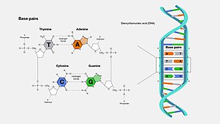
A base pair (bp) is a fundamental unit of double-stranded nucleic acids consisting of two nucleobases bound to each other by hydrogen bonds. They form the building blocks of the DNA double helix and contribute to the folded structure of both DNA and RNA. Dictated by specific hydrogen bonding patterns, "Watson–Crick" base pairs allow the DNA helix to maintain a regular helical structure that is subtly dependent on its nucleotide sequence. The complementary nature of this based-paired structure provides a redundant copy of the genetic information encoded within each strand of DNA. The regular structure and data redundancy provided by the DNA double helix make DNA well suited to the storage of genetic information, while base-pairing between DNA and incoming nucleotides provides the mechanism through which DNA polymerase replicates DNA and RNA polymerase transcribes DNA into RNA. Many DNA-binding proteins can recognize specific base-pairing patterns that identify particular regulatory regions of genes.

<span class="mw-page-title-main">DNA</span> Molecule that carries genetic information

Deoxyribonucleic acid is a polymer composed of two polynucleotide chains that coil around each other to form a double helix. The polymer carries genetic instructions for the development, functioning, growth and reproduction of all known organisms and many viruses. DNA and ribonucleic acid (RNA) are nucleic acids. Alongside proteins, lipids and complex carbohydrates (polysaccharides), nucleic acids are one of the four major types of macromolecules that are essential for all known forms of life.

<span class="mw-page-title-main">Nucleic acid</span> Class of large biomolecules essential to all known life

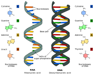
Nucleic acids are large biomolecules that are crucial in all cells and viruses. They are composed of nucleotides, which are the monomer components: a 5-carbon sugar, a phosphate group and a nitrogenous base. The two main classes of nucleic acids are deoxyribonucleic acid (DNA) and ribonucleic acid (RNA). If the sugar is ribose, the polymer is RNA; if the sugar is deoxyribose, a variant of ribose, the polymer is DNA.

DNA topoisomerases are enzymes that catalyze changes in the topological state of DNA, interconverting relaxed and supercoiled forms, linked (catenated) and unlinked species, and knotted and unknotted DNA. Topological issues in DNA arise due to the intertwined nature of its double-helical structure, which, for example, can lead to overwinding of the DNA duplex during DNA replication and transcription. If left unchanged, this torsion would eventually stop the DNA or RNA polymerases involved in these processes from continuing along the DNA helix. A second topological challenge results from the linking or tangling of DNA during replication. Left unresolved, links between replicated DNA will impede cell division. The DNA topoisomerases prevent and correct these types of topological problems. They do this by binding to DNA and cutting the sugar-phosphate backbone of either one or both of the DNA strands. This transient break allows the DNA to be untangled or unwound, and, at the end of these processes, the DNA backbone is resealed. Since the overall chemical composition and connectivity of the DNA do not change, the DNA substrate and product are chemical isomers, differing only in their topology.

<span class="mw-page-title-main">Chargaff's rules</span> Two rules about the percentage of A, C, G, and T in DNA strands

Chargaff's rules state that in the DNA of any species and any organism, the amount of guanine should be equal to the amount of cytosine and the amount of adenine should be equal to the amount of thymine. Further, a 1:1 stoichiometric ratio of purine and pyrimidine bases should exist. This pattern is found in both strands of the DNA. They were discovered by Austrian-born chemist Erwin Chargaff in the late 1940s.

<span class="mw-page-title-main">Hoogsteen base pair</span>

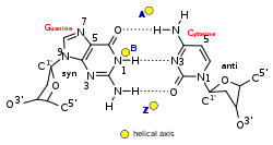
A Hoogsteen base pair is a variation of base-pairing in nucleic acids such as the A•T pair. In this manner, two nucleobases, one on each strand, can be held together by hydrogen bonds in the major groove. A Hoogsteen base pair applies the N7 position of the purine base and C6 amino group, which bind the Watson–Crick (N3–C4) face of the pyrimidine base.

<span class="mw-page-title-main">Triple-stranded DNA</span> DNA structure

Triple-stranded DNA is a DNA structure in which three oligonucleotides wind around each other and form a triple helix. In triple-stranded DNA, the third strand binds to a B-form DNA double helix by forming Hoogsteen base pairs or reversed Hoogsteen hydrogen bonds.

<span class="mw-page-title-main">Molecular Structure of Nucleic Acids: A Structure for Deoxyribose Nucleic Acid</span> 1953 scientific paper on the helical structure of DNA by James Watson and Francis Crick

"Molecular Structure of Nucleic Acids: A Structure for Deoxyribose Nucleic Acid" was the first article published to describe the discovery of the double helix structure of DNA, using X-ray diffraction and the mathematics of a helix transform. It was published by Francis Crick and James D. Watson in the scientific journal Nature on pages 737–738 of its 171st volume.

The history of molecular biology begins in the 1930s with the convergence of various, previously distinct biological and physical disciplines: biochemistry, genetics, microbiology, virology and physics. With the hope of understanding life at its most fundamental level, numerous physicists and chemists also took an interest in what would become molecular biology.

<span class="mw-page-title-main">DNA supercoil</span> Amount of twist in a particular DNA strand

DNA supercoiling refers to the amount of twist in a particular DNA strand, which determines the amount of strain on it. A given strand may be "positively supercoiled" or "negatively supercoiled". The amount of a strand's supercoiling affects a number of biological processes, such as compacting DNA and regulating access to the genetic code. Certain enzymes, such as topoisomerases, change the amount of DNA supercoiling to facilitate functions such as DNA replication and transcription. The amount of supercoiling in a given strand is described by a mathematical formula that compares it to a reference state known as "relaxed B-form" DNA.

Nucleic acid thermodynamics is the study of how temperature affects the nucleic acid structure of double-stranded DNA (dsDNA). The melting temperature (Tm) is defined as the temperature at which half of the DNA strands are in the random coil or single-stranded (ssDNA) state. Tm depends on the length of the DNA molecule and its specific nucleotide sequence. DNA, when in a state where its two strands are dissociated, is referred to as having been denatured by the high temperature.

<span class="mw-page-title-main">A-DNA</span> Potential conformation of DNA

A-DNA is one of the possible double helical structures which DNA can adopt. A-DNA is thought to be one of three biologically active double helical structures along with B-DNA and Z-DNA. It is a right-handed double helix fairly similar to the more common B-DNA form, but with a shorter, more compact helical structure whose base pairs are not perpendicular to the helix-axis as in B-DNA. It was discovered by Rosalind Franklin, who also named the A and B forms. She showed that DNA is driven into the A form when under dehydrating conditions. Such conditions are commonly used to form crystals, and many DNA crystal structures are in the A form. The same helical conformation occurs in double-stranded RNAs, and in DNA-RNA hybrid double helices.

<span class="mw-page-title-main">Molecular models of DNA</span>

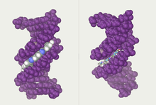
Molecular models of DNA structures are representations of the molecular geometry and topology of deoxyribonucleic acid (DNA) molecules using one of several means, with the aim of simplifying and presenting the essential, physical and chemical, properties of DNA molecular structures either in vivo or in vitro. These representations include closely packed spheres made of plastic, metal wires for skeletal models, graphic computations and animations by computers, artistic rendering. Computer molecular models also allow animations and molecular dynamics simulations that are very important for understanding how DNA functions in vivo.

<span class="mw-page-title-main">Nucleic acid tertiary structure</span> Three-dimensional shape of a nucleic acid polymer

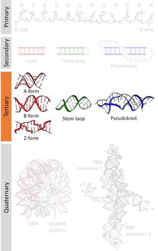
Nucleic acid tertiary structure is the three-dimensional shape of a nucleic acid polymer. RNA and DNA molecules are capable of diverse functions ranging from molecular recognition to catalysis. Such functions require a precise three-dimensional structure. While such structures are diverse and seemingly complex, they are composed of recurring, easily recognizable tertiary structural motifs that serve as molecular building blocks. Some of the most common motifs for RNA and DNA tertiary structure are described below, but this information is based on a limited number of solved structures. Many more tertiary structural motifs will be revealed as new RNA and DNA molecules are structurally characterized.

<span class="mw-page-title-main">Nucleic acid structure</span> Biomolecular structure of nucleic acids such as DNA and RNA

Nucleic acid structure refers to the structure of nucleic acids such as DNA and RNA. Chemically speaking, DNA and RNA are very similar. Nucleic acid structure is often divided into four different levels: primary, secondary, tertiary, and quaternary.

<span class="mw-page-title-main">Nucleic acid secondary structure</span>

Nucleic acid secondary structure is the basepairing interactions within a single nucleic acid polymer or between two polymers. It can be represented as a list of bases which are paired in a nucleic acid molecule. The secondary structures of biological DNAs and RNAs tend to be different: biological DNA mostly exists as fully base paired double helices, while biological RNA is single stranded and often forms complex and intricate base-pairing interactions due to its increased ability to form hydrogen bonds stemming from the extra hydroxyl group in the ribose sugar.

<span class="mw-page-title-main">Triple helix</span> Set of three congruent geometrical helices with the same axis

In the fields of geometry and biochemistry, a triple helix is a set of three congruent geometrical helices with the same axis, differing by a translation along the axis. This means that each of the helices keeps the same distance from the central axis. As with a single helix, a triple helix may be characterized by its pitch, diameter, and handedness. Examples of triple helices include triplex DNA, triplex RNA, the collagen helix, and collagen-like proteins.

Nucleic acid NMR is the use of nuclear magnetic resonance spectroscopy to obtain information about the structure and dynamics of nucleic acid molecules, such as DNA or RNA. It is useful for molecules of up to 100 nucleotides, and as of 2003, nearly half of all known RNA structures had been determined by NMR spectroscopy.

<span class="mw-page-title-main">Obsolete models of DNA structure</span>

In addition to the variety of verified DNA structures, there have been a range of proposed DNA models that have either been disproven, or lack evidence.

Non-canonical base pairs are planar hydrogen bonded pairs of nucleobases, having hydrogen bonding patterns which differ from the patterns observed in Watson-Crick base pairs, as in the classic double helical DNA. The structures of polynucleotide strands of both DNA and RNA molecules can be understood in terms of sugar-phosphate backbones consisting of phosphodiester-linked D 2’ deoxyribofuranose sugar moieties, with purine or pyrimidine nucleobases covalently linked to them. Here, the N9 atoms of the purines, guanine and adenine, and the N1 atoms of the pyrimidines, cytosine and thymine, respectively, form glycosidic linkages with the C1’ atom of the sugars. These nucleobases can be schematically represented as triangles with one of their vertices linked to the sugar, and the three sides accounting for three edges through which they can form hydrogen bonds with other moieties, including with other nucleobases. The side opposite to the sugar linked vertex is traditionally called the Watson-Crick edge, since they are involved in forming the Watson-Crick base pairs which constitute building blocks of double helical DNA. The two sides adjacent to the sugar-linked vertex are referred to, respectively, as the Sugar and Hoogsteen edges.

References

  1. Kabai, Sándor (2007). "Double Helix". The Wolfram Demonstrations Project.
  2. Cobb, Matthew; Comfort, Nathaniel (2023). "What Rosalind Franklin truly contributed to the discovery of DNA's structure". Nature. 616 (7958): 657–660. Bibcode:2023Natur.616..657C. doi:10.1038/d41586-023-01313-5. PMID   37100935.
  3. 1 2 Alberts; et al. (1994). The Molecular Biology of the Cell. New York: Garland Science. ISBN   978-0-8153-4105-5.
  4. Wang JC (1979). "Helical repeat of DNA in solution". PNAS. 76 (1): 200–203. Bibcode:1979PNAS...76..200W. doi: 10.1073/pnas.76.1.200 . PMC   382905 . PMID   284332.
  5. 1 2 Pabo C, Sauer R (1984). "Protein-DNA recognition". Annu Rev Biochem. 53: 293–321. doi:10.1146/annurev.bi.53.070184.001453. PMID   6236744.
  6. James Watson and Francis Crick (1953). "A structure for deoxyribose nucleic acid" (PDF). Nature . 171 (4356): 737–738. Bibcode:1953Natur.171..737W. doi:10.1038/171737a0. PMID   13054692. S2CID   4253007.
  7. Crick F, Watson JD (1954). "The Complementary Structure of Deoxyribonucleic Acid". Proceedings of the Royal Society of London. 223, Series A (1152): 80–96. Bibcode:1954RSPSA.223...80C. doi: 10.1098/rspa.1954.0101 .
  8. "Due credit". Nature. 496 (7445): 270. 18 April 2013. doi: 10.1038/496270a . PMID   23607133.
  9. Witkowski J (2019). "The forgotten scientists who paved the way to the double helix". Nature. 568 (7752): 308–309. Bibcode:2019Natur.568..308W. doi: 10.1038/d41586-019-01176-9 .
  10. Wilkins MH, Stokes AR, Wilson HR (1953). "Molecular Structure of Deoxypentose Nucleic Acids" (PDF). Nature. 171 (4356): 738–740. Bibcode:1953Natur.171..738W. doi:10.1038/171738a0. PMID   13054693. S2CID   4280080.
  11. Elson D, Chargaff E (1952). "On the deoxyribonucleic acid content of sea urchin gametes". Experientia. 8 (4): 143–145. doi:10.1007/BF02170221. PMID   14945441. S2CID   36803326.
  12. Chargaff E, Lipshitz R, Green C (1952). "Composition of the deoxypentose nucleic acids of four genera of sea-urchin". J Biol Chem. 195 (1): 155–160. doi: 10.1016/S0021-9258(19)50884-5 . PMID   14938364.
  13. Chargaff E, Lipshitz R, Green C, Hodes ME (1951). "The composition of the deoxyribonucleic acid of salmon sperm". J Biol Chem. 192 (1): 223–230. doi: 10.1016/S0021-9258(18)55924-X . PMID   14917668.
  14. Chargaff E (1951). "Some recent studies on the composition and structure of nucleic acids". J Cell Physiol Suppl. 38 (Suppl).
  15. Magasanik B, Vischer E, Doniger R, Elson D, Chargaff E (1950). "The separation and estimation of ribonucleotides in minute quantities". J Biol Chem. 186 (1): 37–50. doi: 10.1016/S0021-9258(18)56284-0 . PMID   14778802.
  16. Chargaff E (1950). "Chemical specificity of nucleic acids and mechanism of their enzymatic degradation". Experientia. 6 (6): 201–209. doi:10.1007/BF02173653. PMID   15421335. S2CID   2522535.
  17. Pauling L, Corey RB (Feb 1953). "A proposed structure for the nucleic acids". Proc Natl Acad Sci U S A. 39 (2): 84–97. Bibcode:1953PNAS...39...84P. doi: 10.1073/pnas.39.2.84 . PMC   1063734 . PMID   16578429.
  18. "Nobel Prize - List of All Nobel Laureates".
  19. Breslauer KJ, Frank R, Blöcker H, Marky LA (1986). "Predicting DNA duplex stability from the base sequence". PNAS. 83 (11): 3746–3750. Bibcode:1986PNAS...83.3746B. doi: 10.1073/pnas.83.11.3746 . PMC   323600 . PMID   3459152.
  20. Owczarzy, Richard (2008-08-28). "DNA melting temperature - How to calculate it?". High-throughput DNA biophysics. owczarzy.net. Archived from the original on 2015-04-30. Retrieved 2008-10-02.
  21. Raq, Bio (2016). Chromosome 16: PV92 PCR Informatics Kit (1st ed.). United States: Biotechnology Explorer. p. 104.
  22. "Chapter 9: DNA Replication – Chemistry" . Retrieved 2022-06-10.
  23. Alberts, Bruce; Johnson, Alexander; Lewis, Julian; Raff, Martin; Roberts, Keith; Walter, Peter (2002). "DNA Replication Mechanisms". Molecular Biology of the Cell. 4th Edition.
  24. Dickerson RE (1989). "Definitions and nomenclature of nucleic acid structure components". Nucleic Acids Res. 17 (5): 1797–1803. doi:10.1093/nar/17.5.1797. PMC   317523 . PMID   2928107.
  25. Lu XJ, Olson WK (1999). "Resolving the discrepancies among nucleic acid conformational analyses". J Mol Biol. 285 (4): 1563–1575. doi:10.1006/jmbi.1998.2390. PMID   9917397.
  26. Olson WK, Bansal M, Burley SK, Dickerson RE, Gerstein M, Harvey SC, Heinemann U, Lu XJ, Neidle S, Shakked Z, Sklenar H, Suzuki M, Tung CS, Westhof E, Wolberger C, Berman HM (2001). "A standard reference frame for the description of nucleic acid base-pair geometry". J Mol Biol. 313 (1): 229–237. doi:10.1006/jmbi.2001.4987. PMID   11601858.
  27. Richmond; Davey, CA; et al. (2003). "The structure of DNA in the nucleosome core". Nature. 423 (6936): 145–150. Bibcode:2003Natur.423..145R. doi:10.1038/nature01595. PMID   12736678. S2CID   205209705.
  28. Vargason JM, Eichman BF, Ho PS (2000). "The extended and eccentric E-DNA structure induced by cytosine methylation or bromination". Nature Structural Biology. 7 (9): 758–761. doi:10.1038/78985. PMID   10966645. S2CID   4420623.
  29. Hayashi G, Hagihara M, Nakatani K (2005). "Application of L-DNA as a molecular tag". Nucleic Acids Symp Ser (Oxf). 49 (1): 261–262. doi: 10.1093/nass/49.1.261 . PMID   17150733.
  30. 1 2 Allemand JF, Bensimon D, Lavery R, Croquette V (1998). "Stretched and overwound DNA forms a Pauling-like structure with exposed bases". PNAS. 95 (24): 14152–14157. Bibcode:1998PNAS...9514152A. doi: 10.1073/pnas.95.24.14152 . PMC   24342 . PMID   9826669.
  31. List of 55 fiber structures Archived 2007-05-26 at the Wayback Machine
  32. Bansal M (2003). "DNA structure: Revisiting the Watson-Crick double helix". Current Science. 85 (11): 1556–1563.
  33. Ghosh A, Bansal M (2003). "A glossary of DNA structures from A to Z". Acta Crystallogr D. 59 (4): 620–626. doi:10.1107/S0907444903003251. PMID   12657780.
  34. Rich A, Norheim A, Wang AH (1984). "The chemistry and biology of left-handed Z-DNA". Annual Review of Biochemistry. 53: 791–846. doi:10.1146/annurev.bi.53.070184.004043. PMID   6383204.
  35. Sinden, Richard R (1994-01-15). DNA structure and function (1st ed.). Academic Press. p. 398. ISBN   0-12-645750-6.
  36. Ho PS (1994-09-27). "The non-B-DNA structure of d(CA/TG)n does not differ from that of Z-DNA". Proc Natl Acad Sci USA. 91 (20): 9549–9553. Bibcode:1994PNAS...91.9549H. doi: 10.1073/pnas.91.20.9549 . PMC   44850 . PMID   7937803.
  37. "Double Helix". Genome.gov. Retrieved 2022-06-10.
  38. Wing R, Drew H, Takano T, Broka C, Tanaka S, Itakura K, Dickerson R (1980). "Crystal structure analysis of a complete turn of B-DNA". Nature. 287 (5784): 755–8. Bibcode:1980Natur.287..755W. doi:10.1038/287755a0. PMID   7432492. S2CID   4315465.
  39. Neidle, Stephen; Sanderson, Mark (2022), "DNA structure as observed in fibres and crystals", Principles of Nucleic Acid Structure, Elsevier, pp. 53–108, doi:10.1016/B978-0-12-819677-9.00007-X, ISBN   9780128196779, S2CID   239504252 , retrieved 2022-06-10
  40. Stokes, T. D. (1982). "The double helix and the warped zipper—an exemplary tale". Social Studies of Science. 12 (2): 207–240. doi:10.1177/030631282012002002. PMID   11620855. S2CID   29369576.
  41. Gautham, N. (25 May 2004). "Response to 'Variety in DNA secondary structure'" (PDF). Current Science. 86 (10): 1352–1353. Retrieved 25 May 2012. However, the discovery of topoisomerases took "the sting" out of the topological objection to the plectonaemic double helix. The more recent solution of the single crystal X-ray structure of the nucleosome core particle showed nearly 150 base pairs of the DNA (i.e., about 15 complete turns), with a structure that is in all essential respects the same as the Watson–Crick model. This dealt a death blow to the idea that other forms of DNA, particularly double helical DNA, exist as anything other than local or transient structures.[ permanent dead link ]
  42. Drozdetski AV, Mukhopadhyay A, Onufriev AV (November 2019). "Strongly Bent Double-Stranded DNA: Reconciling Theory and Experiment". Frontiers in Physics. 7: 195. arXiv: 1907.01585 . Bibcode:2019FrP.....7..195O. doi: 10.3389/fphy.2019.00195 . PMC   7323118 . PMID   32601596.
  43. Protozanova E, Yakovchuk P, Frank-Kamenetskii MD (2004). "Stacked–Unstacked Equilibrium at the Nick Site of DNA". J Mol Biol. 342 (3): 775–785. doi:10.1016/j.jmb.2004.07.075. PMID   15342236.
  44. Shimada J, Yamakawa H (1984). "Ring-Closure Probabilities for Twisted Wormlike Chains. Application to DNA". Macromolecules. 17 (4): 4660–4672. Bibcode:1984MaMol..17..689S. doi:10.1021/ma00134a028.
  45. 1 2 Harrison RM, Romano F, Ouldridge TE, Louis AA, Doye JP (2019). "Identifying Physical Causes of Apparent Enhanced Cyclization of Short DNA Molecules with a Coarse-Grained Model". Journal of Chemical Theory and Computation. 15 (8): 4660–4672. doi: 10.1021/acs.jctc.9b00112 . PMC   6694408 . PMID   31282669.
  46. Travers, Andrew (2005). "DNA Dynamics: Bubble 'n' Flip for DNA Cyclisation?". Current Biology. 15 (10): R377–R379. doi: 10.1016/j.cub.2005.05.007 . PMID   15916938. S2CID   10568179.
  47. Konrad MW, Bolonick JW (1996). "Molecular dynamics simulation of DNA stretching is consistent with the tension observed for extension and strand separation and predicts a novel ladder structure". Journal of the American Chemical Society. 118 (45): 10989–10994. doi:10.1021/ja961751x.
  48. Roe DR, Chaka AM (2009). "Structural basis of pathway-dependent force profiles in stretched DNA". Journal of Physical Chemistry B. 113 (46): 15364–15371. doi:10.1021/jp906749j. PMID   19845321.
  49. Bosaeus N, Reymer A, Beke-Somfai T, Brown T, Takahashi M, Wittung-Stafshede P, Rocha S, Nordén B (2017). "A stretched conformation of DNA with a biological role?". Quarterly Reviews of Biophysics. 50: e11. doi: 10.1017/S0033583517000099 . PMID   29233223.
  50. Taghavi A, van Der Schoot P, Berryman JT (2017). "DNA partitions into triplets under tension in the presence of organic cations, with sequence evolutionary age predicting the stability of the triplet phase". Quarterly Reviews of Biophysics. 50: e15. doi: 10.1017/S0033583517000130 . PMID   29233227.
  51. Crick FH (1976). "Linking numbers and nucleosomes". Proc Natl Acad Sci USA. 73 (8): 2639–43. Bibcode:1976PNAS...73.2639C. doi: 10.1073/pnas.73.8.2639 . PMC   430703 . PMID   1066673.
  52. Prunell A (1998). "A topological approach to nucleosome structure and dynamics: the linking number paradox and other issues". Biophys J. 74 (5): 2531–2544. Bibcode:1998BpJ....74.2531P. doi:10.1016/S0006-3495(98)77961-5. PMC   1299595 . PMID   9591679.
  53. Luger K, Mader AW, Richmond RK, Sargent DF, Richmond TJ (1997). "Crystal structure of the nucleosome core particle at 2.8 A resolution". Nature. 389 (6648): 251–260. Bibcode:1997Natur.389..251L. doi:10.1038/38444. PMID   9305837. S2CID   4328827.
  54. Davey CA, Sargent DF, Luger K, Maeder AW, Richmond TJ (2002). "Solvent mediated interactions in the structure of the nucleosome core particle at 1.9 Å resolution". Journal of Molecular Biology. 319 (5): 1097–1113. doi:10.1016/S0022-2836(02)00386-8. PMID   12079350.