Protein quaternary structure

Last updated
The image above contains clickable links
This diagram (which is interactive) of protein structure uses PCNA as an example. (PDB: 1AXC ) Protein structure (4)-en.svg
The image above contains clickable links Interactive icon.svg
The image above contains clickable links
This diagram (which is interactive) of protein structure uses PCNA as an example. ( PDB: 1AXC )

Protein quaternary structure [lower-alpha 1] is the fourth (and highest) classification level of protein structure. Protein quaternary structure refers to the structure of proteins which are themselves composed of two or more smaller protein chains (also referred to as subunits). Protein quaternary structure describes the number and arrangement of multiple folded protein subunits in a multi-subunit complex. It includes organizations from simple dimers to large homooligomers and complexes with defined or variable numbers of subunits. [1] In contrast to the first three levels of protein structure, not all proteins will have a quaternary structure since some proteins function as single units. Protein quaternary structure can also refer to biomolecular complexes of proteins with nucleic acids and other cofactors.

Contents

Description and examples

Many proteins are actually assemblies of multiple polypeptide chains. The quaternary structure refers to the number and arrangement of the protein subunits with respect to one another. [2] Examples of proteins with quaternary structure include hemoglobin, DNA polymerase, ribosomes, antibodies, and ion channels.

Enzymes composed of subunits with diverse functions are sometimes called holoenzymes, in which some parts may be known as regulatory subunits and the functional core is known as the catalytic subunit. Other assemblies referred to instead as multiprotein complexes also possess quaternary structure. Examples include nucleosomes and microtubules. Changes in quaternary structure can occur through conformational changes within individual subunits or through reorientation of the subunits relative to each other. It is through such changes, which underlie cooperativity and allostery in "multimeric" enzymes, that many proteins undergo regulation and perform their physiological function.

The above definition follows a classical approach to biochemistry, established at times when the distinction between a protein and a functional, proteinaceous unit was difficult to elucidate. More recently, people refer to protein–protein interaction when discussing quaternary structure of proteins and consider all assemblies of proteins as protein complexes.

Nomenclature

The quaternary structure of this protein complex would be described as a homo-trimer because it is composed of three identical smaller protein subunits (also designated as monomers or protomers). 1axc tricolor.png
The quaternary structure of this protein complex would be described as a homo-trimer because it is composed of three identical smaller protein subunits (also designated as monomers or protomers).

The number of subunits in an oligomeric complex is described using names that end in -mer (Greek for "part, subunit"). Formal and Greco-Latinate names are generally used for the first ten types and can be used for up to twenty subunits, whereas higher order complexes are usually described by the number of subunits, followed by -meric.

  • 13 = tridecamer
  • 14 = tetradecamer
  • 15 = pentadecamer*
  • 16 = hexadecamer
  • 17 = heptadecamer*
  • 18 = octadecamer
  • 19 = nonadecamer
  • 20 = eicosamer
  • 21 = 21-mer
  • 22 = 22-mer
  • 23 = 23-mer*
  • etc.
*No known examples

The smallest unit forming a homo-oligomer, i.e. one protein chain or subunit, is designated as a monomer, subunit or protomer. The latter term was originally devised to specify the smallest unit of hetero-oligomeric proteins, but is also applied to homo-oligomeric proteins in current literature. The subunits usually arrange in cyclic symmetry to form closed point group symmetries.

Although complexes higher than octamers are rarely observed for most proteins, there are some important exceptions. Viral capsids are often composed of multiples of 60 proteins. Several molecular machines are also found in the cell, such as the proteasome (four heptameric rings = 28 subunits), the transcription complex and the spliceosome. The ribosome is probably the largest molecular machine, and is composed of many RNA and protein molecules.

In some cases, proteins form complexes that then assemble into even larger complexes. In such cases, one uses the nomenclature, e.g., "dimer of dimers" or "trimer of dimers". This may suggest that the complex might dissociate into smaller sub-complexes before dissociating into monomers. This usually implies that the complex consists of different oligomerisation interfaces. For example, a tetrameric protein may have one four-fold rotation axis, i.e. point group symmetry 4 or C4. In this case the four interfaces between the subunits are identical. It may also have point group symmetry 222 or D2. This tetramer has different interfaces and the tetramer can dissociate into two identical homodimers. Tetramers of 222 symmetry are "dimer of dimers". Hexamers of 32 point group symmetry are "trimer of dimers" or "dimer of trimers". Thus, the nomenclature "dimer of dimers" is used to specify the point group symmetry or arrangement of the oligomer, independent of information relating to its dissociation properties.

Another distinction often made when referring to oligomers is whether they are homomeric or heteromeric, referring to whether the smaller protein subunits that come together to make the protein complex are the same (homomeric) or different (heteromeric) from each other. For example, two identical protein monomers would come together to form a homo-dimer, whereas two different protein monomers would create a hetero-dimer.

Structure Determination

Protein quaternary structure can be determined using a variety of experimental techniques that require a sample of protein in a variety of experimental conditions. The experiments often provide an estimate of the mass of the native protein and, together with knowledge of the masses and/or stoichiometry of the subunits, allow the quaternary structure to be predicted with a given accuracy. It is not always possible to obtain a precise determination of the subunit composition for a variety of reasons.

The number of subunits in a protein complex can often be determined by measuring the hydrodynamic molecular volume or mass of the intact complex, which requires native solution conditions. For folded proteins, the mass can be inferred from its volume using the partial specific volume of 0.73 ml/g. However, volume measurements are less certain than mass measurements, since unfolded proteins appear to have a much larger volume than folded proteins; additional experiments are required to determine whether a protein is unfolded or has formed an oligomer.

Common techniques used to study protein quaternary structure

Direct mass measurement of intact complexes

Direct size measurement of intact complexes

Indirect size measurement of intact complexes

Methods that measure the mass or volume under unfolding conditions (such as MALDI-TOF mass spectrometry and SDS-PAGE) are generally not useful, since non-native conditions usually cause the complex to dissociate into monomers. However, these may sometimes be applicable; for example, the experimenter may apply SDS-PAGE after first treating the intact complex with chemical cross-link reagents.

Structure Prediction

Some bioinformatics methods have been developed for predicting the quaternary structural attributes of proteins based on their sequence information by using various modes of pseudo amino acid composition. [2] [8] [9]

Protein folding prediction programs used to predict protein tertiary structure have also been expanding to better predict protein quaternary structure. One such development is AlphaFold-Multimer [10] built upon the AlphaFold model for predicting protein tertiary structure.

Role in Cell Signaling

Protein quaternary structure also plays an important role in certain cell signaling pathways. The G-protein coupled receptor pathway involves a heterotrimeric protein known as a G-protein. G-proteins contain three distinct subunits known as the G-alpha, G-beta, and G-gamma subunits. When the G-protein is activated, it binds to the G-protein coupled receptor protein and the cell signaling pathway is initiated. Another example is the receptor tyrosine kinase (RTK) pathway, which is initiated by the dimerization of two receptor tyrosine kinase monomers. When the dimer is formed, the two kinases can phosphorylate each other and initiate a cell signaling pathway. [11]

Protein–protein interactions

Proteins are capable of forming very tight but also only transient complexes. For example, ribonuclease inhibitor binds to ribonuclease A with a roughly 20 fM dissociation constant. Other proteins have evolved to bind specifically to unusual moieties on another protein, e.g., biotin groups (avidin), phosphorylated tyrosines (SH2 domains) or proline-rich segments (SH3 domains). Protein–protein interactions can be engineered to favor certain oligomerization states. [12]

Intragenic complementation

When multiple copies of a polypeptide encoded by a gene form a quaternary complex, this protein structure is referred to as a multimer. [13] When a multimer is formed from polypeptides produced by two different mutant alleles of a particular gene, the mixed multimer may exhibit greater functional activity than the unmixed multimers formed by each of the mutants alone. In such a case, the phenomenon is referred to as intragenic complementation (also called inter-allelic complementation). Intragenic complementation appears to be common and has been studied in many different genes in a variety of organisms including the fungi Neurospora crassa , Saccharomyces cerevisiae and Schizosaccharomyces pombe ; the bacterium Salmonella typhimurium; the virus bacteriophage T4, [14] an RNA virus, [15] and humans. [16] The intermolecular forces likely responsible for self-recognition and multimer formation were discussed by Jehle. [17]

Assembly

Direct interaction of two nascent proteins emerging from nearby ribosomes appears to be a general mechanism for oligomer formation. [18] Hundreds of protein oligomers were identified that assemble in human cells by such an interaction. [18] The most prevalent form of interaction was between the N-terminal regions of the interacting proteins. Dimer formation appears to be able to occur independently of dedicated assembly machines.

See also

Notes

  1. Here quaternary means "fourth-level structure", not "four-way interaction". Etymologically quartary is correct: quaternary is derived from Latin distributive numbers, and follows binary and ternary; while quartary is derived from Latin ordinal numbers, and follows secondary and tertiary. However, quaternary is standard in biology.

Related Research Articles

<span class="mw-page-title-main">Allosteric regulation</span> Regulation of enzyme activity

In biochemistry, allosteric regulation is the regulation of an enzyme by binding an effector molecule at a site other than the enzyme's active site.

In chemistry, dimerization refers to the process of joining two identical or similar molecular entities by bonds. The resulting bonds can be either strong or weak. Many symmetrical chemical species are described as dimers, even when the monomer is unknown or highly unstable.

<span class="mw-page-title-main">Protein complex</span> Type of stable macromolecular complex

A protein complex or multiprotein complex is a group of two or more associated polypeptide chains. Protein complexes are distinct from multidomain enzymes, in which multiple catalytic domains are found in a single polypeptide chain.

<span class="mw-page-title-main">Insulin receptor</span> Mammalian protein found in Homo sapiens

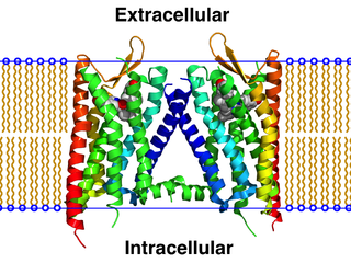
The insulin receptor (IR) is a transmembrane receptor that is activated by insulin, IGF-I, IGF-II and belongs to the large class of receptor tyrosine kinase. Metabolically, the insulin receptor plays a key role in the regulation of glucose homeostasis; a functional process that under degenerate conditions may result in a range of clinical manifestations including diabetes and cancer. Insulin signalling controls access to blood glucose in body cells. When insulin falls, especially in those with high insulin sensitivity, body cells begin only to have access to lipids that do not require transport across the membrane. So, in this way, insulin is the key regulator of fat metabolism as well. Biochemically, the insulin receptor is encoded by a single gene INSR, from which alternate splicing during transcription results in either IR-A or IR-B isoforms. Downstream post-translational events of either isoform result in the formation of a proteolytically cleaved α and β subunit, which upon combination are ultimately capable of homo or hetero-dimerisation to produce the ≈320 kDa disulfide-linked transmembrane insulin receptor.

<span class="mw-page-title-main">Protein structure</span> Three-dimensional arrangement of atoms in an amino acid-chain molecule

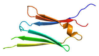
Protein structure is the three-dimensional arrangement of atoms in an amino acid-chain molecule. Proteins are polymers – specifically polypeptides – formed from sequences of amino acids, which are the monomers of the polymer. A single amino acid monomer may also be called a residue, which indicates a repeating unit of a polymer. Proteins form by amino acids undergoing condensation reactions, in which the amino acids lose one water molecule per reaction in order to attach to one another with a peptide bond. By convention, a chain under 30 amino acids is often identified as a peptide, rather than a protein. To be able to perform their biological function, proteins fold into one or more specific spatial conformations driven by a number of non-covalent interactions, such as hydrogen bonding, ionic interactions, Van der Waals forces, and hydrophobic packing. To understand the functions of proteins at a molecular level, it is often necessary to determine their three-dimensional structure. This is the topic of the scientific field of structural biology, which employs techniques such as X-ray crystallography, NMR spectroscopy, cryo-electron microscopy (cryo-EM) and dual polarisation interferometry, to determine the structure of proteins.

<span class="mw-page-title-main">Streptavidin</span> Protein in Streptomyces avidinii

Streptavidin is a 52 kDa protein (tetramer) purified from the bacterium Streptomyces avidinii. Streptavidin homo-tetramers have an extraordinarily high affinity for biotin. With a dissociation constant (Kd) on the order of ≈10−14 mol/L, the binding of biotin to streptavidin is one of the strongest non-covalent interactions known in nature. Streptavidin is used extensively in molecular biology and bionanotechnology due to the streptavidin-biotin complex's resistance to organic solvents, denaturants, detergents, proteolytic enzymes, and extremes of temperature and pH.

<span class="mw-page-title-main">Protein–protein interaction</span> Physical interactions and constructions between multiple proteins

Protein–protein interactions (PPIs) are physical contacts of high specificity established between two or more protein molecules as a result of biochemical events steered by interactions that include electrostatic forces, hydrogen bonding and the hydrophobic effect. Many are physical contacts with molecular associations between chains that occur in a cell or in a living organism in a specific biomolecular context.

A dodecameric protein has a quaternary structure consisting of 12 protein subunits in a complex. Dodecameric complexes can have a number of subunit 'topologies', but typically only a few of the theoretically possible subunit arrangements are observed in protein structures.

A tetrameric protein is a protein with a quaternary structure of four subunits (tetrameric). Homotetramers have four identical subunits, and heterotetramers are complexes of different subunits. A tetramer can be assembled as dimer of dimers with two homodimer subunits, or two heterodimer subunits.

<span class="mw-page-title-main">Argininosuccinate lyase</span> Mammalian protein found in Homo sapiens

The enzyme argininosuccinate lyase (EC 4.3.2.1, ASL, argininosuccinase; systematic name 2-(N ω-L-arginino)succinate arginine-lyase (fumarate-forming)) catalyzes the reversible breakdown of argininosuccinate:

<span class="mw-page-title-main">Protein trimer</span> Macromolecular complex formed by three macromolecules

In biochemistry, a protein trimer is a macromolecular complex formed by three, usually non-covalently bound, macromolecules like proteins or nucleic acids. A homotrimer would be formed by three identical molecules. A heterotrimer would be formed by three different macromolecules. Type II Collagen is an example of homotrimeric protein, while Type I collagen is an AAB-type heterotrimeric protein.

<span class="mw-page-title-main">Hsp27</span> Protein-coding gene in the species Homo sapiens

Heat shock protein 27 (Hsp27) also known as heat shock protein beta-1 (HSPB1) is a protein that in humans is encoded by the HSPB1 gene.

<span class="mw-page-title-main">Propionyl-CoA carboxylase</span>

Propionyl-CoA carboxylase (EC 6.4.1.3, PCC) catalyses the carboxylation reaction of propionyl-CoA in the mitochondrial matrix. PCC has been classified both as a ligase and a lyase. The enzyme is biotin-dependent. The product of the reaction is (S)-methylmalonyl CoA.

<span class="mw-page-title-main">Malate dehydrogenase 2</span> Enzyme that oxidizes malate to oxaloacetate in Krebs cycle

Malate dehydrogenase, mitochondrial also known as malate dehydrogenase 2 is an enzyme that in humans is encoded by the MDH2 gene.

<span class="mw-page-title-main">Morpheein</span> Model of protein allosteric regulation

Morpheeins are proteins that can form two or more different homo-oligomers, but must come apart and change shape to convert between forms. The alternate shape may reassemble to a different oligomer. The shape of the subunit dictates which oligomer is formed. Each oligomer has a finite number of subunits (stoichiometry). Morpheeins can interconvert between forms under physiological conditions and can exist as an equilibrium of different oligomers. These oligomers are physiologically relevant and are not misfolded protein; this distinguishes morpheeins from prions and amyloid. The different oligomers have distinct functionality. Interconversion of morpheein forms can be a structural basis for allosteric regulation, an idea noted many years ago, and later revived. A mutation that shifts the normal equilibrium of morpheein forms can serve as the basis for a conformational disease. Features of morpheeins can be exploited for drug discovery. The dice image represents a morpheein equilibrium containing two different monomeric shapes that dictate assembly to a tetramer or a pentamer. The one protein that is established to function as a morpheein is porphobilinogen synthase, though there are suggestions throughout the literature that other proteins may function as morpheeins.

<span class="mw-page-title-main">G beta-gamma complex</span>

The G beta-gamma complex (Gβγ) is a tightly bound dimeric protein complex, composed of one Gβ and one Gγ subunit, and is a component of heterotrimeric G proteins. Heterotrimeric G proteins, also called guanosine nucleotide-binding proteins, consist of three subunits, called alpha, beta, and gamma subunits, or Gα, Gβ, and Gγ. When a G protein-coupled receptor (GPCR) is activated, Gα dissociates from Gβγ, allowing both subunits to perform their respective downstream signaling effects. One of the major functions of Gβγ is the inhibition of the Gα subunit.

<span class="mw-page-title-main">Protein dimer</span> Macromolecular complex formed by two, usually non-covalently bound, macromolecules

In biochemistry, a protein dimer is a macromolecular complex or multimer formed by two protein monomers, or single proteins, which are usually non-covalently bound. Many macromolecules, such as proteins or nucleic acids, form dimers. The word dimer has roots meaning "two parts", di- + -mer. A protein dimer is a type of protein quaternary structure.

<span class="mw-page-title-main">Nucleic acid quaternary structure</span>

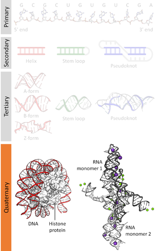
Nucleic acidquaternary structure refers to the interactions between separate nucleic acid molecules, or between nucleic acid molecules and proteins. The concept is analogous to protein quaternary structure, but as the analogy is not perfect, the term is used to refer to a number of different concepts in nucleic acids and is less commonly encountered. Similarly other biomolecules such as proteins, nucleic acids have four levels of structural arrangement: primary, secondary, tertiary, and quaternary structure. Primary structure is the linear sequence of nucleotides, secondary structure involves small local folding motifs, and tertiary structure is the 3D folded shape of nucleic acid molecule. In general, quaternary structure refers to 3D interactions between multiple subunits. In the case of nucleic acids, quaternary structure refers to interactions between multiple nucleic acid molecules or between nucleic acids and proteins. Nucleic acid quaternary structure is important for understanding DNA, RNA, and gene expression because quaternary structure can impact function. For example, when DNA is packed into heterochromatin, therefore exhibiting a type of quaternary structure, gene transcription will be inhibited.

<span class="mw-page-title-main">Autophosphorylation</span>

Autophosphorylation is a type of post-translational modification of proteins. It is generally defined as the phosphorylation of the kinase by itself. In eukaryotes, this process occurs by the addition of a phosphate group to serine, threonine or tyrosine residues within protein kinases, normally to regulate the catalytic activity. Autophosphorylation may occur when a kinases' own active site catalyzes the phosphorylation reaction, or when another kinase of the same type provides the active site that carries out the chemistry. The latter often occurs when kinase molecules dimerize. In general, the phosphate groups introduced are gamma phosphates from nucleoside triphosphates, most commonly ATP.

<span class="mw-page-title-main">GPCR oligomer</span> Class of protein complexes

A GPCR oligomer is a protein complex that consists of a small number of G protein-coupled receptors (GPCRs). It is held together by covalent bonds or by intermolecular forces. The subunits within this complex are called protomers, while unconnected receptors are called monomers. Receptor homomers consist of identical protomers, while heteromers consist of different protomers.

References

  1. Berg JM, Tymoczko JL, Stryer L (2002). "Section 3.5Quaternary Structure: Polypeptide Chains Can Assemble Into Multisubunit Structures". Biochemistry (5. ed., 4. print. ed.). New York, NY [u.a.]: W. H. Freeman. ISBN   0-7167-3051-0.
  2. 1 2 Chou KC, Cai YD (November 2003). "Predicting protein quaternary structure by pseudo amino acid composition". Proteins. 53 (2): 282–289. doi:10.1002/prot.10500. PMID   14517979. S2CID   23979933.
  3. Stiving AQ, VanAernum ZL, Busch F, Harvey SR, Sarni SH, Wysocki VH (January 2019). "Surface-Induced Dissociation: An Effective Method for Characterization of Protein Quaternary Structure". review. Analytical Chemistry. 91 (1): 190–209. doi:10.1021/acs.analchem.8b05071. PMC   6571034 . PMID   30412666.
  4. 1 2 Milligan G, Bouvier M (June 2005). "Methods to monitor the quaternary structure of G protein-coupled receptors". review. The FEBS Journal. 272 (12): 2914–2925. doi:10.1111/j.1742-4658.2005.04731.x. PMID   15955052. S2CID   23274563.
  5. Raicu V, Singh DR (November 2013). "FRET spectrometry: a new tool for the determination of protein quaternary structure in living cells". primary. Biophysical Journal. 105 (9): 1937–1945. Bibcode:2013BpJ...105.1937R. doi:10.1016/j.bpj.2013.09.015. PMC   3824708 . PMID   24209838.
  6. Prischi F, Pastore A (2016). "Application of Nuclear Magnetic Resonance and Hybrid Methods to Structure Determination of Complex Systems". Advanced Technologies for Protein Complex Production and Characterization. review. Advances in Experimental Medicine and Biology. Vol. 896. pp. 351–368. doi:10.1007/978-3-319-27216-0_22. ISBN   978-3-319-27214-6. PMID   27165336.
  7. Wells JN, Marsh JA (2018). "Experimental Characterization of Protein Complex Structure, Dynamics, and Assembly". Protein Complex Assembly. review. Methods in Molecular Biology. Vol. 1764. pp. 3–27. doi:10.1007/978-1-4939-7759-8_1. ISBN   978-1-4939-7758-1. PMID   29605905. Section 4: Nuclear Magnetic Resonance Spectroscopy
  8. Zhang SW, Chen W, Yang F, Pan Q (October 2008). "Using Chou's pseudo amino acid composition to predict protein quaternary structure: a sequence-segmented PseAAC approach". Amino Acids. 35 (3): 591–598. doi:10.1007/s00726-008-0086-x. PMID   18427713. S2CID   689955.
  9. Xiao X, Wang P, Chou KC (2009). "Predicting protein quaternary structural attribute by hybridizing functional domain composition and pseudo amino acid composition". Journal of Applied Crystallography. 42: 169–173. doi:10.1107/S0021889809002751.
  10. Evans R, O'Neill M, Pritzel A, Antropova N, Senior AW, Green T, et al. (4 October 2021). "Protein complex prediction with AlphaFold-Multimer". bioRxiv: 2021.10.04.463034. doi:10.1101/2021.10.04.463034. S2CID   238413014.
  11. Heldin CH (January 1995). "Dimerization of cell surface receptors in signal transduction". Cell. 80 (2): 213–223. doi: 10.1016/0092-8674(95)90404-2 . PMID   7834741. S2CID   18925209.
  12. Ardejani MS, Chok XL, Foo CJ, Orner BP (May 2013). "Complete shift of ferritin oligomerization toward nanocage assembly via engineered protein-protein interactions". Chemical Communications. 49 (34): 3528–3530. doi:10.1039/C3CC40886H. PMID   23511498.
  13. Crick FH, Orgel LE (January 1964). "The theory of inter-allelic complementation". Journal of Molecular Biology. 8: 161–165. doi:10.1016/s0022-2836(64)80156-x. PMID   14149958.
  14. Bernstein H, Edgar RS, Denhardt GH (June 1965). "Intragenic complementation among temperature sensitive mutants of bacteriophage T4D". Genetics. 51 (6): 987–1002. doi:10.1093/genetics/51.6.987. PMC   1210828 . PMID   14337770.
  15. Smallwood S, Cevik B, Moyer SA (December 2002). "Intragenic complementation and oligomerization of the L subunit of the sendai virus RNA polymerase". Virology. 304 (2): 235–245. doi: 10.1006/viro.2002.1720 . PMID   12504565.
  16. Rodríguez-Pombo P, Pérez-Cerdá C, Pérez B, Desviat LR, Sánchez-Pulido L, Ugarte M (June 2005). "Towards a model to explain the intragenic complementation in the heteromultimeric protein propionyl-CoA carboxylase". Biochimica et Biophysica Acta (BBA) - Molecular Basis of Disease. 1740 (3): 489–498. doi: 10.1016/j.bbadis.2004.10.009 . PMID   15949719.
  17. Jehle H (September 1963). "Intermolecular forces and biological specificity". Proceedings of the National Academy of Sciences of the United States of America. 50 (3): 516–524. Bibcode:1963PNAS...50..516J. doi: 10.1073/pnas.50.3.516 . PMC   221211 . PMID   16578546.
  18. 1 2 Bertolini M, Fenzl K, Kats I, Wruck F, Tippmann F, Schmitt J, et al. (January 2021). "Interactions between nascent proteins translated by adjacent ribosomes drive homomer assembly". primary. Science. 371 (6524): 57–64. Bibcode:2021Sci...371...57B. doi:10.1126/science.abc7151. PMC   7613021 . PMID   33384371. S2CID   229935047.