Related Research Articles

Cooper's ligaments are connective tissue in the breast that help maintain structural integrity. They are named for Astley Cooper, who first described them in 1840. Their anatomy can be revealed using Transmission diffraction tomography.
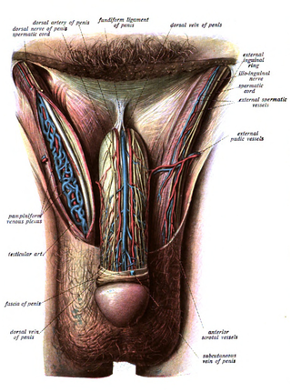
The fundiform ligament or fundiform ligament of the penis is a specialization or thickening of the superficial (Scarpa's) fascia extending from the linea alba of the lower abdominal wall.
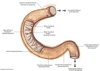
The suspensory muscle of duodenum is a thin muscle connecting the junction between the duodenum and jejunum, as well as the duodenojejunal flexure to connective tissue surrounding the superior mesenteric and coeliac arteries. The suspensory muscle most often connects to both the third and fourth parts of the duodenum, as well as the duodenojejunal flexure, although the attachment is quite variable.
A suspensory ligament is a ligament that supports a body part, especially an organ.

The paired gubernacula, also called the caudal genital ligament, are embryonic structures which begin as undifferentiated mesenchyme attaching to the caudal end of the gonads.

The external iliac arteries are two major arteries which bifurcate off the common iliac arteries anterior to the sacroiliac joint of the pelvis.

The lumbar plexus is a web of nerves in the lumbar region of the body which forms part of the larger lumbosacral plexus. It is formed by the divisions of the first four lumbar nerves (L1-L4) and from contributions of the subcostal nerve (T12), which is the last thoracic nerve. Additionally, the ventral rami of the fourth lumbar nerve pass communicating branches, the lumbosacral trunk, to the sacral plexus. The nerves of the lumbar plexus pass in front of the hip joint and mainly support the anterior part of the thigh.

The perforating cutaneous nerve is a cutaneous nerve of the sacral plexus that provides sensory innervation to the skin of the buttocks.

The renal plexus is a complex network of nerves formed by filaments from the celiac ganglia and plexus, aorticorenal ganglia, lower thoracic splanchnic nerves and first lumbar splanchnic nerve and aortic plexus.

The ovarian artery is an artery that supplies oxygenated blood to the ovary in females. It arises from the abdominal aorta below the renal artery. It can be found within the suspensory ligament of the ovary, anterior to the ovarian vein and ureter.

The broad ligament of the uterus is the wide fold of peritoneum that connects the sides of the uterus to the walls and floor of the pelvis.
The suspensory ligament of the ovary, also infundibulopelvic ligament, is a fold of peritoneum that extends out from the ovary to the wall of the pelvis.

The suspensory ligament of the penis is attached to the pubic symphysis, which holds the penis close to the pubic bone and supports it when erect. The ligament does not directly connect to the corpus cavernosum penis, but may still play a role in erectile dysfunction. The ligament can be surgically lengthened in a procedure known as ligamentolysis, which is a form of penis enlargement.

In the anatomy of the eye, the ciliary processes are formed by the inward folding of the various layers of the choroid, viz. the choroid proper and the lamina basalis, and are received between corresponding foldings of the suspensory ligament of the lens.

The dorsal nerve of the penis is the deepest of three divisions of the pudendal nerve; it accompanies the internal pudendal artery along the ramus of the ischium; it then runs forward along the margin of the inferior ramus of the pubis, between the superior and inferior layers of the fascia of the urogenital diaphragm.

The ovarian vein, the female gonadal vein, carries deoxygenated blood from its corresponding ovary to inferior vena cava or one of its tributaries. It is the female equivalent of the testicular vein, and is the venous counterpart of the ovarian artery. It can be found in the suspensory ligament of the ovary.
The ovarian ligament is a fibrous ligament that connects the ovary to the lateral surface of the uterus.

In human male anatomy, the dorsal veins of the penis are blood vessels that drain the shaft, the skin and the glans of the human penis. They are typically located in the midline on the dorsal aspect of the penis and they comprise the superficial dorsal veinof the penis, that lies in the subcutaneous tissue of the shaft, and the deep dorsal veinof the penis, that lies beneath the deep fascia.

The pampiniform plexus is a venous plexus – a network of many small veins found in the human male spermatic cord, and the suspensory ligament of the ovary. In the male, it is formed by the union of multiple testicular veins from the back of the testis and tributaries from the epididymis.

The following outline is provided as an overview of and topical guide to human anatomy:
References
![]() This article incorporates text in the public domain from page 987 of the 20th edition of Gray's Anatomy (1918)
This article incorporates text in the public domain from page 987 of the 20th edition of Gray's Anatomy (1918)
- ↑ suspensory+ligament+of+ovary Archived 2016-08-15 at the Wayback Machine at eMedicine Dictionary