A spinal nerve is a mixed nerve, which carries motor, sensory, and autonomic signals between the spinal cord and the body. In the human body there are 31 pairs of spinal nerves, one on each side of the vertebral column. These are grouped into the corresponding cervical, thoracic, lumbar, sacral and coccygeal regions of the spine. There are eight pairs of cervical nerves, twelve pairs of thoracic nerves, five pairs of lumbar nerves, five pairs of sacral nerves, and one pair of coccygeal nerves. The spinal nerves are part of the peripheral nervous system.

A nerve plexus is a plexus of intersecting nerves. A nerve plexus is composed of afferent and efferent fibers that arise from the merging of the anterior rami of spinal nerves and blood vessels. There are five spinal nerve plexuses, except in the thoracic region, as well as other forms of autonomic plexuses, many of which are a part of the enteric nervous system. The nerves that arise from the plexuses have both sensory and motor functions. These functions include muscle contraction, the maintenance of body coordination and control, and the reaction to sensations such as heat, cold, pain, and pressure. There are several plexuses in the body, including:
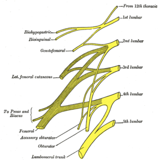
The lumbar nerves are the five pairs of spinal nerves emerging from the lumbar vertebrae. They are divided into posterior and anterior divisions.

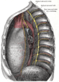
The sympathetic trunks are a paired bundle of nerve fibers that run from the base of the skull to the coccyx. They are a major component of the sympathetic nervous system.

The white ramus communicans from Latin ramus (branch) and communicans (communicating) is the preganglionic sympathetic outflow nerve tract from the spinal cord.
Each spinal nerve receives a branch called a gray ramus communicans from the adjacent paravertebral ganglion of the sympathetic trunk. The gray rami communicantes contain postganglionic nerve fibers of the sympathetic nervous system and are composed of largely unmyelinated neurons. This is in contrast to the white rami communicantes, in which heavily myelinated neurons give the rami their white appearance.
Ramus communicans is the Latin term used for a nerve which connects two other nerves, and can be translated as "communicating branch".

The sympathetic ganglia, or paravertebral ganglia, are autonomic ganglia of the sympathetic nervous system. Ganglia are 20,000 to 30,000 afferent and efferent nerve cell bodies that run along on either side of the spinal cord. Afferent nerve cell bodies bring information from the body to the brain and spinal cord, while efferent nerve cell bodies bring information from the brain and spinal cord to the rest of the body. The cell bodies create long sympathetic chains that are on either side of the spinal cord. They also form para- or pre-vertebral ganglia of gross anatomy.

The deep petrosal nerve is a post-ganglionic branch of the (sympathetic) internal carotid (nervous) plexus that enters the cranial cavity through the carotid canal, then passes perpendicular to the carotid canal in the cartilaginous substance which fills the foramen lacerum to unite with the (parasympathetic) greater petrosal nerve to form the nerve of pterygoid canal.

The lateral grey column is one of the three grey columns of the spinal cord ; the others being the anterior and posterior grey columns. The lateral grey column is primarily involved with activity in the sympathetic division of the autonomic motor system. It projects to the side as a triangular field in the thoracic and upper lumbar regions of the postero-lateral part of the anterior grey column.

Thoracic splanchnic nerves are splanchnic nerves that arise from the sympathetic trunk in the thorax and travel inferiorly to provide sympathetic supply to the abdomen. The nerves contain preganglionic sympathetic fibers and general visceral afferent fibers.
The dorsal ramus of spinal nerve, posterior ramus of spinal nerve, or posterior primary division is the posterior division of a spinal nerve. The dorsal rami provide motor innervation to the deep muscles of the back, and sensory innervation to the skin of the posterior portion of the head, neck and back.

The middle cervical ganglion is the smallest of the three cervical sympathetic ganglia. It presumably represents the merging of the sympathetic ganglia of cervical segments C5–C6. It is usually situated at the level of the sixth cervical vertebra.
The inferior cervical ganglion is one of the three cervical sympathetic ganglia. It is situated between the base of the transverse process of the last cervical vertebra and the neck of the first rib, on the medial side of the costocervical artery.

Sacral splanchnic nerves are splanchnic nerves that connect the inferior hypogastric plexus to the sympathetic trunk in the pelvis.
The general visceral afferent (GVA) fibers conduct sensory impulses from the internal organs, glands, and blood vessels to the central nervous system. They are considered to be part of the visceral nervous system, which is closely related to the autonomic nervous system, but 'visceral nervous system' and 'autonomic nervous system' are not direct synonyms and care should be taken when using these terms. Unlike the efferent fibers of the autonomic nervous system, the afferent fibers are not classified as either sympathetic or parasympathetic.
The lumbar ganglia are paravertebral ganglia located in the inferior portion of the sympathetic trunk. The lumbar portion of the sympathetic trunk typically has 4 lumbar ganglia. The lumbar splanchnic nerves arise from the ganglia here, and contribute sympathetic efferent fibers to the nearby plexuses. The first two lumbar ganglia have both white and gray rami communicates.
The sacral ganglia are paravertebral ganglia of the sympathetic trunk.:39 As the sympathetic trunk heads inferiorly down the sacrum, it turns medially. There are generally four or five sacral ganglia. In addition to gray rami communicantes, the ganglia send off sacral splanchnic nerves to join the inferior hypogastric plexus. Near the coccyx, the right and left sympathetic trunks join to form the ganglion impar.

The following diagram is provided as an overview of and topical guide to the human nervous system:
This page is based on this
Wikipedia article Text is available under the
CC BY-SA 4.0 license; additional terms may apply.
Images, videos and audio are available under their respective licenses.