Phakomatosis

Last updated
Phakomatoses
Other namesNeurocutaneous syndromes
Specialty Medicine, Neurology, Neurosurgery, Medical Genetics, Dermatology, Psychology, Psychiatry and more
Symptoms Dermal, ocular and CNS benign and malignant tumors. Various additional potential complications.
Complications Numerous potential complications including cosmetic, intellectual disability, epilepsy, organ failure and more.
Usual onsetChildhood (most commonly)
DurationLifelong
CausesGenetic causes
TreatmentHighly variable. Many require lifelong surveillance and various treatments depending on the particular syndrome and presentation.

Phakomatoses, also known as neurocutaneous syndromes, are a group of multisystemic diseases that most prominently affect structures primarily derived from the ectoderm such as the central nervous system, skin and eyes. The majority of phakomatoses are single-gene disorders that may be inherited in an autosomal dominant, autosomal recessive or X-linked pattern. Presentations may vary dramatically between patients with the same particular syndrome due to mosaicism, variable expressivity, and penetrance. [1]

Contents

Many phakomatoses are caused by mutations which alter functioning of the RAS–mitogen-activated protein kinase (MAPK) pathway that regulates cellular growth, differentiation, proliferation and death. [2] This results in a tendency for individuals with these mutations to develop various types of benign or malignant tumors depending on the particular mutation. The presence of these tumors may result in functional and/or cosmetic problems depending on their type and location.

History

The term phakomatosis originated in 1923, when the Dutch ophthalmologist van der Hoeve [3] used the term phakoma to refer to a "mother spot" or birthmark, [3] a physical characteristic common to patients with tuberous sclerosis and neurofibromatosis that he examined. [3] The term phakomatoses was derived from phakos, [4] the Greek term for 'birthmark'. [4] He originally used the phrase to describe two diseases: neurofibromatosis and tuberous sclerosis. [4] This term later became imprecise when van der Hoeve also used it to include those with Sturge-Weber syndrome as they do not have similar lesions on the skin. In addition, the term phakomatosis makes no reference to the central nervous system involvement. The term neurocutaneous syndrome was subsequently defined by the Russian-American neurologist Paul Ivan Yakovlev and psychiatrist Riley H. Guthrie and it is currently more commonly used. [2]

The first clinical description was made in “Monstrorum Historia” by an Italian physician and naturalist named Ulisse Aldrovandi who described a patient with probable neurofibromatosis type I in 1592. He described a short man with a large tumor which was likely a plexiform neurofibroma. [2] However, there are artistic or descriptive representations which have been speculated to depict individuals with phakomatoses as far back as the Hellenistic period and ancient Egypt. [5] [6]

Over time, the number of neurocutaneous syndromes have increased and there are several dozen that have been characterized.

Types

There are a large number of neurocutaneous syndromes that exceed the scope of this article. Therefore, characteristics of a few of the more common types are summarized.

Neurofibromatosis Type I (von Recklinghausen disease)

Main symptoms of neurofibromatosis type I. Symptoms of neurofibromatosis type 1 - censored version.png
Main symptoms of neurofibromatosis type I.
Diagnostic criteria of neurofibromatosis type I, requiring at least 2 of the mentioned items. Diagnostic criteria of neurofibromatosis type I.jpg
Diagnostic criteria of neurofibromatosis type I, requiring at least 2 of the mentioned items.

Neurofibromatosis type 1 is the most common phakomatosis and it affects approximately 1 in 2500-3000 live births. [9] It is a genetic disorder due to a germline mutation in the NF1 gene. This gene encodes a protein called neurofibromin that is involved in controlling cellular growth. [10] Malfunction of the gene results in multisystem manifestations involving the skin, central nervous system, peripheral nervous system, eyes and musculoskeletal system. The condition is inherited in an autosomal dominant manner. However, approximately one-half of patients with this condition have no family history and the mutation occurs spontaneously. In most instances, neurofibromatosis type 1 can be diagnosed clinically according to consensus criteria and genetic testing is only used in atypical presentations or for family planning decisions. [11]

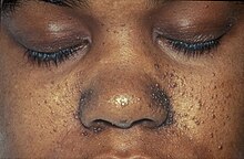
Café au lait spots are one of the most characteristic features of neurofibromatosis type 1. They are hyperpigmented lesions of the skin that increase in number and size during the first years of life. They are present in almost all patients but do not have malignant potential. [12] [13] Freckling in the axillary and inguinal regions are another common presenting feature seen in as many as 90% of patients during childhood. [12] Lisch nodules (benign hamartomas of the iris) are seen in almost all patients but they do not cause any visual or ocular impairment.

Neurofibromas are benign nerve sheath tumors that occur in peripheral nerves. These typically develop during the teenage years. Neurofibromas do not become malignant but can cause cosmetic concerns as well as local pruritus. When present in the spine they can affect nerve roots and result in both motor and sensory defects. [13] Surgery may be required in some cases. On the contrary, plexiform neurofibromas arise from multiple nerve fascicles and malignant transformation occurs in approximately 10% of cases. [14] They may result in significant morbidity as they may cause organ compression, vascular occlusions, bone destruction, pain and cosmetic issues. Plexiform neurofibromas are seen in 30-50% of patients. [12]

Optic pathway gliomas are seen in 15-20% of patients with neurofibromatosis type 1. [12] They most often arise during childhood. They often do not cause additional morbidity but may in up to one-half of patients. [15] Neurocognitive impairment, attention-deficit hyperactivity disorderautism spectrum disorder and behavioral disorders are also commonly seen in patients with neurofibromatosis type 1. [16] Epilepsy is seen in 4-7% of patients. [17]

Musculoskeletal system manifestations can develop in patients with neurofibromatosis type 1. Common findings include sphenoid wing dysplasia, osteopenia, osteoporosis, anterior chest wall deformities as well as scoliosis. Patients are at a much greater risk for fractures than the general population. [13]

Vascular abnormalities are also frequently encountered in patients with neurofibromatosis type 1. These may include renal artery stenosis, pulmonary artery stenosis, cerebral artery stenosis and aneurysms. [13] Complications may include myocardial infarction and stroke. [18]  

Relative to the general population the risk for certain types of cancer is increased significantly. For instance, the risk for brain tumors and breast cancer is increased by 5 times. [13]

Neurofibromatosis Type II

Figure of various morbidities associated with neurofibromatosis type II. Neurofibromatosis type II tumor types.png
Figure of various morbidities associated with neurofibromatosis type II.

Neurofibromatosis type 2 is an autosomal dominant condition that affects approximately 1 in 35,000-40,000 people. [20] It is caused by mutations in the NF2 gene on chromosome 22 which has a high penetrance though most patients do not present with symptoms until adulthood. [21] Approximately half of patients have de novo mutations and as many as 59.7% are mosaic. [22] [23] Patients who present in childhood tend to have a more severe phenotype. [24]

Bilateral vestibular schwannomas are the most characteristic finding and in the vast majority of cases are benign. However, they are responsible for significant morbidity and are responsible for the most common presenting symptom of hearing loss in adults. [25] They can also less commonly present with dizziness or balance impairment. It is estimated that schwannomas occur in over 90% of patients. [26]

Meningiomas are the second most common tumor in NF2. Approximately half of patients have an intracranial meningioma and extramedullary spinal meningiomas occur in one-fifth of patients. [25] Relative to the general population, meningiomas in NF2 tend to occur at a younger age, are more likely to be multiple and are associated with increased mortality. [27]

Spinal cord ependymomas occur in 20-50% of patients but they are asymptomatic in the majority of cases. [25] In cases that they are symptomatic the specific presentation will depend upon where they are located. Potential symptoms include back pain, weakness and sensory changes.

Peripheral neuropathy eventually occurs in most patients with NF2. In some cases, a mononeuropathy affecting the facial nerve can occur as the first presenting symptom of NF2. [25] Other potential manifestations include focal amyotrophy, mononeuropathy multiplex or a severe generalized polyneuropathy in 3-5% of patients. [22]

Ophthalmologic manifestations are also common with 60-80% of patients developing cataracts. [25] Optic nerve meningiomas and retinal hamartomas can result in vision loss.

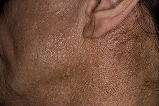
Approximately, 70% of patients will have cutaneous manifestations but only 10% have more than 10 lesions. [25] The majority of skin lesions are schwannomas but neurofibromas can also occur.

Tuberous sclerosis (Bourneville syndrome)

A case of tuberous sclerosis showing facial angiofibromas in characteristic butterfly pattern. Herbert L. Fred, MD and Hendrik A. van Dijk Patient with facial angiofibromas caused by tuberous sclerosis.jpg
A case of tuberous sclerosis showing facial angiofibromas in characteristic butterfly pattern. Herbert L. Fred, MD and Hendrik A. van Dijk
Symptoms and signs of tuberous sclerosis Symptoms and signs of tuberous sclerosis.png
Symptoms and signs of tuberous sclerosis

Tuberous sclerosis complex (TSC) is a multisystemic disorder due to autosomal dominant mutations in either TSC1 or TSC2 which results in the impaired inhibition of the mechanistic target of rapamycin (mTOR) signaling pathway. [28] This leads to impaired regulation of cellular proliferation, survival, homeostasis, migration and other critical functions. [29] The most typically affected organs include the brain, skin, kidney, heart and lung. The incidence of TSC is approximately 1 in 6,000 live births. [30] Similar to other neurocutaneous disorders there is variable penetrance and expressivity. [28] TSC1 mutations tend to have a less severe phenotype and are more likely to be familial. [28] A major development in the treatment of this condition occurred in 2010s when the FDA approved mTOR inhibitors for the treatment of several manifestations of TSC.  

Epilepsy is among the most common manifestations of TSC and it occurs 80-90% of patients. [31] It usually presents during the first few years of life and is medically refractory in two-thirds of patients. [32] Approximately, one-third of patients have infantile spasms. [32] There are several types of brain lesions that can be found in TSC including subependymal nodules (SENs), cortical tubers and subependymal giant cell astrocytomas (SEGAs). SENs and cortical tubers occur in approximately 80% and 90% of patients, respectively. [33] SEGAs occur in 10-15% of patients and are a major potential cause of morbidity and potentially mortality. [34] They tend to present during the first 20 years of life. TSC-Associated Neuropsychiatric Disorder (TAND) refers to the behavioral, intellectual and psychiatric manifestations of TSC including autism spectrum disorder, attention deficit hyperactivity disorder, intellectual disability, depression and anxiety. Approximately 90% of patients will have at least 1 symptom of TAND. [35]

Lymphangioleiomyomatosis (LAM) occurs in the lung and may result in pneumothorax, cystic lung destruction and pleural effusions. Symptoms which occur as a result may include fatigue, chest pain and shortness of breath. It occurs in approximately 30-40% of women with TSC, up to 80% by the age of 40, and it is much less common in men. [36]

Renal angiomyolipomas and cysts are the most common manifestations of TSC involving the kidney. Renal disease is among the most common causes of early death in TSC. One study found that renal lesions were present in 80% of patients by a mean age of 10.5 years. [37] Renal cell carcinoma occurs in 2-5% of patients with TSC at a mean age of 28–30 years. [38]

Cardiac rhabdomyomas are benign hamartomas and are the most common cardiac manifestations of TSC. It is found in approximately two-third of newborns with TSC. [39] Most of the time they do not cause symptoms and spontaneously regress. In a minority of cases, they may result in heart failure, arrhythmias, and murmurs. [39]

Dermatological manifestations occur in almost all patients and may include facial angiofibromas, confetti skin lesions, ungual fibromas, shagreen patches, hypomelanotic macules and fibrous cephalic plaques.[13] None of these tend to result in significant complications however facial angiofibromas may cause significant cosmetic concerns. [40]

Sturge–Weber Syndrome

Port wine stains of an 8-year-old female with Sturge-Weber Syndrome Port wine stains of an 8-year-old female with Sturge-Weber Syndrome.png
Port wine stains of an 8-year-old female with Sturge-Weber Syndrome

Sturge-Weber syndrome occurs in approximately 1 in 20,000-50,000 live births and is caused by a somatic activating mutation in GNAQ. [41] [42] Normally, GNAQ is involved in cell growth signal transmission. [42] It is classically characterized by a facial port-wine stain in the ophthalmic division of the trigeminal nerve, glaucoma and leptomeningeal angioma. [41] However, the clinical presentation can vary significantly depending on the timing of the somatic mutation ranging from an isolated port-wine stain to complete Sturge-Weber syndrome. [41]

The characteristic port-wine stain, also called nevus flammeus, is caused by a capillary or venular malformation. It is present from birth while the extent and size of it is associated with the risk of leptomeningeal and ophthalmologic involvement. [43] The greatest risk is associated with port-wine stains that appear to involve the entire V1 distribution followed by partial V1 involvement. [43] There is ontroversy as to whether or not the distribution of port-wine stains truly follows trigeminal nerve branches per se. [44] Port-wine stains are most often unilateral but can be bilateral. Typically, brain and eye involvement occur on the same side as the port-wine stain. Over time, port-wine stains can develop soft tissue or bone hypertrophy, proliferative nodules, and progressive ectasia which can lead to significant disfigurement. [45]

The two most common ocular manifestations include glaucoma and choroidal hemagioma. Glaucoma is estimated to occur in 30-70% with a bimodal peak occurring at the time of birth in 60% and between childhood and adolescence in 40%. [46] The most frequent form of glaucoma is open-angle however closed-angle glaucoma may also occur. [46] Congenital onset glaucoma is associated with other changes of the eye including megalocornea and buphthalmos. [47] The choroidal hemangioma occurs in 40-50% of patients. [48] They are usually asymptomatic but they may become thickened over time and may be associated with an increased risk for glaucoma. [46] [49]

Seizures typically develop within the first two years of life and occur in 75-100% of patients. [50] [41] Onset of seizures occur in 95% by the age of 5 years. [51] Status epilepticus occurs in approximately 50% of patients. [52] Approximately 25% of patients develop drug-resistant epilepsy. [53] In these cases, surgery may be pursued with the two main approaches being lesionectomy and hemispherectomy. [53]

Neurological impairment can accrue gradually over time and may occur in the context of stroke-like episodes which may be triggered by seizures or head injuries. [50] Intellectual disability occurs in approximately one-half of patients. [50] In addition, patients with Sturge-Weber syndrome have an increased prevalence of depression, endocrinological abnormalities, headaches with migraine-like features, ADHD and behavioral problems. [54] Cortically mediated visual field defects and hemiparesis occur in approximately one-third and one-half of patients, respectively. [50]

Von Hippel–Lindau syndrome (hemangiomatosis)

Locations of the main types of cysts and tumors in Von Hippel-Lindau disease. Cysts and tumors in Von Hippel-Lindau disease.png
Locations of the main types of cysts and tumors in Von Hippel–Lindau disease.

Von Hippel-Lindau (VHL) disease is an autosomal dominant condition caused by mutations of the VHL gene. [56] Approximately one-in-five cases are de novo rather than familial and it has nearly complete penetrance. [57] VHL occurs in an estimated 1 in 36,000-45,000 live births. [57] VHL normally functions as a tumor suppressor gene and thus when not functioning normally results in the development of benign and malignant tumors as well as cysts. Some of the most common manifestations include hemangioblastomas in the retina and central nervous system, clear cell renal cell carcinomas, pheochromocytomas, endolymphatic sac tumors and pancreatic neuroendocrine tumors. Life expectancy is reduced for individuals with this condition and one study found a median age of death at 52 years. [58]

CNS hemangioblastomas are the cardinal feature of VHL and occur in as many as 80% of cases. [59] The most common locations include the cerebellum, spinal cord and brainstem. [59] These tumors are benign however they may cause significant morbidity as well as mortality due to mass effect. [60] Tumor growth rates can be highly variable. Treatment options include resection and radiation. [60]

Retinal hemangioblastomas occur in approximately half of patients and are often the first presenting manifestation of VHL. [61] One longitudinal study that followed patients for a mean period of 7.3 years found that in individuals with unilateral disease 100% had bilateral involvement by the age of 56 years. [62] Blindness or severe visual impairment occurs in less than 10% of patients. [63] [64] Laser photocoagulation and cryotherapy are the most common surgical treatments. [59]

Renal cell carcinoma is a major cause of death in patients with VHL and it occurs in 70% of patients. [59] Lesions larger than 3 cm are associated with a risk of metastasis and thus present a recommended threshold for resection. [65] [66] Nephron sparing surgery allows for preservation of kidney function and 10-year survival rates up to 81%. [67]

Additional manifestations of VHL may include pheochromocytomas in as many as 16% of patients with VHL. [68] They are most often unilateral but can be bilateral or multifocal. Approximately 5% are malignant. [59]

Pancreatic involvement occurs in 77% of patients with VHL. Asymptomatic cysts consist of the majority of cases. Neuroendocrine tumors occur in approximately 15% of cases. [69] Less than 10% with neuroendocrine tumors will develop metastases. [70]

Genetics

The majority of neurocutaneous syndromes are single-gene disorders however they are caused by different genes and have different inheritance patterns. For instance, neurofibromatosis 1 and 2, Tuberous sclerosis complex, Von Hippel-Lindau syndrome and Legius syndrome are inherited in an autosomal dominant manner. Incontinentia pigmenti is X-linked dominant and Sturge-Weber syndrome is sporadic. Some neurocutaneous disorders are found exclusively as mosaics such as Sturge-Weber syndrome and Proteus syndrome. Others such as neurofibromatosis type 1 and 2 as well as tuberous sclerosis complex can potentially be mosaics but may not be. [71] Mosaicism may be suspected in cases with either a mild or incomplete presentation of a neurocutaneous disorder. A definitive diagnosis is most likely to be obtained if testing of affected tissues is possible. [72] [73]

Patients and families of those affected by neurocutaneous disorders often benefit from genetic counseling. It may provide an opportunity to provide a better understanding of the potential risk to future offspring as well as to improve coping with the various implications of the condition. If desired, a prenatal diagnosis can be obtained by either amniocentesis or chorionic villus sampling. Another potential option is preimplantation genetic diagnosis where an embryo that does not have the mutation can be selectively implanted into the mother. [74]

Diagnosis

Many neurocutaneous syndromes have established diagnostic criteria which may facilitate a diagnosis without necessarily requiring genetic testing. For instance, neurofibromatosis types 1 and 2, tuberous sclerosis complex and incontinentia pigmenti have formal diagnostic criteria. [75] Whereas other syndromes such as Noonan syndrome with multiple lentigines, for instance, do not. In cases where conditions that do not have diagnostic criteria are suspected, genetic testing is more likely to be necessary. Diagnostic criteria are not perfect as sensitivity and specificity is not 100%. [76] Similarly, genetic testing can produce false negatives. For example, genetic testing is positive in only 75-90% of cases of tuberous sclerosis complex. [77] Thus, clinicians must apply clinical judgement when evaluating an individual suspected to have a neurocutaneous syndrome.

Treatment

The treatment of each neurocutaneous syndrome is unique. For some neurocutaneous syndromes such as neurofibromatosis 1 and tuberous sclerosis complex there are guidelines with recommendations for surveillance and management. [78] [79] For less common syndromes such guidelines are not yet available. Surveillance is a necessity for many neurocutaneous syndromes because new manifestations may develop over time which may only be detected with specific and focused testing. Thus, patients may be advised to obtain particular evaluations (e.g., MRI, ophthalmologic or dermatological examinations) within recommended intervals over time with the aim of detecting new manifestations of the syndrome early on.

Most of the currently available treatments for neurocutaneous syndromes do not address the underlying genetic cause. For example, epilepsy surgery in tuberous sclerosis complex or vestibular schwannoma surgery in neurofibromatosis type 2. However, there are some treatments that do address the underlying cause such as the use of mTOR inhibitors in tuberous sclerosis complex. There are currently significant efforts underway to develop additional treatments that address the underlying causes of neurocutaneous syndromes. [80]

Neurocutaneous syndromes are complex, lifelong conditions that have the potential to affect many different organ systems over time. Thus, patients may benefit from a multidisciplinary approach for care. Integrated, multidisciplinary clinics have developed in an effort to optimize the long-term care for patients with neurocutaneous syndromes. [81] Specialties represented at these clinics may include genetics, neurology, ophthalmology, hematology-oncology, neurosurgery, psychiatry, dermatology and more.

Related Research Articles

<span class="mw-page-title-main">Von Hippel–Lindau disease</span> Medical condition

Von Hippel–Lindau disease (VHL), also known as VonHippel–Lindau syndrome, is a rare genetic disorder with multisystem involvement. It is characterized by visceral cysts and benign tumors with potential for subsequent malignant transformation. It is a type of phakomatosis that results from a mutation in the Von Hippel–Lindau tumor suppressor gene on chromosome 3p25.3.

<span class="mw-page-title-main">Neurofibromatosis</span> Medical condition

Neurofibromatosis (NF) refers to a group of three distinct genetic conditions in which tumors grow in the nervous system. The tumors are non-cancerous (benign) and often involve the skin or surrounding bone. Although symptoms are often mild, each condition presents differently. Neurofibromatosis type I (NF1) is typically characterized by café au lait spots, neurofibromas, scoliosis, and headaches. Neurofibromatosis type II (NF2), on the other hand, may present with early-onset hearing loss, cataracts, tinnitus, difficulty walking or maintain balance, and muscle atrophy. The third type is called schwannomatosis and often presents in early adulthood with widespread pain, numbness, or tingling due to nerve compression.

<span class="mw-page-title-main">Multiple endocrine neoplasia</span> Group of genetic conditions

Multiple endocrine neoplasia is a condition which encompasses several distinct syndromes featuring tumors of endocrine glands, each with its own characteristic pattern. In some cases, the tumors are malignant, in others, benign. Benign or malignant tumors of nonendocrine tissues occur as components of some of these tumor syndromes.

<span class="mw-page-title-main">Tuberous sclerosis</span> Genetic condition causing non-cancerous tumours

Tuberous sclerosis complex (TSC) is a rare multisystem autosomal dominant genetic disease that causes non-cancerous tumours to grow in the brain and on other vital organs such as the kidneys, heart, liver, eyes, lungs and skin. A combination of symptoms may include seizures, intellectual disability, developmental delay, behavioral problems, skin abnormalities, lung disease, and kidney disease.

Spinal tumors are neoplasms located in either the vertebral column or the spinal cord. There are three main types of spinal tumors classified based on their location: extradural and intradural. Extradural tumors are located outside the dura mater lining and are most commonly metastatic. Intradural tumors are located inside the dura mater lining and are further subdivided into intramedullary and extramedullary tumors. Intradural-intramedullary tumors are located within the dura and spinal cord parenchyma, while intradural-extramedullary tumors are located within the dura but outside the spinal cord parenchyma. The most common presenting symptom of spinal tumors is nocturnal back pain. Other common symptoms include muscle weakness, sensory loss, and difficulty walking. Loss of bowel and bladder control may occur during the later stages of the disease.

<span class="mw-page-title-main">Benign tumor</span> Mass of cells which cannot spread throughout the body

A benign tumor is a mass of cells (tumor) that does not invade neighboring tissue or metastasize. Compared to malignant (cancerous) tumors, benign tumors generally have a slower growth rate. Benign tumors have relatively well differentiated cells. They are often surrounded by an outer surface or stay contained within the epithelium. Common examples of benign tumors include moles and uterine fibroids.

<span class="mw-page-title-main">Birt–Hogg–Dubé syndrome</span> Rare autosomal dominant cancer syndrome

Birt–Hogg–Dubé syndrome (BHD), also Hornstein–Birt–Hogg–Dubé syndrome, Hornstein–Knickenberg syndrome, and fibrofolliculomas with trichodiscomas and acrochordons is a human, adult onset, autosomal dominant genetic disorder caused by a mutation in the folliculin (FLCN) gene. It can cause susceptibility to kidney cancer, renal and pulmonary cysts, and noncancerous tumors of the hair follicles, called fibrofolliculomas. The symptoms seen in each family are unique, and can include any combination of the three symptoms. Fibrofolliculomas are the most common manifestation, found on the face and upper trunk in over 80% of people with BHD over the age of 40. Pulmonary cysts are equally common (84%) and 24% of people with BHD eventually experience a collapsed lung. Kidney tumors, both cancerous and benign, occur in 14–34% of people with BHD; the associated kidney cancers are often rare hybrid tumors.

<span class="mw-page-title-main">Neurofibromatosis type I</span> Type of neurofibromatosis disease

Neurofibromatosis type I (NF-1), or von Recklinghausen syndrome, is a complex multi-system human disorder caused by the mutation of neurofibromin 1 (NF-1), a gene on chromosome 17 that is responsible for production of a protein (neurofibromin) which is needed for normal function in many human cell types. NF-1 causes tumors along the nervous system which can grow anywhere on the body. NF-1 is one of the most common genetic disorders and is not limited to any person's race or sex. NF-1 is an autosomal dominant disorder, which means that mutation or deletion of one copy of the NF-1 gene is sufficient for the development of NF-1, although presentation varies widely and is often different even between relatives affected by NF-1.

<span class="mw-page-title-main">Hamartoma</span> Tumour-like overgrowth due to a systemic genetic condition

A hamartoma is a mostly benign, local malformation of cells that resembles a neoplasm of local tissue but is usually due to an overgrowth of multiple aberrant cells, with a basis in a systemic genetic condition, rather than a growth descended from a single mutated cell (monoclonality), as would typically define a benign neoplasm/tumor. Despite this, many hamartomas are found to have clonal chromosomal aberrations that are acquired through somatic mutations, and on this basis the term hamartoma is sometimes considered synonymous with neoplasm. Hamartomas are by definition benign, slow-growing or self-limiting, though the underlying condition may still predispose the individual towards malignancies.

<span class="mw-page-title-main">Angiomatosis</span> Medical condition

Angiomatosis is a non-neoplastic condition characterised by nests of proliferating capillaries arranged in a lobular pattern, displacing adjacent muscle and fat. It consists of many angiomas.

<span class="mw-page-title-main">Hemangioblastoma</span> Medical condition

Hemangioblastomas, or haemangioblastomas, are vascular tumors of the central nervous system that originate from the vascular system, usually during middle age. Sometimes, these tumors occur in other sites such as the spinal cord and retina. They may be associated with other diseases such as polycythemia, pancreatic cysts and Von Hippel–Lindau syndrome. Hemangioblastomas are most commonly composed of stromal cells in small blood vessels and usually occur in the cerebellum, brainstem or spinal cord. They are classed as grade I tumors under the World Health Organization's classification system.

<span class="mw-page-title-main">Von Hippel–Lindau tumor suppressor</span> Mammalian protein found in Homo sapiens

The Von Hippel–Lindau tumor suppressor also known as pVHL is a protein that, in humans, is encoded by the VHL gene. Mutations of the VHL gene are associated with Von Hippel–Lindau disease, which is characterized by hemangioblastomas of the brain, spinal cord and retina. It is also associated with kidney and pancreatic lesions.

<span class="mw-page-title-main">Timeline of tuberous sclerosis</span>

The history of tuberous sclerosis (TSC) research spans less than 200 years. TSC is a rare, multi-system genetic disease that can cause benign tumours to grow on the brain or other vital organs such as the kidneys, heart, eyes, lungs, and skin. A combination of symptoms may include seizures, developmental delay, behavioural problems and skin abnormalities, as well as lung and kidney disease. TSC is caused by mutations on either of two genes, TSC1 and TSC2, which encode for the proteins hamartin and tuberin respectively. These proteins act as tumour growth suppressors and regulate cell proliferation and differentiation. Originally regarded as a rare pathological curiosity, it is now an important focus of research into tumour formation and suppression.

<span class="mw-page-title-main">Adrenal gland disorder</span> Medical condition

Adrenal gland disorders are conditions that interfere with the normal functioning of the adrenal glands. Your body produces too much or too little of one or more hormones when you have an adrenal gland dysfunction. The type of issue you have and the degree to which it affects your body's hormone levels determine the symptoms.

<span class="mw-page-title-main">Koenen's tumor</span> Medical condition

Koenen's tumor (KT), also commonly termed periungual angiofibroma, is a subtype of the angiofibromas. Angiofibromas are benign papule, nodule, and/or tumor lesions that are separated into various subtypes based primarily on the characteristic locations of their lesions. KTs are angiofibromas that develop in and under the toenails and/or fingernails. KTs were once considered as the same as another subtype of the angiofibromas viz., acral angiofibromas. While the literature may still sometimes regard KTs as acral angiofibromas, acral angiofibromas are characteristically located in areas close to but not in the toenails and fingernails as well as in the soles of the feet and palms of the hands. KTs are here regarded as distinct from acral angiofibromas.

<span class="mw-page-title-main">Legius syndrome</span> Medical condition

Legius syndrome (LS) is an autosomal dominant condition characterized by cafe au lait spots. It was first described in 2007 and is often mistaken for neurofibromatosis type I. It is caused by mutations in the SPRED1 gene. It is also known as neurofibromatosis type 1-like syndrome.

<span class="mw-page-title-main">Endolymphatic sac tumor</span>

An endolymphatic sac tumor (ELST) is a very uncommon papillary epithelial neoplasm arising within the endolymphatic sac or endolymphatic duct. This tumor shows a very high association with Von Hippel–Lindau syndrome (VHL).

<span class="mw-page-title-main">Hereditary cancer syndrome</span> Inherited genetic condition that predisposes a person to cancer

A hereditary cancer syndrome is a genetic disorder in which inherited genetic mutations in one or more genes predispose the affected individuals to the development of cancer and may also cause early onset of these cancers. Hereditary cancer syndromes often show not only a high lifetime risk of developing cancer, but also the development of multiple independent primary tumors.

The Pacak-Zhuang syndrome is a recently described disease manifestation in females that includes multiple paragangliomas or pheochromocytomas and somatostatinomas, both neuroendocrine tumors, and secondary polycythemia associated with high erythropoietin levels. Paragangliomas in these patients are mainly localized to the abdomen whereas somatostatinomas are found in the second portion of the duodenum, as shown by imaging or biochemistry. This syndrome is of special interest as finding more than one type of neuroendocrine tumor in one individual is unusual. Such co-occurrences are usually seen in patients carrying hereditary syndromes like multiple endocrine neoplasia (MEN), neurofibromatosis 1 (NF1), or von Hippel-Lindau (VHL) disease.

References

  1. Gürsoy, Semra; Erçal, Derya (2018-10-10). "Genetic Evaluation of Common Neurocutaneous Syndromes". Pediatric Neurology. 89: 3–10. doi:10.1016/j.pediatrneurol.2018.08.006. ISSN   1873-5150. PMID   30424961. S2CID   53302650.
  2. 1 2 3 Ruggieri, Martino; Praticò, Andrea D. (2015-12-17). "Mosaic Neurocutaneous Disorders and Their Causes". Seminars in Pediatric Neurology. 22 (4): 207–233. doi:10.1016/j.spen.2015.11.001. ISSN   1558-0776. PMID   26706010.
  3. 1 2 3 Fernández-Guarino M, Boixeda P, de Las Heras E, Aboin S, García-Millán C, Olasolo PJ (January 2008). "Phakomatosis pigmentovascularis: Clinical findings in 15 patients and review of the literature". Journal of the American Academy of Dermatology. 58 (1): 88–93. doi:10.1016/j.jaad.2007.08.012. PMID   18045734.
  4. 1 2 3 Huson SM, Korf BR (2013). "The Phakomatoses". Emery and Rimoin's Principles and Practice of Medical Genetics. Elsevier. pp. 1–45. doi:10.1016/b978-0-12-383834-6.00128-2. ISBN   978-0-12-383834-6.
  5. Ruggieri M, Gentile AE, Ferrara V, Papi M, Praticò AD, Mudry A, et al. (June 2021). "Neurocutaneous syndromes in art and antiquities". American Journal of Medical Genetics. Part C, Seminars in Medical Genetics. 187 (2): 224–234. doi:10.1002/ajmg.c.31917. PMC   8252443 . PMID   34013593.
  6. Ruggieri, Martino; Praticò, Andrea D.; Caltabiano, Rosario; Polizzi, Agata (2018-02-01). "Early history of the different forms of neurofibromatosis from ancient Egypt to the British Empire and beyond: First descriptions, medical curiosities, misconceptions, landmarks, and the persons behind the syndromes". American Journal of Medical Genetics Part A. 176 (3): 515–550. doi: 10.1002/ajmg.a.38486 . ISSN   1552-4825. PMID   29388340. S2CID   26042790.
  7. - Source for main symptoms: "Neurofibromatosis". Mayo Clinic. 2021-01-21.
    - Image by Mikael Häggström, MD, using source images by various authors.
  8. Graphical abstract from: Legius E, Messiaen L, Wolkenstein P, Pancza P, Avery RA, Berman Y; et al. (2021). "Revised diagnostic criteria for neurofibromatosis type 1 and Legius syndrome: an international consensus recommendation". Genet Med. 23 (8): 1506–1513. doi:10.1038/s41436-021-01170-5. PMC   8354850 . PMID   34012067.{{cite journal}}: CS1 maint: multiple names: authors list (link)
    - "User License: Creative Commons Attribution (CC BY 4.0)"
  9. Lammert M, Friedman JM, Kluwe L, Mautner VF (January 2005). "Prevalence of neurofibromatosis 1 in German children at elementary school enrollment". Archives of Dermatology. 141 (1): 71–74. doi: 10.1001/archderm.141.1.71 . PMID   15655144.
  10. Gutmann DH, Parada LF, Silva AJ, Ratner N (October 2012). "Neurofibromatosis type 1: modeling CNS dysfunction". The Journal of Neuroscience. 32 (41): 14087–14093. doi:10.1523/jneurosci.3242-12.2012. PMC   3477849 . PMID   23055477. S2CID   1747782.
  11. Tadini G, Brems H, Legius E (2020). "Proposal of New Diagnostic Criteria". Multidisciplinary Approach to Neurofibromatosis Type 1. Cham: Springer International Publishing. pp. 309–313. doi:10.1007/978-3-319-92450-2_21. ISBN   978-3-319-92449-6. S2CID   226576109.
  12. 1 2 3 4 Tabata MM, Li S, Knight P, Bakker A, Sarin KY (August 2020). "Phenotypic heterogeneity of neurofibromatosis type 1 in a large international registry". JCI Insight. 5 (16). doi:10.1172/jci.insight.136262. PMC   7455126 . PMID   32814709. S2CID   221202508.
  13. 1 2 3 4 5 Hirbe AC, Gutmann DH (August 2014). "Neurofibromatosis type 1: a multidisciplinary approach to care". The Lancet. Neurology. 13 (8): 834–843. doi: 10.1016/s1474-4422(14)70063-8 . PMID   25030515. S2CID   9813111.
  14. Raue F (2012-12-11). "Faculty Opinions recommendation of Survival meta-analyses for >1800 malignant peripheral nerve sheath tumor patients with and without neurofibromatosis type 1". doi: 10.3410/f.717964914.793467319 .{{cite journal}}: Cite journal requires |journal= (help)
  15. SergottRC (2007-03-23). "Optic pathway gliomas in neurofibromatosis-1: controversies and recommendations". Yearbook of Ophthalmology. 2007: 200–201. doi:10.1016/s0084-392x(08)70156-4. ISSN   0084-392X.
  16. Torres Nupan MM, Velez Van Meerbeke A, López Cabra CA, Herrera Gomez PM (2017). "Cognitive and Behavioral Disorders in Children with Neurofibromatosis Type 1". Frontiers in Pediatrics. 5: 227. doi: 10.3389/fped.2017.00227 . PMC   5670111 . PMID   29164079.
  17. Ostendorf AP, Gutmann DH, Weisenberg JL (October 2013). "Epilepsy in individuals with neurofibromatosis type 1". Epilepsia. 54 (10): 1810–1814. doi:10.1111/epi.12348. PMID   24032542. S2CID   1603461.
  18. Terry AR, Jordan JT, Schwamm L, Plotkin SR (January 2016). "Increased Risk of Cerebrovascular Disease Among Patients With Neurofibromatosis Type 1: Population-Based Approach". Stroke. 47 (1): 60–65. doi: 10.1161/strokeaha.115.011406 . PMID   26645253. S2CID   3253811.
  19. Bachir S, Shah S, Shapiro S, Koehler A, Mahammedi A, Samy RN; et al. (2021). "Neurofibromatosis Type 2 (NF2) and the Implications for Vestibular Schwannoma and Meningioma Pathogenesis". Int J Mol Sci. 22 (2): 690. doi: 10.3390/ijms22020690 . PMC   7828193 . PMID   33445724.{{cite journal}}: CS1 maint: multiple names: authors list (link)
  20. Evans DG, Howard E, Giblin C, Clancy T, Spencer H, Huson SM, Lalloo F (February 2010). "Birth incidence and prevalence of tumor-prone syndromes: estimates from a UK family genetic register service". American Journal of Medical Genetics. Part A. 152A (2): 327–332. doi: 10.1002/ajmg.a.33139 . PMID   20082463. S2CID   25884728.
  21. Evans DG, Huson SM, Donnai D, Neary W, Blair V, Newton V, et al. (December 1992). "A genetic study of type 2 neurofibromatosis in the United Kingdom. II. Guidelines for genetic counselling". Journal of Medical Genetics. 29 (12): 847–852. doi:10.1136/jmg.29.12.847. PMC   1016199 . PMID   1479599. S2CID   10548080.
  22. 1 2 Evans DG (June 2009). "Neurofibromatosis type 2 (NF2): a clinical and molecular review". Orphanet Journal of Rare Diseases. 4 (1): 16. doi: 10.1186/1750-1172-4-16 . PMC   2708144 . PMID   19545378. S2CID   15410658.
  23. Evans DG, Hartley CL, Smith PT, King AT, Bowers NL, Tobi S, et al. (January 2020). "Incidence of mosaicism in 1055 de novo NF2 cases: much higher than previous estimates with high utility of next-generation sequencing". Genetics in Medicine. 22 (1): 53–59. doi: 10.1038/s41436-019-0598-7 . PMID   31273341. S2CID   195795815.
  24. Halliday D, Emmanouil B, Vassallo G, Lascelles K, Nicholson J, Chandratre S, et al. (August 2019). "Trends in phenotype in the English paediatric neurofibromatosis type 2 cohort stratified by genetic severity". Clinical Genetics. 96 (2): 151–162. doi:10.1111/cge.13551. hdl: 10026.1/15073 . PMID   30993672. S2CID   119105041.
  25. 1 2 3 4 5 6 Asthagiri AR, Parry DM, Butman JA, Kim HJ, Tsilou ET, Zhuang Z, Lonser RR (June 2009). "Neurofibromatosis type 2". Lancet. 373 (9679): 1974–1986. doi:10.1016/S0140-6736(09)60259-2. PMC   4748851 . PMID   19476995.
  26. Dirks MS, Butman JA, Kim HJ, Wu T, Morgan K, Tran AP, et al. (July 2012). "Long-term natural history of neurofibromatosis Type 2-associated intracranial tumors". Journal of Neurosurgery. 117 (1): 109–117. doi:10.3171/2012.3.jns111649. PMC   4749021 . PMID   22503123.
  27. Baser ME, Friedman JM, Aeschliman D, Joe H, Wallace AJ, Ramsden RT, Evans DG (October 2002). "Predictors of the risk of mortality in neurofibromatosis 2". American Journal of Human Genetics. 71 (4): 715–723. doi:10.1086/342716. PMC   378530 . PMID   12235555. S2CID   23250053.
  28. 1 2 3 Henske, Elizabeth P.; Jóźwiak, Sergiusz; Kingswood, J. Christopher; Sampson, Julian R.; Thiele, Elizabeth A. (2016-05-26). "Tuberous sclerosis complex". Nature Reviews. Disease Primers. 2: 16035. doi:10.1038/nrdp.2016.35. ISSN   2056-676X. PMID   27226234. S2CID   68051262.
  29. Zoncu, Roberto; Efeyan, Alejo; Sabatini, David M. (2010-12-15). "mTOR: from growth signal integration to cancer, diabetes and ageing". Nature Reviews. Molecular Cell Biology. 12 (1): 21–35. doi:10.1038/nrm3025. ISSN   1471-0080. PMC   3390257 . PMID   21157483.
  30. Curatolo, Paolo; Bombardieri, Roberta; Jozwiak, Sergiusz (2008-08-23). "Tuberous sclerosis". The Lancet. 372 (9639): 657–668. doi:10.1016/S0140-6736(08)61279-9. ISSN   0140-6736. PMID   18722871. S2CID   28589689.
  31. Stafstrom, Carl E.; Staedtke, Verena; Comi, Anne M. (2017). "Epilepsy Mechanisms in Neurocutaneous Disorders: Tuberous Sclerosis Complex, Neurofibromatosis Type 1, and Sturge-Weber Syndrome". Frontiers in Neurology. 8: 87. doi: 10.3389/fneur.2017.00087 . ISSN   1664-2295. PMC   5355446 . PMID   28367137.
  32. 1 2 Chu-Shore, Catherine J.; Major, Philippe; Camposano, Susana; Muzykewicz, David; Thiele, Elizabeth A. (2010-07-01). "The natural history of epilepsy in tuberous sclerosis complex". Epilepsia. 51 (7): 1236–1241. doi:10.1111/j.1528-1167.2009.02474.x. ISSN   1528-1167. PMC   3065368 . PMID   20041940.
  33. Northrup, Hope; Krueger, Darcy A.; International Tuberous Sclerosis Complex Consensus Group (2013-10-01). "Tuberous sclerosis complex diagnostic criteria update: recommendations of the 2012 International Tuberous Sclerosis Complex Consensus Conference". Pediatric Neurology. 49 (4): 243–254. doi:10.1016/j.pediatrneurol.2013.08.001. ISSN   1873-5150. PMC   4080684 . PMID   24053982.
  34. Roth, Jonathan; Roach, E. Steve; Bartels, Ute; Jóźwiak, Sergiusz; Koenig, Mary Kay; Weiner, Howard L.; Franz, David N.; Wang, Henry Z. (2013-10-17). "Subependymal giant cell astrocytoma: diagnosis, screening, and treatment. Recommendations from the International Tuberous Sclerosis Complex Consensus Conference 2012". Pediatric Neurology. 49 (6): 439–444. doi: 10.1016/j.pediatrneurol.2013.08.017 . ISSN   1873-5150. PMID   24138953.
  35. de Vries, Petrus J.; Whittemore, Vicky H.; Leclezio, Loren; Byars, Anna W.; Dunn, David; Ess, Kevin C.; Hook, Dena; King, Bryan H.; Sahin, Mustafa; Jansen, Anna (2015-01-01). "Tuberous sclerosis associated neuropsychiatric disorders (TAND) and the TAND Checklist". Pediatric Neurology. 52 (1): 25–35. doi:10.1016/j.pediatrneurol.2014.10.004. ISSN   1873-5150. PMC   4427347 . PMID   25532776.
  36. Adriaensen, M. E. a. P. M.; Schaefer-Prokop, C. M.; Duyndam, D. a. C.; Zonnenberg, B. A.; Prokop, M. (2011-07-01). "Radiological evidence of lymphangioleiomyomatosis in female and male patients with tuberous sclerosis complex". Clinical Radiology. 66 (7): 625–628. doi:10.1016/j.crad.2011.02.009. ISSN   1365-229X. PMID   21459371.
  37. Ewalt, D. H.; Sheffield, E.; Sparagana, S. P.; Delgado, M. R.; Roach, E. S. (1998-07-01). "Renal lesion growth in children with tuberous sclerosis complex". The Journal of Urology. 160 (1): 141–145. doi:10.1016/S0022-5347(01)63072-6. ISSN   0022-5347. PMID   9628635.
  38. Borkowska, Julita; Schwartz, Robert A.; Kotulska, Katarzyna; Jozwiak, Sergiusz (2011-01-01). "Tuberous sclerosis complex: tumors and tumorigenesis". International Journal of Dermatology. 50 (1): 13–20. doi: 10.1111/j.1365-4632.2010.04727.x . ISSN   1365-4632. PMID   21182496. S2CID   34212098.
  39. 1 2 Jóźwiak, Sergiusz; Kotulska, Katarzyna; Kasprzyk-Obara, Jolanta; Domańska-Pakieła, Dorota; Tomyn-Drabik, Małgorzata; Roberts, Penelope; Kwiatkowski, David (2006-10-01). "Clinical and genotype studies of cardiac tumors in 154 patients with tuberous sclerosis complex". Pediatrics. 118 (4): e1146–1151. doi:10.1542/peds.2006-0504. ISSN   1098-4275. PMID   16940165. S2CID   25133016.
  40. Teng, Joyce M. C.; Cowen, Edward W.; Wataya-Kaneda, Mari; Gosnell, Elizabeth S.; Witman, Patricia M.; Hebert, Adelaide A.; Mlynarczyk, Greg; Soltani, Keyoumars; Darling, Thomas N. (2014-10-01). "Dermatologic and dental aspects of the 2012 International Tuberous Sclerosis Complex Consensus Statements". JAMA Dermatology. 150 (10): 1095–1101. doi:10.1001/jamadermatol.2014.938. ISSN   2168-6084. PMID   25029267.
  41. 1 2 3 4 Sudarsanam, Annapurna; Ardern-Holmes, Simone L. (2013-11-25). "Sturge–Weber syndrome: From the past to the present". European Journal of Paediatric Neurology. 18 (3): 257–266. doi:10.1016/j.ejpn.2013.10.003. ISSN   1090-3798. PMID   24275166.
  42. 1 2 Shirley, Matthew D.; Tang, Hao; Gallione, Carol J.; Baugher, Joseph D.; Frelin, Laurence P.; Cohen, Bernard; North, Paula E.; Marchuk, Douglas A.; Comi, Anne M.; Pevsner, Jonathan (2013-05-23). "Sturge-Weber syndrome and port-wine stains caused by somatic mutation in GNAQ". The New England Journal of Medicine. 368 (21): 1971–1979. doi:10.1056/NEJMoa1213507. ISSN   1533-4406. PMC   3749068 . PMID   23656586.
  43. 1 2 Ch'ng, Sydney; Tan, Swee T. (2007-07-09). "Facial port-wine stains - clinical stratification and risks of neuro-ocular involvement". Journal of Plastic, Reconstructive & Aesthetic Surgery: JPRAS. 61 (8): 889–893. doi:10.1016/j.bjps.2007.05.011. ISSN   1878-0539. PMID   17604243.
  44. Waelchli, R.; Aylett, S.E.; Robinson, K.; Chong, W.K.; Martinez, A.E.; Kinsler, V.A. (2014-06-27). "New vascular classification of port-wine stains: improving prediction of Sturge–Weber risk". British Journal of Dermatology. 171 (4): 861–867. doi:10.1111/bjd.13203. ISSN   0007-0963. PMC   4284033 . PMID   24976116.
  45. Sabeti, Sara; Ball, Karen L.; Burkhart, Craig; Eichenfield, Lawrence; Fernandez Faith, Esteban; Frieden, Ilona J.; Geronemus, Roy; Gupta, Deepti; Krakowski, Andrew C.; Levy, Moise L.; Metry, Denise (2021-01-01). "Consensus Statement for the Management and Treatment of Port-Wine Birthmarks in Sturge-Weber Syndrome". JAMA Dermatology. 157 (1): 98–104. doi:10.1001/jamadermatol.2020.4226. ISSN   2168-6068. PMC   8547264 . PMID   33175124.
  46. 1 2 3 Lambiase, Alessandro; Mantelli, Flavio; Bruscolini, Alice; La Cava, Maurizio; Abdolrahimzadeh, Solmaz (2016-05-13). "Ocular manifestations of Sturge-Weber syndrome: pathogenesis, diagnosis, and management". Clinical Ophthalmology. 10: 871–878. doi: 10.2147/opth.s101963 . ISSN   1177-5483. PMC   4874637 . PMID   27257371.
  47. Silverstein, Marlee; Salvin, Jonathan (2019-09-01). "Ocular manifestations of Sturge–Weber syndrome". Current Opinion in Ophthalmology. 30 (5): 301–305. doi:10.1097/icu.0000000000000597. ISSN   1040-8738. PMID   31313748. S2CID   197423353.
  48. Higueros, E.; Roe, E.; Granell, E.; Baselga, E. (2017-06-01). "Sturge-Weber Syndrome: A Review". Actas Dermo-Sifiliograficas. 108 (5): 407–417. doi:10.1016/j.ad.2016.09.022. ISSN   1578-2190. PMID   28126187.
  49. Singh, Arun D.; Kaiser, Peter K.; Sears, Jonathan E. (2005-03-01). "Choroidal hemangioma". Ophthalmology Clinics of North America. 18 (1): 151–161, ix. doi:10.1016/j.ohc.2004.07.004. ISSN   0896-1549. PMID   15763200.
  50. 1 2 3 4 Powell, Sebastian; Fosi, Tangunu; Sloneem, Jenny; Hawkins, Christina; Richardson, Hanna; Aylett, Sarah (2021-09-01). "Neurological presentations and cognitive outcome in Sturge-Weber syndrome". European Journal of Paediatric Neurology. 34: 21–32. doi:10.1016/j.ejpn.2021.07.005. ISSN   1090-3798. PMID   34293629.
  51. Sujansky, Eva; Conradi, Susan (1995-05-22). "Outcome of Sturge-Weber syndrome in 52 adults". American Journal of Medical Genetics. 57 (1): 35–45. doi:10.1002/ajmg.1320570110. ISSN   0148-7299. PMID   7645596.
  52. Sugano, Hidenori; Iimura, Yasushi; Igarashi, Ayuko; Nakazawa, Mika; Suzuki, Hiroharu; Mitsuhashi, Takumi; Nakajima, Madoka; Higo, Takuma; Ueda, Tetsuya; Nakanishi, Hajime; Niijima, Shinichi (2021-04-01). "Extent of Leptomeningeal Capillary Malformation is Associated With Severity of Epilepsy in Sturge-Weber Syndrome". Pediatric Neurology. 117: 64–71. doi: 10.1016/j.pediatrneurol.2020.12.012 . ISSN   0887-8994. PMID   33677229. S2CID   232140769.
  53. 1 2 Frank, Nicole Alexandra; Greuter, Ladina; Dill, Patricia Elsa; Guzman, Raphael; Soleman, Jehuda (2022-05-01). "Focal lesionectomy as surgical treatment of epilepsy in patients with Sturge-Weber syndrome: a case-based systematic review and meta-analysis". Neurosurgical Focus. 52 (5): E4. doi: 10.3171/2022.2.focus21788 . ISSN   1092-0684. PMID   35535828. S2CID   248595880.
  54. Pascual-Castroviejo, Ignacio; Pascual-Pascual, Samuel-Ignacio; Velazquez-Fragua, Ramón; Viaño, Juán (2008-07-01). "Sturge-Weber syndrome: study of 55 patients". The Canadian Journal of Neurological Sciences. Le Journal Canadien des Sciences Neurologiques. 35 (3): 301–307. doi: 10.1017/s0317167100008878 . ISSN   0317-1671. PMID   18714797. S2CID   35525783.
  55. Leung RS, Biswas SV, Duncan M, Rankin S (2008). "Imaging features of von Hippel-Lindau disease". Radiographics. 28 (1): 65–79, quiz 323. doi:10.1148/rg.281075052. PMID   18203931.{{cite journal}}: CS1 maint: multiple names: authors list (link)
  56. Kaelin, William G. (2002-09-01). "Molecular basis of the VHL hereditary cancer syndrome". Nature Reviews Cancer. 2 (9): 673–682. doi:10.1038/nrc885. ISSN   1474-175X. PMID   12209156. S2CID   20186415.
  57. 1 2 Sgambati, M.T.; Stolle, C.; Choyke, P.L.; Walther, M.M.; Zbar, B.; Linehan, W.M.; Glenn, G.M. (2000-01-01). "Mosaicism in von Hippel–Lindau Disease: Lessons from Kindreds with Germline Mutations Identified in Offspring with Mosaic Parents". The American Journal of Human Genetics. 66 (1): 84–91. doi:10.1086/302726. ISSN   0002-9297. PMC   1288351 . PMID   10631138.
  58. Wilding, Anna; Ingham, Sarah Louise; Lalloo, Fiona; Clancy, Tara; Huson, Susan M; Moran, Anthony; Evans, D Gareth (2012-02-23). "Life expectancy in hereditary cancer predisposing diseases: an observational study". Journal of Medical Genetics. 49 (4): 264–269. doi:10.1136/jmedgenet-2011-100562. ISSN   0022-2593. PMID   22362873. S2CID   41306596.
  59. 1 2 3 4 5 Maher, Eamonn R.; Neumann, Hartmut Ph; Richard, Stéphane (2011-03-09). "von Hippel-Lindau disease: a clinical and scientific review". European Journal of Human Genetics. 19 (6): 617–623. doi:10.1038/ejhg.2010.175. ISSN   1476-5438. PMC   3110036 . PMID   21386872.
  60. 1 2 Lonser, Russell R.; Butman, John A.; Huntoon, Kristin; Asthagiri, Ashok R.; Wu, Tianxia; Bakhtian, Kamran D.; Chew, Emily Y.; Zhuang, Zhengping; Linehan, W. Marston; Oldfield, Edward H. (2014-05-01). "Prospective natural history study of central nervous system hemangioblastomas in von Hippel-Lindau disease". Journal of Neurosurgery. 120 (5): 1055–1062. doi:10.3171/2014.1.JNS131431. ISSN   1933-0693. PMC   4762041 . PMID   24579662.
  61. Dollfus, Hélène; Massin, Pascale; Taupin, Pierre; Nemeth, Catherine; Amara, Sandrine; Giraud, Sophie; Béroud, Christophe; Dureau, Pascal; Gaudric, Alain; Landais, Paul; Richard, Stéphane (2002-09-01). "Retinal hemangioblastoma in von Hippel-Lindau disease: a clinical and molecular study". Investigative Ophthalmology & Visual Science. 43 (9): 3067–3074. ISSN   0146-0404. PMID   12202531.
  62. Kreusel, Klaus-Martin; Bechrakis, Nikolaos E.; Krause, Lothar; Neumann, Hartmut P. H.; Foerster, Michael H. (2006-08-01). "Retinal angiomatosis in von Hippel-Lindau disease: a longitudinal ophthalmologic study". Ophthalmology. 113 (8): 1418–1424. doi:10.1016/j.ophtha.2006.02.059. ISSN   1549-4713. PMID   16769118.
  63. Wittström, Elisabeth; Nordling, Margareta; Andréasson, Sten (2014-02-20). "Genotype-phenotype correlations, and retinal function and structure in von Hippel-Lindau disease". Ophthalmic Genetics. 35 (2): 91–106. doi:10.3109/13816810.2014.886265. ISSN   1744-5094. PMID   24555745. S2CID   20618502.
  64. Toy, Brian C.; Agrón, Elvira; Nigam, Divya; Chew, Emily Y.; Wong, Wai T. (2012-12-01). "Longitudinal analysis of retinal hemangioblastomatosis and visual function in ocular von Hippel-Lindau disease". Ophthalmology. 119 (12): 2622–2630. doi:10.1016/j.ophtha.2012.06.026. ISSN   1549-4713. PMC   3504630 . PMID   22906772.
  65. Walther, M. M.; Reiter, R.; Keiser, H. R.; Choyke, P. L.; Venzon, D.; Hurley, K.; Gnarra, J. R.; Reynolds, J. C.; Glenn, G. M.; Zbar, B.; Linehan, W. M. (1999-09-01). "Clinical and genetic characterization of pheochromocytoma in von Hippel-Lindau families: comparison with sporadic pheochromocytoma gives insight into natural history of pheochromocytoma". The Journal of Urology. 162 (3 Pt 1): 659–664. doi:10.1097/00005392-199909010-00004. ISSN   0022-5347. PMID   10458336.
  66. Duffey, Branden G.; Choyke, Peter L.; Glenn, Gladys; Grubb, Robert L.; Venzon, David; Linehan, W. Marston; Walther, McClellan M. (2004-07-01). "The relationship between renal tumor size and metastases in patients with von Hippel-Lindau disease". The Journal of Urology. 172 (1): 63–65. doi:10.1097/01.ju.0000132127.79974.3f. ISSN   0022-5347. PMID   15201738.
  67. Steinbach, F.; Novick, A. C.; Zincke, H.; Miller, D. P.; Williams, R. D.; Lund, G.; Skinner, D. G.; Esrig, D.; Richie, J. P.; deKernion, J. B. (1995-06-01). "Treatment of renal cell carcinoma in von Hippel-Lindau disease: a multicenter study". The Journal of Urology. 153 (6): 1812–1816. doi:10.1016/S0022-5347(01)67318-X. ISSN   0022-5347. PMID   7752324.
  68. Binderup, Marie Louise Mølgaard; Bisgaard, Marie Luise; Harbud, Vibeke; Møller, Hans Ulrik; Gimsing, Steen; Friis-Hansen, Lennart; Hansen, Thomas van Overeem; Bagi, Per; Knigge, Ulrich; Kosteljanetz, Michael; Bøgeskov, Lars (2013-12-01). "Von Hippel-Lindau disease (vHL). National clinical guideline for diagnosis and surveillance in Denmark. 3rd edition". Danish Medical Journal. 60 (12): B4763. ISSN   2245-1919. PMID   24355456.
  69. Charlesworth, Michael; Verbeke, Caroline S.; Falk, Gavin A.; Walsh, Matthew; Smith, Andrew M.; Morris-Stiff, Gareth (2012-02-28). "Pancreatic lesions in von Hippel-Lindau disease? A systematic review and meta-synthesis of the literature". Journal of Gastrointestinal Surgery. 16 (7): 1422–1428. doi:10.1007/s11605-012-1847-0. ISSN   1873-4626. PMID   22370733. S2CID   2090370.
  70. Blansfield, Joseph A.; Choyke, Lynda; Morita, Shane Y.; Choyke, Peter L.; Pingpank, James F.; Alexander, H. Richard; Seidel, Geoffrey; Shutack, Yvonne; Yuldasheva, Nargiza; Eugeni, Michelle; Bartlett, David L. (2007-12-01). "Clinical, genetic and radiographic analysis of 108 patients with von Hippel-Lindau disease (VHL) manifested by pancreatic neuroendocrine neoplasms (PNETs)". Surgery. 142 (6): 814–818, discussion 818.e1–2. doi:10.1016/j.surg.2007.09.012. ISSN   1532-7361. PMC   6771023 . PMID   18063061.
  71. Radtke, Heather B.; Lalor, Leah E.; Basel, Donald G.; Siegel, Dawn H. (2020-11-18). "Clinical Implications of Mosaicism and Low-Level Mosaicism in Neurocutaneous Disorders". Current Genetic Medicine Reports. 8 (4): 132–139. doi:10.1007/s40142-020-00193-9. ISSN   2167-4876. S2CID   226985730.
  72. Lalonde, Emilie; Ebrahimzadeh, Jessica; Rafferty, Keith; Richards-Yutz, Jennifer; Grant, Richard; Toorens, Erik; Marie Rosado, Jennifer; Schindewolf, Erica; Ganguly, Tapan; Kalish, Jennifer M.; Deardorff, Matthew A. (2019-02-13). "Molecular diagnosis of somatic overgrowth conditions: A single-center experience". Molecular Genetics & Genomic Medicine. 7 (3): e536. doi:10.1002/mgg3.536. ISSN   2324-9269. PMC   6418364 . PMID   30761771.
  73. McNulty, Samantha N.; Evenson, Michael J.; Corliss, Meagan M.; Love-Gregory, Latisha D.; Schroeder, Molly C.; Cao, Yang; Lee, Yi-Shan; Drolet, Beth A.; Neidich, Julie A.; Cottrell, Catherine E.; Heusel, Jonathan W. (2019-10-03). "Diagnostic Utility of Next-Generation Sequencing for Disorders of Somatic Mosaicism: A Five-Year Cumulative Cohort". The American Journal of Human Genetics. 105 (4): 734–746. doi:10.1016/j.ajhg.2019.09.002. ISSN   0002-9297. PMC   6817554 . PMID   31585106.
  74. Spits, C.; De Rycke, M.; Van Ranst, N.; Joris, H.; Verpoest, W.; Lissens, W.; Devroey, P.; Van Steirteghem, A.; Liebaers, I.; Sermon, K. (2005-05-01). "Preimplantation genetic diagnosis for neurofibromatosis type 1". Molecular Human Reproduction. 11 (5): 381–387. doi: 10.1093/molehr/gah170 . ISSN   1360-9947. PMID   15833774.
  75. Bodemer, C.; Diociaiuti, A.; Hadj-Rabia, S.; Robert, M.P.; Desguerre, I.; Manière, M.-C.; Dure-Molla, M.; De Liso, P.; Federici, M.; Galeotti, A.; Fusco, F. (2020-07-17). "Multidisciplinary consensus recommendations from a European network for the diagnosis and practical management of patients with incontinentia pigmenti". Journal of the European Academy of Dermatology and Venereology. 34 (7): 1415–1424. doi: 10.1111/jdv.16403 . ISSN   0926-9959. PMID   32678511. S2CID   220610122.
  76. Fox, Jonah; Ben-Shachar, Shay; Uliel, Shimrit; Svirsky, Ran; Saitsu, Hirotomo; Matsumoto, Naomichi; Fattal-Valevski, Aviva (2017-01-27). "Rare familial TSC2 gene mutation associated with atypical phenotype presentation of Tuberous Sclerosis Complex". American Journal of Medical Genetics Part A. 173 (3): 744–748. doi:10.1002/ajmg.a.38027. ISSN   1552-4825. PMID   28127866. S2CID   6584102.
  77. Northrup, Hope; Krueger, Darcy A.; International Tuberous Sclerosis Complex Consensus Group (2013-10-01). "Tuberous sclerosis complex diagnostic criteria update: recommendations of the 2012 International Tuberous Sclerosis Complex Consensus Conference". Pediatric Neurology. 49 (4): 243–254. doi:10.1016/j.pediatrneurol.2013.08.001. ISSN   1873-5150. PMC   4080684 . PMID   24053982.
  78. Stewart, Douglas R.; Korf, Bruce R.; Nathanson, Katherine L.; Stevenson, David A.; Yohay, Kaleb (2018-07-01). "Care of adults with neurofibromatosis type 1: a clinical practice resource of the American College of Medical Genetics and Genomics (ACMG)". Genetics in Medicine. 20 (7): 671–682. doi: 10.1038/gim.2018.28 . ISSN   1098-3600. PMID   30006586. S2CID   49721557.
  79. Amin, S.; Kingswood, J. C.; Bolton, P. F.; Elmslie, F.; Gale, D. P.; Harland, C.; Johnson, S. R.; Parker, A.; Sampson, J. R.; Smeaton, M.; Wright, I. (2019-03-01). "The UK guidelines for management and surveillance of Tuberous Sclerosis Complex". QJM: Monthly Journal of the Association of Physicians. 112 (3): 171–182. doi: 10.1093/qjmed/hcy215 . ISSN   1460-2393. PMID   30247655.
  80. Wilson, Britney N.; John, Ann M.; Handler, Marc Zachary; Schwartz, Robert A. (2021-06-01). "Neurofibromatosis type 1: New developments in genetics and treatment". Journal of the American Academy of Dermatology. 84 (6): 1667–1676. doi:10.1016/j.jaad.2020.07.105. ISSN   0190-9622. PMID   32771543. S2CID   221092583.
  81. Grossen, Audrey; Gavula, Theresa; Chrusciel, Deepti; Evans, Alexander; McNall-Knapp, Rene; Taylor, Ashley; Fossey, Benay; Brakefield, Margaret; Carter, Carrick; Schwartz, Nadine; Gross, Naina (2022-05-01). "Multidisciplinary neurocutaneous syndrome clinics: a systematic review and institutional experience". Neurosurgical Focus. 52 (5): E2. doi: 10.3171/2022.2.focus21776 . ISSN   1092-0684. PMID   35535824. S2CID   248499994.