Bone fracture

Last updated

Bone fracture
Other namesbroken bone, bone break
Broken fixed arm.jpg
Internal and external views of an arm with a compound fracture, both before and after surgery
Specialty Orthopedics
Diagnostic method X-ray, MRI

A bone fracture (abbreviated FRX or Fx, Fx, or #) is a medical condition in which there is a partial or complete break in the continuity of any bone in the body. In more severe cases, the bone may be broken into several fragments, known as a comminuted fracture. [1] A bone fracture may be the result of high force impact or stress, or a minimal trauma injury as a result of certain medical conditions that weaken the bones, such as osteoporosis, osteopenia, bone cancer, or osteogenesis imperfecta, where the fracture is then properly termed a pathologic fracture. [2]

Contents

Signs and symptoms

Although bone tissue contains no pain receptors, a bone fracture is painful for several reasons: [3]

Damage to adjacent structures such as nerves, muscles or blood vessels, spinal cord, and nerve roots (for spine fractures), or cranial contents (for skull fractures) may cause other specific signs and symptoms. [4]

Complications

An old fracture with nonunion of the fracture fragments Nonunion2010.JPG
An old fracture with nonunion of the fracture fragments

Some fractures may lead to serious complications including a condition known as compartment syndrome. If not treated, eventually, compartment syndrome may require amputation of the affected limb. Other complications may include non-union, where the fractured bone fails to heal, or malunion, where the fractured bone heals in a deformed manner. One form of malunion is the malrotation of a bone, which is especially common after femoral and tibial fractures. [5] Complications of fractures may be classified into three broad groups, depending upon their time of occurrence. These are as follows –

  1. Immediate complications – occurs at the time of the fracture.
  2. Early complications – occurring in the initial few days after the fracture.
  3. Late complications – occurring a long time after the fracture.


ImmediateEarlyLate
Systemic
  • Hypovolaemic shock
SystemicImperfect union of the fracture
Local
  • Injury to major vessels
  • Injury to muscles and tendons
  • Injury to joints
  • Injury to viscera
Local
  • Infection
  • Compartment syndrome
Others
  • Avascular necrosis
  • Shortening
  • Joint stiffness
  • Sudeck's dystrophy
  • Osteomyelitis
  • Ischaemic contracture
  • Myositis ossificans
  • Osteoarthritis

Pathophysiology

Stages in Fracture Repair: The healing of a bone fracture follows a series of progressive steps: (a) A fracture hematoma forms. (b) Internal and external calli form. (c) Cartilage of the calli is replaced by trabecular bone. (d) Remodeling occurs. 613 Stages of Fracture Repair.jpg
Stages in Fracture Repair: The healing of a bone fracture follows a series of progressive steps: (a) A fracture hematoma forms. (b) Internal and external calli form. (c) Cartilage of the calli is replaced by trabecular bone. (d) Remodeling occurs.

The natural process of healing a fracture starts when the injured bone and surrounding tissues bleed, forming a fracture hematoma. The blood coagulates to form a blood clot situated between the broken fragments. [6] Within a few days, blood vessels grow into the jelly-like matrix of the blood clot. The new blood vessels bring phagocytes to the area, which gradually removes the non-viable material. The blood vessels also bring fibroblasts in the walls of the vessels and these multiply and produce collagen fibres. In this way, the blood clot is replaced by a matrix of collagen. Collagen's rubbery consistency allows bone fragments to move only a small amount unless severe or persistent force is applied.[ citation needed ]

At this stage, some of the fibroblasts begin to lay down bone matrix in the form of collagen monomers. These monomers spontaneously assemble to form the bone matrix, for which bone crystals (calcium hydroxyapatite) are deposited in amongst, in the form of insoluble crystals. This mineralization of the collagen matrix stiffens it and transforms it into bone. In fact, bone is a mineralized collagen matrix; if the mineral is dissolved out of bone, it becomes rubbery. Healing bone callus on average is sufficiently mineralized to show up on X-ray within 6 weeks in adults and less in children. This initial "woven" bone does not have the strong mechanical properties of mature bone. By a process of remodelling, the woven bone is replaced by mature "lamellar" bone. The whole process may take up to 18 months, but in adults, the strength of the healing bone is usually 80% of normal by 3 months after the injury.[ citation needed ]

Several factors may help or hinder the bone healing process. For example, tobacco smoking hinders the process of bone healing, [7] and adequate nutrition (including calcium intake) will help the bone healing process. Weight-bearing stress on bone, after the bone has healed sufficiently to bear the weight, also builds bone strength.

Although there are theoretical concerns about NSAIDs slowing the rate of healing, there is not enough evidence to warrant withholding the use of this type analgesic in simple fractures. [8]

Effects of smoking

Smokers generally have lower bone density than non-smokers, so they have a much higher risk of fractures. There is also evidence that smoking delays bone healing. [9]

Diagnosis

Radiography to identify possible fractures after a knee injury Xraymachine.JPG
Radiography to identify possible fractures after a knee injury

A bone fracture may be diagnosed based on the history given and the physical examination performed. Radiographic imaging often is performed to confirm the diagnosis. Under certain circumstances, radiographic examination of the nearby joints is indicated in order to exclude dislocations and fracture-dislocations. In situations where projectional radiography alone is insufficient, Computed Tomography (CT) or Magnetic Resonance Imaging (MRI) may be indicated.[ citation needed ]

Classification

Compare healthy bone with different types of fractures:
(a) closed fracture
(b) open fracture
(c) transverse fracture
(d) spiral fracture
(e) comminuted fracture
(f) impacted fracture
(g) greenstick fracture
(h) oblique fracture 612 Types of Fractures.jpg
Compare healthy bone with different types of fractures:
   (a) closed fracture
   (b) open fracture
   (c) transverse fracture
   (d) spiral fracture
   (e) comminuted fracture
   (f) impacted fracture
   (g) greenstick fracture
   (h) oblique fracture
Open ankle fracture with luxation Offene Luxation.jpg
Open ankle fracture with luxation
Periprosthetic fracture of left femur Periprosthetic fracture of left femur, case 1, before treatment.jpg
Periprosthetic fracture of left femur

In orthopedic medicine, fractures are classified in various ways. Historically they are named after the physician who first described the fracture conditions, however, there are more systematic classifications as well.[ citation needed ]

They may be divided into stable versus unstable depending on the likelihood that they may shift further.[ citation needed ]

Mechanism

  • Traumatic fracture – a fracture due to sustained trauma. e.g., fractures caused by a fall, road traffic accident, fight, etc.
  • Pathologic fracture – a fracture through a bone that has been made weak by some underlying disease is called pathological fracture. e.g., a fracture through a bone weakened by metastasis. Osteoporosis is the most common cause of pathological fracture.
  • Periprosthetic fracture – a fracture at the point of mechanical weakness at the end of an implant.

Soft-tissue involvement

  • Closed/simple fractures are those in which the overlying skin is intact [10]
  • Open/compound fractures involve wounds that communicate with the fracture, or where fracture hematoma is exposed, and may thus expose bone to contamination. Open injuries carry a higher risk of infection. Reports indicate an incidence of infection after internal fixation of closed fracture of 1-2%, rising to 30% in open fractures. [11]
    • Clean fracture
    • Contaminated fracture

Displacement

  • Non-displaced
  • Displaced
    • Translated, or ad latus, with sideways displacement. [12]
    • Angulated
    • Rotated
    • Shortened, a reduction in overall bone length when displaced fracture fragments overlap

Fracture pattern

  • Linear fracture – a fracture that is parallel to the bone's long axis
  • Transverse fracture – a fracture that is at a right angle to the bone's long axis
  • Oblique fracture – a fracture that is diagonal to a bone's long axis (more than 30°)
  • Spiral fracture – a fracture where at least one part of the bone has been twisted
  • Compression fracture/wedge fracture – usually occurs in the vertebrae, for example when the front portion of a vertebra in the spine collapses due to osteoporosis (a medical condition which causes bones to become brittle and susceptible to fracture, with or without trauma)
  • Impacted fracture – a fracture caused when bone fragments are driven into each other
  • Avulsion fracture – a fracture where a fragment of bone is separated from the main mass

Fragments

  • Incomplete fracture – a fracture in which the bone fragments are still partially joined, in such cases, there is a crack in the osseous tissue that does not completely traverse the width of the bone.
  • Complete fracture – a fracture in which bone fragments separate completely.
  • Comminuted fracture – a fracture in which the bone has broken into several pieces.

Anatomical location

An anatomical classification may begin with specifying the involved body part, such as the head or arm, followed by more specific localization. Fractures that have additional definition criteria than merely localization often may be classified as subtypes of fractures, such as a Holstein-Lewis fracture being a subtype of a humerus fracture. Most typical examples in an orthopaedic classification given in the previous section cannot be classified appropriately into any specific part of an anatomical classification, however, as they may apply to multiple anatomical fracture sites.

OTA/AO classification

The Orthopaedic Trauma Association Committee for Coding and Classification published its classification system [20] in 1996, adopting a similar system to the 1987 AO Foundation system. [21] In 2007, they extended their system, [22] unifying the two systems regarding wrist, hand, foot, and ankle fractures.

Classifications named after people

A number of classifications are named after the person (eponymous) who developed it.

Prevention

Both high- and low-force trauma can cause bone fracture injuries. [29] [30] Preventive efforts to reduce motor vehicle crashes, the most common cause of high-force trauma, include reducing distractions while driving. [31] Common distractions are driving under the influence and texting or calling while driving, both of which lead to an approximate 6-fold increase in crashes. [31] Wearing a seatbelt can also reduce the likelihood of injury in a collision. [31] 30 km/h or 20 mph speed limits (as opposed to the more common intracity 50 km/h / 30 mph) also drastically reduce the risk of accident, serious injury and even death in crashes between motor vehicles and humans. Vision Zero aims to reduce traffic deaths to zero through better traffic design and other measures and to drastically reduce traffic injuries which would prevent many bone fractures.

A common cause of low-force trauma is an at-home fall. [29] [30] When considering preventative efforts, the National Institute of Health (NIH) examines ways to reduce the likelihood of falling, the force of the fall, and bone fragility. [32] To prevent at-home falls they suggest keeping cords out of high-traffic areas where someone could trip, installing handrails and keeping stairways well-lit, and installing an assistive bar near the bathtub in the washroom for support. [32] To reduce the impact of a fall the NIH recommends to try falling straight down on your buttocks or onto your hands. [32]

Some sports have a relatively high risk of bone fractures as a common sports injury. Preventive measures depend to some extent on the specific sport, but learning proper technique, wearing protective gear and having a realistic estimation of one's own capabilities and limitations can all help reduce the risk of bone fracture. In contact sports rules have been put in place to protect athlete health, such as the prohibition of unnecessary roughness in American football.

Taking calcium and vitamin D supplements can help strengthen your bones. [32] Vitamin D supplements combined with additional calcium marginally reduces the risk of hip fractures and other types of fracture in older adults; however, vitamin D supplementation alone did not reduce the risk of fractures. [33]

Patterns

PhotoTypeDescriptionCausesEffects
In the fingertip. More images Laengsfraktur D4-Endglied - Verlauf 25M - CR ap - 001.jpg
In the fingertip. More images
Linear fractureParallel to the bone's long axis
more images Kind of fractures - Closed fracture -- Smart-Servier.png
more images
Transverse fractureAt a right angle to the bone's long axisMay occur when the bone is bent, [34] and snaps in the middle.
Kind of fractures - Oblique -- Smart-Servier.png
Oblique fractureDiagonal to a bone's long axis (more than 30°)
more images Armbreak - Spiral Fracture.jpg
more images
Spiral fracture or torsion fractureAt least one part of the bone has been twisted (image shows an arm-wrestler)Torsion on the bone [34] May rotate, and must be reduced to heal properly
more images Blausen 0250 CompressionFracture Vertebrae.png
more images
Compression fracture/wedge fracture Usually occurs in the vertebrae, for example when the front portion of a vertebra in the spine collapses due to osteoporosis (a medical condition which causes bones to become brittle and susceptible to fracture, with or without trauma)
Impacted fractureBone fragments are driven into each other
more images MalletFinger.PNG
more images
Avulsion fracture A fragment of bone is separated from the main mass (image shows a Busch fracture)
more images Nagelkranzfraktur.jpg
more images
Comminuted fracture The bone is shatteredoften from crushing injuries [34]

Treatment

X-ray showing the proximal portion of a fractured tibia with an intramedullary nail K-Knie-z2.jpg
X-ray showing the proximal portion of a fractured tibia with an intramedullary nail
The surgical treatment of mandibular angle fracture; fixation of the bone fragments by the plates, the principles of osteosynthesis are stability (immobility of the fragments that creates the conditions for bones coalescence) and functionality The patriotic open osteosynthesis.jpg
The surgical treatment of mandibular angle fracture; fixation of the bone fragments by the plates, the principles of osteosynthesis are stability (immobility of the fragments that creates the conditions for bones coalescence) and functionality
Proximal femur nail with locking and stabilisation screws for treatment of femur fractures of left thigh Proximal femur nail.jpg
Proximal femur nail with locking and stabilisation screws for treatment of femur fractures of left thigh

Treatment of bone fractures are broadly classified as surgical or conservative, the latter basically referring to any non-surgical procedure, such as pain management, immobilization or other non-surgical stabilization. A similar classification is open versus closed treatment, in which open treatment refers to any treatment in which the fracture site is opened surgically, regardless of whether the fracture is an open or closed fracture. [35]

Pain management

In arm fractures in children, ibuprofen has been found to be as effective as a combination of paracetamol and codeine. [36] In the EMS setting it might be applicable to administer 1mg/kg of iv ketamine to achieve a dissociated state.

Immobilization

Since bone healing is a natural process that will occur most often, fracture treatment aims to ensure the best possible function of the injured part after healing. Bone fractures typically are treated by restoring the fractured pieces of bone to their natural positions (if necessary), and maintaining those positions while the bone heals. Often, aligning the bone, called reduction, in a good position and verifying the improved alignment with an X-ray is all that is needed. This process is extremely painful without anaesthesia, about as painful as breaking the bone itself. To this end, a fractured limb usually is immobilized with a plaster or fibreglass cast or splint that holds the bones in position and immobilizes the joints above and below the fracture. When the initial post-fracture oedema or swelling goes down, the fracture may be placed in a removable brace or orthosis. If being treated with surgery, surgical nails, screws, plates, and wires are used to hold the fractured bone together more directly. Alternatively, fractured bones may be treated by the Ilizarov method which is a form of an external fixator.

Occasionally smaller bones, such as phalanges of the toes and fingers, may be treated without the cast, by buddy wrapping them, which serves a similar function to making a cast. A device called a Suzuki frame may be used in cases of deep, complex intra-articular digit fractures. [37] By allowing only limited movement, immobilization helps preserve anatomical alignment while enabling callus formation, toward the target of achieving union.

Splinting results in the same outcome as casting in children who have a distal radius fracture with little shifting. [38]

Surgery

Surgical methods of treating fractures have their own risks and benefits, but usually, surgery is performed only if conservative treatment has failed, is very likely to fail, or is likely to result in a poor functional outcome. [39] With some fractures such as hip fractures (usually caused by osteoporosis), surgery is offered routinely because non-operative treatment results in prolonged immobilisation, which commonly results in complications including chest infections, pressure sores, deconditioning, deep vein thrombosis (DVT), and pulmonary embolism, which are more dangerous than surgery. [40] When a joint surface is damaged by a fracture, surgery is also commonly recommended to make an accurate anatomical reduction and restore the smoothness of the joint.

Infection is especially dangerous in bones, due to the recrudescent nature of bone infections. Bone tissue is predominantly extracellular matrix, rather than living cells, and the few blood vessels needed to support this low metabolism are only able to bring a limited number of immune cells to an injury to fight infection. For this reason, open fractures and osteotomies call for very careful antiseptic procedures and prophylactic use of antibiotics.

Occasionally, bone grafting is used to treat a fracture. [41]

Sometimes bones are reinforced with metal. [42] These implants must be designed and installed with care. Stress shielding occurs when plates or screws carry too large of a portion of the bone's load, causing atrophy. This problem is reduced, but not eliminated, by the use of low-modulus materials, including titanium and its alloys. The heat generated by the friction of installing hardware can accumulate easily and damage bone tissue, reducing the strength of the connections. If dissimilar metals are installed in contact with one another (i.e., a titanium plate with cobalt-chromium alloy or stainless steel screws), galvanic corrosion will result. The metal ions produced can damage the bone locally and may cause systemic effects as well.

Bone stimulation

Bone stimulation with either electromagnetic or ultrasound waves may be suggested as an alternative to surgery to reduce the healing time for non-union fractures. [43] [44] The proposed mechanism of action is by stimulating osteoblasts and other proteins that form bones using these modalities. The evidence supporting the use of ultrasound and shockwave therapy for improving unions is very weak [43] and it is likely that these approaches do not make a clinically significant difference for a delayed union or non-union. [45]

Physical therapy

Physical therapy exercises (either home-based or physiotherapist-led) to improve functional mobility and strength, gait training for hip fractures, and other physical exercise are also often suggested to help recover physical capacities after a fracture has healed. [46] [47]

Children

In children, whose bones are still developing, there are risks of either a growth plate injury or a greenstick fracture.

See also

Related Research Articles

<span class="mw-page-title-main">Colles' fracture</span> Type of distal radius fracture

A Colles' fracture is a type of fracture of the distal forearm in which the broken end of the radius is bent backwards. Symptoms may include pain, swelling, deformity, and bruising. Complications may include damage to the median nerve.

<span class="mw-page-title-main">Joint dislocation</span> Medical injury

A joint dislocation, also called luxation, occurs when there is an abnormal separation in the joint, where two or more bones meet. A partial dislocation is referred to as a subluxation. Dislocations are often caused by sudden trauma on the joint like an impact or fall. A joint dislocation can cause damage to the surrounding ligaments, tendons, muscles, and nerves. Dislocations can occur in any major joint or minor joint. The most common joint dislocation is a shoulder dislocation.

<span class="mw-page-title-main">Distal radius fracture</span> Fracture of the radius bone near the wrist

A distal radius fracture, also known as wrist fracture, is a break of the part of the radius bone which is close to the wrist. Symptoms include pain, bruising, and rapid-onset swelling. The ulna bone may also be broken.

<span class="mw-page-title-main">Clavicle fracture</span> Medical condition

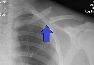
A clavicle fracture, also known as a broken collarbone, is a bone fracture of the clavicle. Symptoms typically include pain at the site of the break and a decreased ability to move the affected arm. Complications can include a collection of air in the pleural space surrounding the lung (pneumothorax), injury to the nerves or blood vessels in the area, and an unpleasant appearance.

<span class="mw-page-title-main">Hip fracture</span> Broken bone in hip joint region

A hip fracture is a break that occurs in the upper part of the femur, at the femoral neck or (rarely) the femoral head. Symptoms may include pain around the hip, particularly with movement, and shortening of the leg. Usually the person cannot walk.

<span class="mw-page-title-main">Avulsion fracture</span> Tearing away of a bone by physical trauma

An avulsion fracture is a bone fracture which occurs when a fragment of bone tears away from the main mass of bone as a result of physical trauma. This can occur at the ligament by the application of forces external to the body or at the tendon by a muscular contraction that is stronger than the forces holding the bone together. Generally muscular avulsion is prevented by the neurological limitations placed on muscle contractions. Highly trained athletes can overcome this neurological inhibition of strength and produce a much greater force output capable of breaking or avulsing a bone.

<span class="mw-page-title-main">Hip dislocation</span> Orthopedic injury

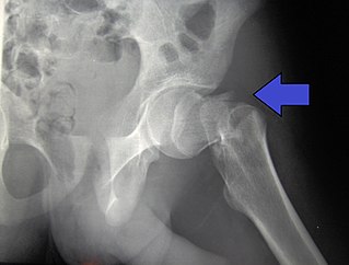
A hip dislocation is when the thighbone (femur) separates from the hip bone (pelvis). Specifically it is when the ball–shaped head of the femur separates from its cup–shaped socket in the hip bone, known as the acetabulum. The joint of the femur and pelvis is very stable, secured by both bony and soft-tissue constraints. With that, dislocation would require significant force which typically results from significant trauma such as from a motor vehicle collision or from a fall from elevation. Hip dislocations can also occur following a hip replacement or from a developmental abnormality known as hip dysplasia.

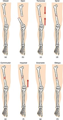
An open fracture, also called a compound fracture, is a type of bone fracture that has an open wound in the skin near the fractured bone. The skin wound is usually caused by the bone breaking through the surface of the skin. An open fracture can be life threatening or limb-threatening due to the risk of a deep infection and/or bleeding. Open fractures are often caused by high energy trauma such as road traffic accidents and are associated with a high degree of damage to the bone and nearby soft tissue. Other potential complications include nerve damage or impaired bone healing, including malunion or nonunion. The severity of open fractures can vary. For diagnosing and classifying open fractures, Gustilo-Anderson open fracture classification is the most commonly used method. This classification system can also be used to guide treatment, and to predict clinical outcomes. Advanced trauma life support is the first line of action in dealing with open fractures and to rule out other life-threatening condition in cases of trauma. The person is also administered antibiotics for at least 24 hours to reduce the risk of an infection.

<span class="mw-page-title-main">Humerus fracture</span> Medical condition

A humerus fracture is a break of the humerus bone in the upper arm. Symptoms may include pain, swelling, and bruising. There may be a decreased ability to move the arm and the person may present holding their elbow. Complications may include injury to an artery or nerve, and compartment syndrome.

<span class="mw-page-title-main">Supracondylar humerus fracture</span> Medical condition

A supracondylar humerus fracture is a fracture of the distal humerus just above the elbow joint. The fracture is usually transverse or oblique and above the medial and lateral condyles and epicondyles. This fracture pattern is relatively rare in adults, but is the most common type of elbow fracture in children. In children, many of these fractures are non-displaced and can be treated with casting. Some are angulated or displaced and are best treated with surgery. In children, most of these fractures can be treated effectively with expectation for full recovery. Some of these injuries can be complicated by poor healing or by associated blood vessel or nerve injuries with serious complications.

The Essex-Lopresti fracture is a fracture of the radial head of the forearm with concomitant dislocation of the distal radio-ulnar joint along with disruption of the thin interosseous membrane which holds them together. The injury is named after Peter Essex-Lopresti who described it in 1951.

The Bosworth fracture is a rare fracture of the distal fibula with an associated fixed posterior dislocation of the proximal fibular fragment which becomes trapped behind the posterior tibial tubercle. The injury is caused by severe external rotation of the ankle. The ankle remains externally rotated after the injury, making interpretation of X-rays difficult which can lead to misdiagnosis and incorrect treatment. The injury is most commonly treated by open reduction internal fixation as closed reduction is made difficult by the entrapment of the fibula behind the tibia.

<span class="mw-page-title-main">Femoral fracture</span> Broken femur, at shaft or distally

A femoral fracture is a bone fracture that involves the femur. They are typically sustained in high-impact trauma, such as car crashes, due to the large amount of force needed to break the bone. Fractures of the diaphysis, or middle of the femur, are managed differently from those at the head, neck, and trochanter; those are conventionally called hip fractures. Thus, mentions of femoral fracture in medicine usually refer implicitly to femoral fractures at the shaft or distally.

<span class="mw-page-title-main">Ulna fracture</span> Medical condition

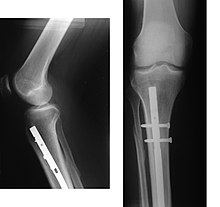
An ulna fracture is a break in the ulna bone, one of the two bones in the forearm. It is often associated with a fracture of the other forearm bone, the radius.

<span class="mw-page-title-main">Tibial plateau fracture</span> Medical condition

A tibial plateau fracture is a break of the upper part of the tibia (shinbone) that involves the knee joint. This could involve the medial, lateral, central, or bicondylar. Symptoms include pain, swelling, and a decreased ability to move the knee. People are generally unable to walk. Complication may include injury to the artery or nerve, arthritis, and compartment syndrome.

<span class="mw-page-title-main">Olecranon fracture</span> Medical condition

Olecranon fracture is a fracture of the bony portion of the elbow. The injury is fairly common and often occurs following a fall or direct trauma to the elbow. The olecranon is the proximal extremity of the ulna which is articulated with the humerus bone and constitutes a part of the elbow articulation. Its location makes it vulnerable to direct trauma.

<span class="mw-page-title-main">Pilon fracture</span> Medical condition

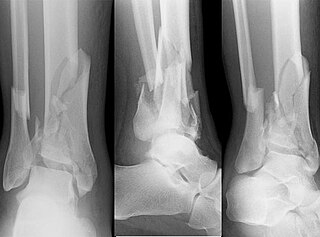
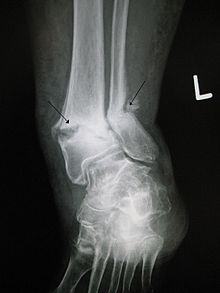
A pilon fracture, is a fracture of the distal part of the tibia, involving its articular surface at the ankle joint. Pilon fractures are caused by rotational or axial forces, mostly as a result of falls from a height or motor vehicle accidents. Pilon fractures are rare, comprising 3 to 10 percent of all fractures of the tibia and 1 percent of all lower extremity fractures, but they involve a large part of the weight-bearing surface of the tibia in the ankle joint. Because of this, they may be difficult to fixate and are historically associated with high rates of complications and poor outcome.

<span class="mw-page-title-main">Index of trauma and orthopaedics articles</span>

Orthopedic surgery is the branch of surgery concerned with conditions involving the musculoskeletal system. Orthopedic surgeons use both surgical and nonsurgical means to treat musculoskeletal injuries, sports injuries, degenerative diseases, infections, bone tumours, and congenital limb deformities. Trauma surgery and traumatology is a sub-specialty dealing with the operative management of fractures, major trauma and the multiply-injured patient.

<span class="mw-page-title-main">Proximal humerus fracture</span> Break of the upper part of the bone of the arm

A proximal humerus fracture is a break of the upper part of the bone of the arm (humerus). Symptoms include pain, swelling, and a decreased ability to move the shoulder. Complications may include axillary nerve or axillary artery injury.

<span class="mw-page-title-main">Broken finger</span> Medical condition

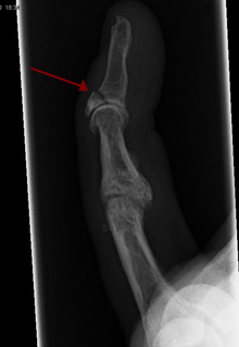
A broken finger or finger fracture is a common type of bone fracture, affecting a finger. Symptoms may include pain, swelling, tenderness, bruising, deformity and reduced ability to move the finger. Although most finger fractures are easy to treat, failing to deal with a fracture appropriately may result in long-term pain and disability.

References

  1. Katherine, Abel (2013). Official CPC Certification Study Guide. American Medical Association. p. 108.
  2. Witmer, Daniel K.; Marshall, Silas T.; Browner, Bruce D. (2016). "Emergency Care of Musculoskeletal Injuries". In Townsend, Courtney M.; Beauchamp, R. Daniel; Evers, B. Mark; Mattox, Kenneth L. (eds.). Sabiston Textbook of Surgery (20th ed.). Elsevier. pp. 462–504. ISBN   978-0-323-40163-0.
  3. MedicineNet – Fracture Archived 2008-12-21 at the Wayback Machine Medical Author: Benjamin C. Wedro, MD, FAAEM.
  4. Danielle Campagne (September 2022). "Overview of Fractures". mdmanuals.com.
  5. "Compartment Syndrome". The Lecturio Medical Concept Library. Retrieved 25 June 2021.
  6. Silva, Joana Cavaco (11 July 2018). "Bone fracture repair: Procedures, risks, and healing time". Medical News Today . William Morrison, M.D. (medical reviewer). Retrieved 21 April 2022.
  7. Sloan, A.; Hussain, I.; Maqsood, M.; Eremin, O.; El-Sheemy, M. (2010). "The effects of smoking on fracture healing". The Surgeon. 8 (2): 111–6. doi:10.1016/j.surge.2009.10.014. PMID   20303894.
  8. Pountos, Ippokratis; Georgouli, Theodora; Calori, Giorgio M.; Giannoudis, Peter V. (2012). "Do Nonsteroidal Anti-Inflammatory Drugs Affect Bone Healing? A Critical Analysis". The Scientific World Journal. 2012: 1–14. doi: 10.1100/2012/606404 . PMC   3259713 . PMID   22272177.
  9. Kanis, J. A.; Johnell, O.; Oden, A.; Johansson, H.; De Laet, C.; Eisman, J. A.; Fujiwara, S.; Kroger, H.; McCloskey, E. V.; Mellstrom, D.; Melton, L. J.; Pols, H.; Reeve, J.; Silman, A.; Tenenhouse, A. (2004). "Smoking and fracture risk: A meta-analysis". Osteoporosis International. 16 (2): 155–62. doi:10.1007/s00198-004-1640-3. PMID   15175845. S2CID   19890259.
  10. "Simple fracture | pathology". Encyclopedia Britannica. Retrieved 19 May 2021.
  11. Metsemakers, WJ; Onsea, J; Neutjens, E; Steffens, E; Schuermans, A; McNally, M; Nijs, S (December 2017). "Prevention of fracture-related infection: a multidisciplinary care package". International Orthopaedics. 41 (12): 2457–2469. doi: 10.1007/s00264-017-3607-y . PMID   28831576. S2CID   12894601.
  12. Roberto Schubert. "Fractures of the extremities (general rules and nomenclature)". Radiopaedia . Retrieved 21 February 2018.
  13. Essex Lopresti fracture Archived 2009-10-01 at the Wayback Machine at Wheeless' Textbook of Orthopaedics online
  14. "Bennett's fracture-subluxation" . GPnotebook.
  15. 1 2 Hunter, Tim B.; Peltier, Leonard F.; Lund, Pamela J. (2000). "Radiologic History Exhibit". RadioGraphics. 20 (3): 819–36. doi:10.1148/radiographics.20.3.g00ma20819. PMID   10835130.
  16. Mellick, Larry B.; Milker, Laura; Egsieker, Erik (1999). "Childhood accidental spiral tibial (CAST) fractures". Pediatric Emergency Care. 15 (5): 307–9. doi:10.1097/00006565-199910000-00001. PMID   10532655.
  17. Perry, C. R.; Rice, S; Rao, A; Burdge, R (1983). "Posterior fracture-dislocation of the distal part of the fibula. Mechanism and staging of injury". The Journal of Bone and Joint Surgery. American Volume. 65 (8): 1149–57. doi:10.2106/00004623-198365080-00016. PMID   6630259.[ permanent dead link ]
  18. TheFreeDictionary Lisfranc's fracture Citing: Mosby's Medical Dictionary, 8th edition. Copyright 2009
  19. Mabry LM, Patti TN, Ross MD, Bleakley CM, Gisselman AS (July 2021). "Isolated Medial Cuneiform Fractures: A Systematic Search and Qualitative Analysis of Case Studies". J Am Podiatr Med Assoc. 111 (4): 1–9. doi:10.7547/20-047. PMID   34478529. S2CID   225705519.{{cite journal}}: CS1 maint: multiple names: authors list (link)
  20. "Fracture and dislocation compendium. Orthopaedic Trauma Association Committee for Coding and Classification". Journal of Orthopaedic Trauma. 10 (Suppl 1): v–ix, 1–154. 1996. PMID   8814583.
  21. Müller ME, Nazarian S, Koch P (1987). Classification AO des fractures. Tome I. Les os longs. Berlin: Springer-Verlag.[ page needed ]
  22. Marsh, J. L.; Slongo, T. F.; Agel, J; Broderick, J. S.; Creevey, W; Decoster, T. A.; Prokuski, L; Sirkin, M. S.; Ziran, B; Henley, B; Audigé, L (2007). "Fracture and dislocation classification compendium - 2007: Orthopaedic Trauma Association classification, database and outcomes committee". Journal of Orthopaedic Trauma. 21 (10 Suppl): S1–133. doi: 10.1097/00005131-200711101-00001 . PMID   18277234. S2CID   24535478.
  23. "Denis classification of spinal fractures" . GPnotebook.
  24. Rüedi, etc. all; Thomas P. Rüedi; Richard E. Buckley; Christopher G. Moran (2007). AO principles of fracture management, Volume 1. Thieme. p. 96. ISBN   978-3-13-117442-0.
  25. "Fractures of the Acetabulum". wheelessonline.com. Archived from the original on 26 September 2009.
  26. Mourad, L (1997). "Neer classification of fractures of the proximal humerus". Orthopedic Nursing. 16 (2): 76. PMID   9155417.
  27. Proximal Humerus Fractures at eMedicine
  28. "Hip Fractures". The Lecturio Medical Concept Library. Retrieved 24 July 2021.
  29. 1 2 "Open Fractures - OrthoInfo - AAOS" . Retrieved 3 December 2018.
  30. 1 2 Court-Brown, Charles M.; Bugler, Kate E.; Clement, Nicholas D.; Duckworth, Andrew D.; McQueen, Margaret M. (June 2012). "The epidemiology of open fractures in adults. A 15-year review". Injury. 43 (6): 891–897. doi:10.1016/j.injury.2011.12.007. ISSN   1879-0267. PMID   22204774.
  31. 1 2 3 Sidwell, Richard; Matar, Maher M.; Sakran, Joseph V. (1 October 2017). "Trauma Education and Prevention". Surgical Clinics of North America. 97 (5): 1185–1197. doi:10.1016/j.suc.2017.06.010. ISSN   0039-6109. PMID   28958365.
  32. 1 2 3 4 "Preventing Falls and Related Fractures | NIH Osteoporosis and Related Bone Diseases National Resource Center". www.bones.nih.gov. Retrieved 3 December 2018.
  33. Avenell, Alison; Mak, Jenson C. S.; O'Connell, Dianne (14 April 2014). "Vitamin D and vitamin D analogues for preventing fractures in post-menopausal women and older men". The Cochrane Database of Systematic Reviews. 2021 (4): CD000227. doi:10.1002/14651858.CD000227.pub4. ISSN   1469-493X. PMC   7032685 . PMID   24729336.
  34. 1 2 3 McDaniel, Dalton J.; Rehman, Uzma H. (2 November 2021). "Phalanx Fractures of the Hand". StatPearls. StatPearls Publishing. PMID   32491557 via PubMed.
  35. "Overview of Bone Fractures". The Lecturio Medical Concept Library. Retrieved 25 July 2021.
  36. Drendel, Amy L.; Gorelick, Marc H.; Weisman, Steven J.; Lyon, Roger; Brousseau, David C.; Kim, Michael K. (2009). "A Randomized Clinical Trial of Ibuprofen Versus Paracetamol with Codeine for Acute Pediatric Arm Fracture Pain". Annals of Emergency Medicine. 54 (4): 553–60. doi:10.1016/j.annemergmed.2009.06.005. PMID   19692147.
  37. Keramidas EG, Miller G (October 2005). "The Suzuki frame for complex intraarticular fractures of the thumb". Plastic and Reconstructive Surgery. 116 (5): 1326–31. doi:10.1097/01.prs.0000181786.39062.0b. PMID   16217475. S2CID   31890854.
  38. Boutis, K.; Willan, A.; Babyn, P.; Goeree, R.; Howard, A. (2010). "Cast versus splint in children with minimally angulated fractures of the distal radius: A randomized controlled trial". Canadian Medical Association Journal. 182 (14): 1507–12. doi:10.1503/cmaj.100119. PMC   2950182 . PMID   20823169.
  39. "Fractures". Johns Hopkins Medicine. 28 February 2020. Retrieved 25 July 2021.
  40. "Hip Fractures". The Lecturio Medical Concept Library. Retrieved 24 July 2021.
  41. Klokkevold PR, Jovanovic SA (2002). "Advanced Implant Surgery and Bone Grafting Techniques". In Newman MG, Takei HM, Carranza FA (eds.). Carranza's Clinical Periodontology (9th ed.). W.B. Saunders. pp. 907–8. ISBN   9780721683317.
  42. "Fractures". Johns Hopkins Medicine. 28 February 2020. Retrieved 25 July 2021.
  43. 1 2 Leighton, R.; Watson, J.T; Giannoudis, P.; Papakostidis, C.; Harrison, A.; Steen, R.G. (May 2017). "Healing of fracture nonunions treated with low-intensity pulsed ultrasound (LIPUS): A systematic review and meta-analysis". Injury. 48 (7): 1339–1347. doi: 10.1016/j.injury.2017.05.016 . PMID   28532896.
  44. Victoria, Galkowski; Petrisor, Brad; Drew, Brian; Dick, David (2009). "Bone stimulation for fracture healing: What′s all the fuss?". Indian Journal of Orthopaedics. 43 (2): 117–20. doi: 10.4103/0019-5413.50844 (inactive 24 April 2024). ISSN   0019-5413. PMC   2762251 . PMID   19838359.{{cite journal}}: CS1 maint: DOI inactive as of April 2024 (link)
  45. Searle, Henry Kc; Lewis, Sharon R.; Coyle, Conor; Welch, Matthew; Griffin, Xavier L. (3 March 2023). "Ultrasound and shockwave therapy for acute fractures in adults". The Cochrane Database of Systematic Reviews. 2023 (3): CD008579. doi:10.1002/14651858.CD008579.pub4. ISSN   1469-493X. PMC   9983300 . PMID   36866917.
  46. Pradhan, Sara; Chiu, Sarah; Burton, Claire; Forsyth, Jacky; Corp, Nadia; Paskins, Zoe; van der Windt, Danielle A.; Babatunde, Opeyemi O. (3 June 2022). "Overall Effects and Moderators of Rehabilitation in Patients With Wrist Fracture: A Systematic Review". Physical Therapy. 102 (6): pzac032. doi: 10.1093/ptj/pzac032 . ISSN   1538-6724. PMID   35421234.
  47. Fairhall, Nicola J.; Dyer, Suzanne M.; Mak, Jenson Cs; Diong, Joanna; Kwok, Wing S.; Sherrington, Catherine (7 September 2022). "Interventions for improving mobility after hip fracture surgery in adults". The Cochrane Database of Systematic Reviews. 2022 (9): CD001704. doi:10.1002/14651858.CD001704.pub5. ISSN   1469-493X. PMC   9451000 . PMID   36070134.