Supracondylar humerus fracture

Last updated
Supracondylar humerus fracture
Supracondylar09.JPG
An elbow X-ray showing a displaced supracondylar fracture in a young child
Specialty Orthopedic

A supracondylar humerus fracture is a fracture of the distal humerus just above the elbow joint. The fracture is usually transverse or oblique and above the medial and lateral condyles and epicondyles. This fracture pattern is relatively rare in adults, but is the most common type of elbow fracture in children. [1] In children, many of these fractures are non-displaced and can be treated with casting. Some are angulated or displaced and are best treated with surgery. In children, most of these fractures can be treated effectively with expectation for full recovery. [2] Some of these injuries can be complicated by poor healing or by associated blood vessel or nerve injuries with serious complications.

Contents

Signs and symptoms

A child will complain of pain and swelling over the elbow immediately post trauma with loss of function of affected upper limb. Late onset of pain (hours after injury) could be due to muscle ischaemia (reduced oxygen supply). This can lead to loss of muscle function. [3]

It is important to check for viability of the affected limb post trauma. Clinical parameters such as temperature of the limb extremities (warm or cold), capillary refilling time, oxygen saturation of the affected limb, presence of distal pulses (radial and ulnar pulses), assessment of peripheral nerves (radial, median, and ulnar nerves), and any wounds which would indicate open fracture. Doppler ultrasonography should be performed to ascertain blood flow of the affected limb if the distal pulses are not palpable. Anterior interosseus branch of the median nerve most often injured in postero-lateral displacement of the distal humerus as the proximal fragment is displaced antero-medially. This is evidenced by the weakness of the hand with a weak "OK" sign on physical examination (Unable to do an "OK" sign; instead a pincer grasp is performed). Radial nerve would be injured if the distal humerus is displaced postero-medially. This is because the proximal fragment will be displaced antero-laterally. Ulnar nerve is most commonly injured in the flexion type of injury because it crosses the elbow below the medial epidcondyle of the humerus. [3]

A puckered, dimple, or an ecchymosis of the skin just anterior to the distal humerus is a sign of difficult reduction because the proximal fragment may have already penetrated the brachialis muscle and the subcutaneous layer of the skin. [3]

Complications

Volkmann's contracture

Swelling and vascular injury following the fracture can lead to the development of the compartment syndrome which leads to long-term complication of Volkmann's contracture (fixed flexion of the elbow, pronation of the forearm, flexion at the wrist, and joint extension of the metacarpophalangeal joint ). Therefore, early surgical reduction is indicated to prevent this type of complication. [3]

Malunion

The distal humerus grows slowly post fracture (only contributes 10 to 20% of the longitudinal growth of the humerus), therefore, there is a high rate of malunion if the supracondylar fracture is not corrected appropriately. Such malunion can result in cubitus varus deformity.[ citation needed ]

Mechanism

Extension type of supracondylar humerus fractures typically result from a fall on to an outstretched hand, usually leading to a forced hyperextension of the elbow. The olecranon acts as a fulcrum which focuses the stress on distal humerus (supracondylar area), predisposing the distal humerus to fracture. The supracondylar area undergoes remodeling at the age of 6 to 7, making this area thin and prone to fractures. Important arteries and nerves (median nerve, radial nerve, brachial artery, and ulnar nerve) are located at the supracondylar area and can give rise to complications if these structures are injured. Most vulnerable structure to get damaged is Median Nerve. [3] Meanwhile, the flexion-type of supracondylar humerus fracture is less common. It occurs by falling on the point of the elbow, or falling with the arm twisted behind the back. This causes anterior dislocation of the proximal fragment of the humerus. [4]

Diagnosis

A supracondylar humerus facture is diagnosed by x-ray and the injured limb will be examined to assess the surrounding soft tissue, neurovascular status, and to identify any other injuries to the affected area. [5] Pain, swelling, and deformity near the elbow or arm area is common and a bleed near the fracture may result in an effusion in the elbow joint. With severe displacement, there may be an anterior dimple from the proximal bone end trapped within the biceps muscle. The skin is usually intact. If there is a laceration that communicates with the fracture site, it is an open fracture, which increases infection risk. For fractures with significant displacement, the bone end can be trapped within the biceps muscle with resulting tension producing an indentation to the skin, which is called a "pucker sign".[ citation needed ]

Vascular system examination

The vascular status must be assessed, including the warmth and perfusion of the hand, the time for capillary refill, and the presence of a palpable radial pulse. Limb vascular status is categorized as "normal," "pulseless with a (warm, pink) perfused hand," or "pulseless–pale (nonperfused)" (see "neurovascular complications" below).

Sensory and motor nerve examination

The neurologic status must be assessed including the sensory and motor function of the radial, ulnar, and median nerves (see "neurovascular complications" below). Neurologic deficits are found in 10-20% of patients. [6] The mostly commonly injured nerve is the median nerve (specifically, the anterior interosseous portion of the median nerve). Injuries to the ulnar and radial nerves are less common.

X-rays

Diagnosis is confirmed by x-ray imaging. Antero-posterior (AP) and lateral view of the elbow joint should be obtained. Any other sites of pain, deformity, or tenderness should warrant an X-ray for that area too. X-ray of the forearm (AP and lateral) should also be obtained for because of the common association of supracondylar fractures with the fractures of the forearm. Ideally, splintage should be used to immobilise the elbow at 20 to 30 degrees flexion in order to prevent further injury of the blood vessels and nerves while doing X-rays. Splinting of fracture site with full flexion or extension of the elbow is not recommended as it can stretch the blood vessels and nerves over the bone fragments or can cause impingement of these structures into the fracture site. [3]

Depending on the child's age, parts of the bone will still be developing and if not yet calcified, will not show up on the X-rays. The capitulum of the humerus is the first to ossify at the age of one year. Head of radius and medial epicondyle of the humerus starts to ossify at 4 to 5 years of age, followed by trochlea of humerus and olecranon of the ulna at 8 to 9 years of age, and lateral epicondyle of the humerus to ossify at 10 years of age. [3]

Anterior X-ray

Baumann's Angle Baumann's Angle.jpg
Baumann's Angle

Carrying angle can be evaluated through AP view of the elbow by looking at the Baumann's angle. [3] There are two definitions of Bowmann's angle:

The first definition of Baumann's angle is an angle between a line parallel to the longitudinal axis of the humeral shaft and a line drawn along the lateral epicondyle.[ citation needed ]

Another definition of Baumann's angle is also known as the humeral-capitellar angle. It is the angle between the line perpendicular to the long axis of the humerus and the growth plate of the lateral condyle. Reported normal values for Baumann's angle range between 9 and 26°. [7] An angle of more than 10° is regarded as acceptable. [7]

Lateral X-ray

On lateral view of the elbow, there are five radiological features should be looked for: tear drop sign, anterior humeral line, coronoid line, fish-tail sign, and fat pad sign/sail sign (anterior and posterior). [3] [8]

Tear drop sign - Tear drop sign is seen on a normal radiograph, but is disturbed in supracondylar fracture. [8]

Anterior humeral line - It is a line drawn down along the front of the humerus on the lateral view and it should pass through the middle third of the capitulum of the humerus. [9] If it passes through the anterior third of the capitulum, it indicates the posterior displacement of distal fragment. [8]

Fat pad sign/sail sign - A non-displaced fracture can be difficult to identify and a fracture line may not be visible on the X-rays. However, the presence of a joint effusion is helpful in identifying a non-displaced fracture. Bleeding from the fracture expands the joint capsule and is visualized on the lateral view as a darker area anteriorly and posteriorly, and is known as the sail sign. [8]

Coronoid line - A line drawn along the anterior border of the coronoid process of the ulna should touch the anterior part of the lateral condyle of the humerus. If lateral condyle appears posterior to this line, it indicates the posterior displacement of lateral condyle. [8]

Fish-tail sign - The distal fragment is rotated away from the proximal fragment, thus the sharp ends of the proximal fragment looks like a shape of a fish-tail. [8]

Classification

Supracondylar fractures: Gartland classification Gartland Classification.jpg
Supracondylar fractures: Gartland classification
TypeDescription [3]
INon-displaced
IIAngulated with intact posterior cortex
IIAAngulation
IIBAngulation with rotation
IIIComplete displacement but have perisosteal (medial/lateral) contact
IIIAMedial periosteal hinge intact. Distal fragment goes posteromedially
IIIBLateral periosteal hinge intact. Distal fragment goes posterolaterally
IV Periostial disruption with instability in both flexion and extension

Management

Treatment options for supracondyl humerus fractures vary depending if the bone is displaced (out of position) or not displaced (see classification section above). [1]

Gartland type I

Undisplaced or minimally displaced fractures can be treated by using an above elbow splint in 90 degrees flexion for 3 weeks. Orthopaedic cast and extreme flexion should be avoided to prevent compartment syndrome and vascular compromise. In case the varus of the fracture site is more than 10 degrees when compared to the normal elbow, closed reduction and percutaneous pinning using X-ray image intensifier inside operating theater is recommended. In one study, for those children who was done percutaneous pinning, immobilisation using a posterior splint and an arm sling has earlier resumption of activity when compared to immobilisation using collar and cuff sling. Both methods gives similar pain scores and activity level at two weeks of treatment. [3]

Gartland type II

Gartland Type II fractures requires closed reduction and casting at 90 degrees flexion. Percutaneous pinning is required if more than 90 degrees flexion is required to maintain the reduction. Closed reduction with percutaneous pinning has low complication rates. Closed reduction can be done by applying traction along the long axis of the humerus with elbow in slight flexion. Full extension of the elbow is not recommended because the neurovascular structures can hook around the proximal fragment of the humerus. If the proximal humerus is suspected to have pierced the brachialis muscle, gradual traction over the proximal humerus should be given instead. After that, reduction can be done through hyperflexion of the elbow can be done with the olecranon pushing anteriorly. If the distal fragment is internally rotated, reduction maneuver can be applied with extra stress applied over medial elbow with pronation of the forearm at the same time. [3]

Gartland type III and IV

Gartland III and IV are unstable and prone to neurovascular injury. Therefore, closed or open reduction together with percutaneous pinning within 24 hours is the preferred method of management with low complication rates. Straight arm lateral traction can be a safe method to deal with Gartland Type III fractures. Although Gartland Type III fractures with posteromedial displacement of distal fragment can be reduced with closed reduction and casting, those with posterolateral displacement should preferably be fixed by percutaneous pinning. [3]

Percutaneous pinning

Percutaneous pinning are usually inserted over the medial or lateral sides of the elbow under X-ray image intensifier guidance. There is 1.8 times higher risk of getting nerve injury when inserting both medial and lateral pins compared to lateral pin insertion alone. However, medial and lateral pins insertions are able to stabilise the fractures more properly than lateral pins alone. Therefore, medial and lateral pins insertion should be done with care to prevent nerve injuries around elbow region. [3]

Percutaneous pinning should be done when close manipulation fails to achieve the reduction, unstable fracture after closed reduction, neurological deficits occurs during or after the manipulation of fracture, and surgical exploration is required to determine the integrity of the blood vessels and nerves. In open fractures, surgical wound debridement should be performed to prevent any infection into the elbow joint. All Type II and III fractures requiring elbow flexion of more than 90° to maintain the reduction needs to be fixed by percutaneous pinning. All Type IV fractures of supracondylar humerus are unstable; therefore, requires percutaneous pinning. Besides, any polytrauma with multiple fractures of the same side requiring surgical intervention is another indication for percutaneous pinning. [3]

Follow up

For routine displaced supracondylar fractures requiring percutaneous pinning, radiographic evaluation and clinical assessment can be delayed until the pin removal. Pins are only removed when there is no tenderness over the elbow region at 3 to 4 weeks. After pin removal, mobilisation of the elbow can begin. [3]

Neurovascular complications

Absence of radial pulse is reported in 6–20% of the supracondylar fracture cases. This is because brachial artery is frequently injured in Gartland Type II and Type III fractures, especially when the distal fragment is displaced postero-laterally (proximal fragment displaced antero-medially). The available evidence suggests that if the child has a median nerve palsy, their neurological recovery is faster, more complete and more probable if an open reduction and exploration of the nerve is performed. [10] If the operating surgeons chooses to perform a closed reduction & percutaneous k-wiring in the presence of a neurological (and/or vascular) deficit and the pulse does not return immediately or the neurological deficit recover, then urgent surgical exploration is indicated. [3] In patients with a "pink but pulseless hand" (absent radial pulse but demonstrable perfusion at extremities) after successful reduction and percutaneous pinning, there is uncertainty about the ideal management and imaging or surgical exploration should be considered [11] given the risk of Volkmann's contracture.

Epidemiology

Supracondylar humerus fractures is commonly found in children between 5 and 7 years (90% of the cases), after the clavicle and forearm fractures. It is more often occurs in males, accounting of 16% of all pediatric fractures and 60% of all paediatric elbow fractures. The mechanism of injury is most commonly due to fall on an outstretch hand. [3] Extension type of injury (70% of all elbow fractures) is more common than the flexion type of injury (1% to 11% of all elbow injuries). [4] Injury often occurs on the non-dominant part of the limb. Flexion type of injury is more commonly found in older children. Open fractures can occur for up to 30% of the cases. [3]

Related Research Articles

<span class="mw-page-title-main">Ulna</span> Medial bone from forearm

The ulna or ulnal bone is a long bone found in the forearm that stretches from the elbow to the wrist, and when in anatomical position, is found on the medial side of the forearm. That is, the ulna is on the same side of the forearm as the little finger. It runs parallel to the radius, the other long bone in the forearm. The ulna is longer and the radius is shorter, but the radius is thicker and the ulna is thinner. Therefore, the ulna is considered to be the smaller bone of the two bones in the lower arm. The corresponding bone in the lower leg is the fibula.

<span class="mw-page-title-main">Humerus</span> Long bone of the upper arm

The humerus is a long bone in the arm that runs from the shoulder to the elbow. It connects the scapula and the two bones of the lower arm, the radius and ulna, and consists of three sections. The humeral upper extremity consists of a rounded head, a narrow neck, and two short processes. The body is cylindrical in its upper portion, and more prismatic below. The lower extremity consists of 2 epicondyles, 2 processes, and 3 fossae. As well as its true anatomical neck, the constriction below the greater and lesser tubercles of the humerus is referred to as its surgical neck due to its tendency to fracture, thus often becoming the focus of surgeons.

<span class="mw-page-title-main">Brachioradialis</span> Muscle of the upper limb

The brachioradialis is a muscle of the forearm that flexes the forearm at the elbow. It is also capable of both pronation and supination, depending on the position of the forearm. It is attached to the distal styloid process of the radius by way of the brachioradialis tendon, and to the lateral supracondylar ridge of the humerus.

<span class="mw-page-title-main">Median nerve</span> Nerve of the upper limb

The median nerve is a nerve in humans and other animals in the upper limb. It is one of the five main nerves originating from the brachial plexus.

The forearm is the region of the upper limb between the elbow and the wrist. The term forearm is used in anatomy to distinguish it from the arm, a word which is used to describe the entire appendage of the upper limb, but which in anatomy, technically, means only the region of the upper arm, whereas the lower "arm" is called the forearm. It is homologous to the region of the leg that lies between the knee and the ankle joints, the crus.

<span class="mw-page-title-main">Ulnar nerve</span> Nerve which runs near the ulna bone

In human anatomy, the ulnar nerve is a nerve that runs near the ulna bone. The ulnar collateral ligament of elbow joint is in relation with the ulnar nerve. The nerve is the largest in the human body unprotected by muscle or bone, so injury is common. This nerve is directly connected to the little finger, and the adjacent half of the ring finger, innervating the palmar aspect of these fingers, including both front and back of the tips, perhaps as far back as the fingernail beds.

<span class="mw-page-title-main">Radius (bone)</span> One of the two long bones of the forearm

The radius or radial bone is one of the two large bones of the forearm, the other being the ulna. It extends from the lateral side of the elbow to the thumb side of the wrist and runs parallel to the ulna. The ulna is longer than the radius, but the radius is thicker. The radius is a long bone, prism-shaped and slightly curved longitudinally.

<span class="mw-page-title-main">Upper limb</span> Consists of the arm, forearm, and hand

The upper limbs or upper extremities are the forelimbs of an upright-postured tetrapod vertebrate, extending from the scapulae and clavicles down to and including the digits, including all the musculatures and ligaments involved with the shoulder, elbow, wrist and knuckle joints. In humans, each upper limb is divided into the arm, forearm and hand, and is primarily used for climbing, lifting and manipulating objects.

<span class="mw-page-title-main">Distal radius fracture</span> Fracture of the radius bone near the wrist

A distal radius fracture, also known as wrist fracture, is a break of the part of the radius bone which is close to the wrist. Symptoms include pain, bruising, and rapid-onset swelling. The ulna bone may also be broken.

<span class="mw-page-title-main">Triceps</span> Muscle on the back of the upper arm

The triceps, or triceps brachii, is a large muscle on the back of the upper limb of many vertebrates. It consists of 3 parts: the medial, lateral, and long head. It is the muscle principally responsible for extension of the elbow joint.

<span class="mw-page-title-main">Cubital fossa</span> The human elbow pit

The cubital fossa,chelidon, grivet or elbow pit, is the area on the anterior side of the upper part between the arm and forearm of a human or other hormid animals. It lies anteriorly to the elbow when in standard anatomical position.

<span class="mw-page-title-main">Flexor carpi ulnaris muscle</span>

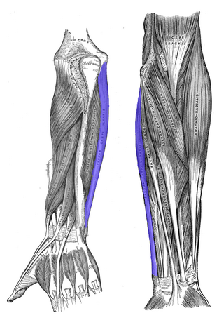
The flexor carpi ulnaris (FCU) is a muscle of the forearm that flexes and adducts at the wrist joint.

The pronator teres is a muscle that, along with the pronator quadratus, serves to pronate the forearm. It has two origins, at the medial humeral supracondylar ridge and the ulnar tuberosity, and inserts near the middle of the radius.

<span class="mw-page-title-main">Dog anatomy</span> Studies of the visible part of a canine

Dog anatomy comprises the anatomical studies of the visible parts of the body of a domestic dog. Details of structures vary tremendously from breed to breed, more than in any other animal species, wild or domesticated, as dogs are highly variable in height and weight. The smallest known adult dog was a Yorkshire Terrier that stood only 6.3 cm (2.5 in) at the shoulder, 9.5 cm (3.7 in) in length along the head and body, and weighed only 113 grams (4.0 oz). The heaviest dog was an English Mastiff named Zorba which weighed 314 pounds (142 kg). The tallest known adult dog is a Great Dane that stands 106.7 cm (42.0 in) at the shoulder.

<span class="mw-page-title-main">Medial epicondyle of the humerus</span> Rounded eminence on the medial side of the humerus

The medial epicondyle of the humerus is an epicondyle of the humerus bone of the upper arm in humans. It is larger and more prominent than the lateral epicondyle and is directed slightly more posteriorly in the anatomical position. In birds, where the arm is somewhat rotated compared to other tetrapods, it is called the ventral epicondyle of the humerus. In comparative anatomy, the more neutral term entepicondyle is used.

<span class="mw-page-title-main">Monteggia fracture</span> Medical condition

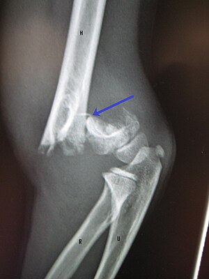
The Monteggia fracture is a fracture of the proximal third of the ulna with dislocation of the proximal head of the radius. It is named after Giovanni Battista Monteggia.

<span class="mw-page-title-main">Fascial compartments of arm</span> Anatomical compartments

The fascial compartments of arm refers to the specific anatomical term of the compartments within the upper segment of the upper limb of the body. The upper limb is divided into two segments, the arm and the forearm. Each of these segments is further divided into two compartments which are formed by deep fascia – tough connective tissue septa (walls). Each compartment encloses specific muscles and nerves.

<span class="mw-page-title-main">Ulnar nerve entrapment</span> Medical condition

Ulnar nerve entrapment is a condition where pressure on the ulnar nerve as it passes through the cubital tunnel causes nerve dysfunction (neuropathy). The symptoms of neuropathy are paresthesia (tingling) and numbness primarily affecting the little finger and ring finger of the hand. Entrapment may occur at any point from the spine at cervical vertebra C7 to the wrist; the most common point of entrapment is in the elbow. Prevention is mostly through correct posture and avoiding repetitive or constant strain. Treatment is usually conservative, including medication, activity modification, and exercise, but may sometimes include surgery. Symptoms can be alleviated by attempts to keep the elbow from flexing while sleeping, such as sticking one’s arm in the pillow case, so the pillow restricts flexion.

<span class="mw-page-title-main">Humerus fracture</span> Medical condition

A humerus fracture is a break of the humerus bone in the upper arm. Symptoms may include pain, swelling, and bruising. There may be a decreased ability to move the arm and the person may present holding their elbow. Complications may include injury to an artery or nerve, and compartment syndrome.

<span class="mw-page-title-main">Elbow</span> Joint between the upper and lower parts of the arm

The elbow is the region between the upper arm and the forearm that surrounds the elbow joint. The elbow includes prominent landmarks such as the olecranon, the cubital fossa, and the lateral and the medial epicondyles of the humerus. The elbow joint is a hinge joint between the arm and the forearm; more specifically between the humerus in the upper arm and the radius and ulna in the forearm which allows the forearm and hand to be moved towards and away from the body. The term elbow is specifically used for humans and other primates, and in other vertebrates forelimb plus joint is used.

References

  1. 1 2 Marson, Ben A.; Ikram, Adeel; Craxford, Simon; Lewis, Sharon R.; Price, Kathryn R.; Ollivere, Benjamin J. (2022-06-09). "Interventions for treating supracondylar elbow fractures in children". The Cochrane Database of Systematic Reviews. 2022 (6): CD013609. doi:10.1002/14651858.CD013609.pub2. ISSN   1469-493X. PMC   9178297 . PMID   35678077.
  2. "OrthoKids - Elbow Fractures". orthokids.org. Retrieved 2017-08-24.
  3. 1 2 3 4 5 6 7 8 9 10 11 12 13 14 15 16 17 18 19 Vineet, Kumar; Ajai, Singh (1 December 2016). "Fracture Supracondylar Humerus: A Review". Journal of Clinical and Diagnostic Research. 10 (12): RE01–RE06. doi:10.7860/JCDR/2016/21647.8942. PMC   5296534 . PMID   28208961.
  4. 1 2 Eira, Kuoppala; Roope, Parvianien; Tytti, Pokka; Minna, Serlo; Juha-Jaakko, Sinikumpu (11 May 2016). "Low incidence of flexion-type supracondylar humerus fractures but high rate of complications". Acta Orthopaedica. 87 (4): 406–411. doi:10.1080/17453674.2016.1176825. PMC   4967285 . PMID   27168001.
  5. Hope, Natalie; Varacallo, Matthew (2023), "Supracondylar Humerus Fractures", StatPearls, Treasure Island (FL): StatPearls Publishing, PMID   32809768 , retrieved 2023-03-23
  6. Terry Canale, S.; Azar, Frederick M.; Beaty, James H. (2016-11-21). Campbell's operative orthopaedics. Azar, Frederick M.,, Canale, S. T. (S. Terry),, Beaty, James H.,, Preceded by: Campbell, Willis C. (Willis Cohoon), 1880-1941. (Thirteenth ed.). Philadelphia, PA. ISBN   978-0323374620. OCLC   962333989.{{cite book}}: CS1 maint: location missing publisher (link)
  7. 1 2 Page 1405 in: S. Terry Canale, James H. Beaty (2012). Campbell's Operative Orthopaedics (12 ed.). Elsevier Health Sciences. ISBN   9780323087186.
  8. 1 2 3 4 5 6 John, Ebnezar; Rakesh, John (31 December 2016). Textbook of orthopaedics. JP Medical Ltd. p. 135. ISBN   9789386056689 . Retrieved 15 April 2018.
  9. 1 2 3 Kilborn, Tracy; Moodley, Halvani; Mears, Stewart (2015). "Elbow your way into reporting paediatric elbow fractures – A simple approach". South African Journal of Radiology. 19 (2). doi: 10.4102/sajr.v19i2.881 . ISSN   2078-6778.
  10. Wilks, Daniel J.; Ye, Xuan; Biggins, Rose; Wang, Kemble K.; Wade, Ryckie G.; McCombe, David (August 2023). "Median Nerve Palsy in Pediatric Supracondylar Humerus Fractures Recovers Faster With Open Than Closed Reduction". Journal of Pediatric Orthopaedics. 43 (7): 407–413. doi:10.1097/BPO.0000000000002424. ISSN   0271-6798.
  11. Griffin, K.J.; Walsh, S.R.; Markar, S.; Tang, T.Y.; Boyle, J.R.; Hayes, P.D. (2008). "The Pink Pulseless Hand: A Review of the Literature Regarding Management of Vascular Complications of Supracondylar Humeral Fractures in Children". European Journal of Vascular and Endovascular Surgery. 36 (6): 697–702. doi: 10.1016/j.ejvs.2008.08.013 . PMID   18851922.

Bibliography