
Trismus is a condition of restricted opening of the mouth. The term was initially used in the setting of tetanus. Trismus may be caused by spasm of the muscles of mastication or a variety of other causes. Temporary trismus occurs much more frequently than permanent trismus. It is known to interfere with eating, speaking, and maintaining proper oral hygiene. This interference, specifically with an inability to swallow properly, results in an increased risk of aspiration. In some instances, trismus presents with altered facial appearance. The condition may be distressing and painful. Examination and treatments requiring access to the oral cavity can be limited, or in some cases impossible, due to the nature of the condition itself.

In the human skull, the zygomatic bone, also called cheekbone or malar bone, is a paired irregular bone which articulates with the maxilla, the temporal bone, the sphenoid bone and the frontal bone. It is situated at the upper and lateral part of the face and forms the prominence of the cheek, part of the lateral wall and floor of the orbit, and parts of the temporal fossa and the infratemporal fossa. It presents a malar and a temporal surface; four processes, and four borders.

In anatomy, the zygomatic arch, or cheek bone, is a part of the skull formed by the zygomatic process of the temporal bone and the temporal process of the zygomatic bone, the two being united by an oblique suture ; the tendon of the temporal muscle passes medial to the arch, to gain insertion into the coronoid process of the mandible (jawbone).

Subconjunctival bleeding, also known as subconjunctival hemorrhage or subconjunctival haemorrhage, is bleeding from a small blood vessel over the whites of the eye. It results in a red spot in the white of the eye. There is generally little to no pain and vision is not affected. Generally only one eye is affected.

The zygomaticotemporal nerve (zygomaticotemporal branch, temporal branch) is a cutaneous (sensory) nerve of the head. It is a branch of the zygomatic nerve (itself a branch of the maxillary nerve (CN V2)). It arises in the orbit and exits the orbit through the zygomaticotemporal foramen in the zygomatic bone to enter the temporal fossa. It is distributed to the skin of the side of the forehead. It also contains a parasympathetic secretomotor component for the lacrimal gland which it confers to the lacrimal nerve (which then delivers it to the gland).

An orbital blowout fracture is a traumatic deformity of the orbital floor or medial wall that typically results from the impact of a blunt object larger than the orbital aperture, or eye socket. Most commonly this results in a herniation of orbital contents through the orbital fractures. The proximity of maxillary and ethmoidal sinus increases the susceptibility of the floor and medial wall for the orbital blowout fracture in these anatomical sites. Most commonly, the inferior orbital wall, or the floor, is likely to collapse, because the bones of the roof and lateral walls are robust. Although the bone forming the medial wall is the thinnest, it is buttressed by the bone separating the ethmoidal air cells. The comparatively thin bone of the floor of the orbit and roof of the maxillary sinus has no support and so the inferior wall collapses mostly. Therefore, medial wall blowout fractures are the second-most common, and superior wall, or roof and lateral wall, blowout fractures are uncommon and rare, respectively. They are characterized by double vision, sunken ocular globes, and loss of sensation of the cheek and upper gums from infraorbital nerve injury.
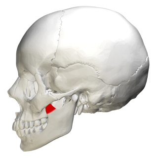
In human anatomy, the mandible's coronoid process is a thin, triangular eminence, which is flattened from side to side and varies in shape and size. Its anterior border is convex and is continuous below with the anterior border of the ramus. Its posterior border is concave and forms the anterior boundary of the mandibular notch. The lateral surface is smooth, and affords insertion to the temporalis and masseter muscles. Its medial surface gives insertion to the temporalis, and presents a ridge which begins near the apex of the process and runs downward and forward to the inner side of the last molar tooth.
Median palatal cysts are uncommon hard palate fissural cysts that are not odontogenic. These lesions are located behind the incisive canal in the midline of the hard palate. The majority of the time, median palatine cysts are asymptomatic and are discovered by coincidence while a patient is being evaluated for a different ailment.

The nose is the first organ of the respiratory system. It is also the principal organ in the olfactory system. The shape of the nose is determined by the nasal bones and the nasal cartilages, including the nasal septum which separates the nostrils and divides the nasal cavity into two.
The buccal fat pad is one of several encapsulated fat masses in the cheek. It is a deep fat pad located on either side of the face between the buccinator muscle and several more superficial muscles. The inferior portion of the buccal fat pad is contained within the buccal space. It should not be confused with the malar fat pad, which is directly below the skin of the cheek. It should also not be confused with jowl fat pads. It is implicated in the formation of hollow cheeks and the nasolabial fold, but not in the formation of jowls.

The Le Fortfractures are a pattern of midface fractures originally described by the French surgeon, René Le Fort, in the early 1900s. He described three distinct fracture patterns. Although not always applicable to modern-day facial fractures, the Le Fort type fracture classification is still utilized today by medical providers to aid in describing facial trauma for communication, documentation, and surgical planning. Several surgical techniques have been established for facial reconstruction following Le Fort fractures, including maxillomandibular fixation (MMF) and open reduction and internal fixation (ORIF). The main goal of any surgical intervention is to re-establish occlusion, or the alignment of upper and lower teeth, to ensure the patient is able to eat. Complications following Le Fort fractures rely on the anatomical structures affected by the inciding injury.

The posterior auricular muscle is a muscle behind the auricle of the outer ear. It arises from the mastoid part of the temporal bone, and inserts into the lower part of the cranial surface of the auricle of the outer ear. It draws the auricle backwards, usually a very slight effect.
Zygoma reduction, also known as cheekbone reduction surgery, is a surgery used to reduce the facial width by excising part of the zygomatic bone and arch. Wide cheekbones are a characteristic facial trait of Asians, whose skull shapes tend to be more brachycephalic in comparison with Caucasian counterparts, whose skull shapes tend to be more dolichocephalic .This surgery is popular among Asians due to their inherent wide cheekbones. Due to the advanced surgical skills of Korean surgeons who perform facial contouring surgeries, the number of Asian people undergoing this surgery is increasing.
A high pressure injection injury is an injury caused by high-pressure injection of oil, grease, diesel fuel, gasoline, solvents, water, or even air, into the body. The most common causes are accidents with grease guns, paint sprayers, and pressure washers, but working on diesel and gasoline engine fuel injection systems as well as pinhole leaks in pressurized hydraulic lines can also cause this injury. Additionally, there is at least one known case of deliberate self-injection with a grease gun.

A femoral fracture is a bone fracture that involves the femur. They are typically sustained in high-impact trauma, such as car crashes, due to the large amount of force needed to break the bone. Fractures of the diaphysis, or middle of the femur, are managed differently from those at the head, neck, and trochanter; those are conventionally called hip fractures. Thus, mentions of femoral fracture in medicine usually refer implicitly to femoral fractures at the shaft or distally.

Cryptotia is the condition where an ear appears to have its upper portion buried underneath the side of the head. The condition also involves underdeveloped scapha and antihelical crura. Cryptotia is also known as buried ear or hidden ear.
Breast hematoma is a collection of blood within the breast. It arises from internal bleeding (hemorrhage) and may arise due to trauma or due to a non-traumatic cause.

The zygomaticomaxillary complex fracture, also known as a quadripod fracture, quadramalar fracture, and formerly referred to as a tripod fracture or trimalar fracture, has four components, three of which are directly related to connections between the zygoma and the face, and the fourth being the orbital floor. Its specific locations are the lateral orbital wall, separation of the maxilla and zygoma at the anterior maxilla, the zygomatic arch, and the orbital floor near the infraorbital canal.
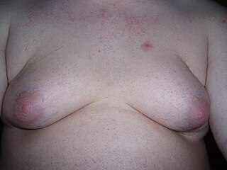
Adipomastia, also known colloquially as fatty breasts, is a condition defined as an excess of skin and/or a flat layer of adipose tissue in the breasts without true gynecomastia. It is commonly present in men with obesity, and is particularly apparent in men who have undergone massive weight loss. A related/synonymous term is pseudogynecomastia. The condition is different and should be distinguished from gynecomastia, which involves female-like protruding fat tissue and/or glandular tissue in a male. The two conditions can usually be distinguished easily by palpation to check for the presence of glandular tissue. Another difference between the conditions is that breast pain/tenderness does not occur in pseudogynecomastia. Sometimes, gynecomastia and pseudogynecomastia are present together; this is related to the fact that fat tissue expresses aromatase, the enzyme responsible for the synthesis of estrogen, and estrogen is produced to a disproportionate extent in men with excessive amounts of fat, resulting in simultaneous glandular enlargement.

Canthotomy is a surgical procedure where the lateral canthus, or corner, of the eye is cut to relieve the fluid pressure inside or behind the eye, known as intraocular pressure (IOC). The procedure is typically done in emergency situations when the intraocular pressure becomes too high, which can damage the optic nerve and lead to blindness if left untreated.
