Related Research Articles
A subluxation is an incomplete or partial dislocation of a joint or organ. According to the World Health Organization, a subluxation is a "significant structural displacement" and is therefore visible on static imaging studies, such as X-rays. Unlike real subluxations, the pseudoscientific concept of a chiropractic "vertebral subluxation" may or may not be visible on x-rays.

A bone fracture is a medical condition in which there is a partial or complete break in the continuity of any bone in the body. In more severe cases, the bone may be broken into several fragments, known as a comminuted fracture. A bone fracture may be the result of high force impact or stress, or a minimal trauma injury as a result of certain medical conditions that weaken the bones, such as osteoporosis, osteopenia, bone cancer, or osteogenesis imperfecta, where the fracture is then properly termed a pathologic fracture.

A joint dislocation, also called luxation, occurs when there is an abnormal separation in the joint, where two or more bones meet. A partial dislocation is referred to as a subluxation. Dislocations are often caused by sudden trauma on the joint like an impact or fall. A joint dislocation can cause damage to the surrounding ligaments, tendons, muscles, and nerves. Dislocations can occur in any major joint or minor joint. The most common joint dislocation is a shoulder dislocation.

A cervical fracture, commonly called a broken neck, is a fracture of any of the seven cervical vertebrae in the neck. Examples of common causes in humans are traffic collisions and diving into shallow water. Abnormal movement of neck bones or pieces of bone can cause a spinal cord injury, resulting in loss of sensation, paralysis, or usually death soon thereafter, primarily via compromising neurological supply to the respiratory muscles as well as innervation to the heart.

A distal radius fracture, also known as wrist fracture, is a break of the part of the radius bone which is close to the wrist. Symptoms include pain, bruising, and rapid-onset swelling. The ulna bone may also be broken.

A Lisfranc injury, also known as Lisfranc fracture, is an injury of the foot in which one or more of the metatarsal bones are displaced from the tarsus.

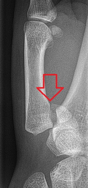
The Galeazzi fracture is a fracture of the distal third of the radius with dislocation of the distal radioulnar joint. It classically involves an isolated fracture of the junction of the distal third and middle third of the radius with associated subluxation or dislocation of the distal radio-ulnar joint; the injury disrupts the forearm axis joint.

A hip dislocation is when the thighbone (femur) separates from the hip bone (pelvis). Specifically it is when the ball–shaped head of the femur separates from its cup–shaped socket in the hip bone, known as the acetabulum. The joint of the femur and pelvis is very stable, secured by both bony and soft-tissue constraints. With that, dislocation would require significant force which typically results from significant trauma such as from a motor vehicle collision or from a fall from elevation. Hip dislocations can also occur following a hip replacement or from a developmental abnormality known as hip dysplasia.

The occipital condyles are undersurface protuberances of the occipital bone in vertebrates, which function in articulation with the superior facets of the atlas vertebra.
Atlanto-occipital dislocation, orthopedic decapitation, or internal decapitation describes ligamentous separation of the spinal column from the skull base. It is possible for a human to survive such an injury; however, 70% of cases result in immediate death. It should not be confused with atlanto-axial dislocation, which describes ligamentous separation between the first and second cervical vertebra.
Bennett fracture is a type of partial broken finger involving the base of the thumb, and extends into the carpometacarpal (CMC) joint.

A patellar dislocation is a knee injury in which the patella (kneecap) slips out of its normal position. Often the knee is partly bent, painful and swollen. The patella is also often felt and seen out of place. Complications may include a patella fracture or arthritis.
The Bosworth fracture is a rare fracture of the distal fibula with an associated fixed posterior dislocation of the proximal fibular fragment which becomes trapped behind the posterior tibial tubercle. The injury is caused by severe external rotation of the ankle. The ankle remains externally rotated after the injury, making interpretation of X-rays difficult which can lead to misdiagnosis and incorrect treatment. The injury is most commonly treated by open reduction internal fixation as closed reduction is made difficult by the entrapment of the fibula behind the tibia.
David Marsh Bosworth was an American orthopedic surgeon and medical educator. He is remembered for describing the Bosworth fracture.
Sir Frank Wild Holdsworth was an English orthopaedic surgeon remembered for pioneering work on rehabilitation of spinal injury patients. He described the Holdsworth fracture of the spine in 1963.
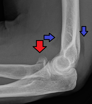
The Hume fracture is an injury of the elbow comprising a fracture of the olecranon with an associated anterior dislocation of the radial head which occurs in children. It was originally described as an undisplaced olecranon fracture, but more recently includes displaced fractures and can be considered a variant of the Monteggia fracture.
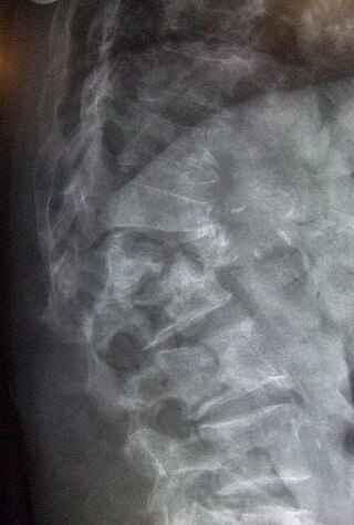
A spinal fracture, also called a vertebral fracture or a broken back, is a fracture affecting the vertebrae of the spinal column. Most types of spinal fracture confer a significant risk of spinal cord injury. After the immediate trauma, there is a risk of spinal cord injury if the fracture is unstable, that is, likely to change alignment without internal or external fixation.

Dislocations occur when two bones that originally met at the joint detach. Dislocations should not be confused with subluxation. Subluxation is when the joint is still partially attached to the bone.
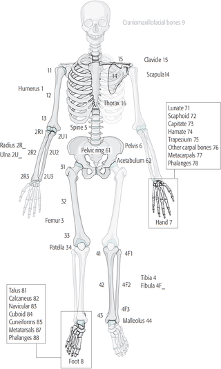
The Müller AO Classification of fractures is a system for classifying bone fractures initially published in 1987 by the AO Foundation as a method of categorizing injuries according to therognosis of the patient's anatomical and functional outcome. "AO" is an initialism for the German "Arbeitsgemeinschaft für Osteosynthesefragen", the predecessor of the AO Foundation.
Radial head fractures are a common type of elbow fracture that typically occurs after a fall on an outstretched arm. They account for approximately one third of all elbow fractures and are frequently associated with other injuries of the elbow. Radial head fractures are diagnosed by a clinical assessment and medical imaging. A radial head fracture is treated according to the severity of the injury and its Mason-Johnston classification. Treatment may be surgical or nonsurgical. Stable isolated fractures typically have excellent outcomes. Unstable fractures with other associated injuries have varying outcomes. Common adverse outcomes include stiffness, pain, poor bone healing, and hardware complications.
References
- ↑ Tim B Hunter; Leonard F Peltier; Pamela J Lund (2000). "Musculoskeletal Eponyms: Who Are Those Guys?". RadioGraphics. 20 (3): 819–36. doi:10.1148/radiographics.20.3.g00ma20819. PMID 10835130.
- ↑ Holdsworth FW (February 1963). "Fractures, dislocations, and fracture-dislocations of the spine". J Bone Joint Surg Br. 45-B (1): 6–20. doi:10.1302/0301-620X.45B1.6. Archived from the original on 2007-12-05. Retrieved 2009-11-05.