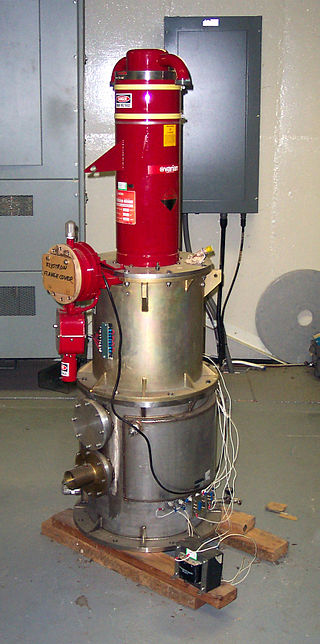
A photoinjector[1] is a type of source for intense electron beams which relies on the photoelectric effect. A laser pulse incident onto the cathode of a photoinjector drives electrons out of it, and into the accelerating field of the electron gun.[2] In comparison with the widespread thermionicelectron gun, photoinjectors produce electron beams of higher brightness, which means more particles packed into smaller volume of phase space (beam emittance). Photoinjectors serve as the main electron source for single-pass synchrotron light sources, such as free-electron lasers[3] and for ultrafast electron diffraction setups.[4] The first RF photoinjector was developed in 1985 at Los Alamos National Laboratory and used as the source for a free-electron-laser experiment.[5][6] High-brightness electron beams produced by photoinjectors are used directly or indirectly to probe the molecular, atomic and nuclear structure of matter for fundamental research, as well as material characterization.
A photoinjector comprises a photocathode, electron gun (AC or DC), power supplies, driving laser system, timing and synchronization system, emittance compensation magnets. It can include vacuum system and cathode fabrication or transport system. It is usually followed by beam diagnostics and higher-energy accelerators.
Animation showing how an AC linear accelerator works. For a photoinjector, the source S is the cathode inside the fractional cell, followed by four booster cells of RF field.
The key component of a photoinjector is a photocathode, which is located inside the cavity of electron gun (usually, a 0.6-fractional cell for optimal distribution of accelerating field). Extracted electron beam suffers from its own space-charge fields that deteriorate the beam brightness. For that reason, photoelectron guns often have one or more full-size booster cells to increase the beam energy and reduce the space-charge effect. The gun's accelerating field is RF (radio-frequency) wave provided by a klystron or other RF power source. For low-energy beams, such as ones used in electron diffraction and microscopy, electrostatic acceleration (DC) is a suitable.
The photoemission on the cathode is initiated by an incident pulse from the driving laser. Depending on the material of the photocathode, the laser wavelength can vary from 1700nm (infrared) down to 100-200nm (ultraviolet). Emission from the cavity wall is possible with laser wavelength of about 250nm for copper walls or cathodes. Semiconductor cathodes are often sensitive to ambient conditions and might require a clean preparation chamber located behind the photoelectron gun. The optical system of the driving laser is often designed to control the pulse structure, and consequently, the distribution of electrons in the extracted bunch. For example, a fs-scale laser pulse with an elliptical transverse profile creates a thin "pancake" electron bunch, that evolves into a uniformly filled ellipsoid under its own space-charge fields.[7] A more sophisticated laser pulse with a comb-like longitudinal profile generates a similarly shaped, comb electron beam.[8][9]
Notes
↑ I. Ben-Zvi, photoinjectors, in A. W. Chao, H.O. Moser and Z. Zhao, Editors, "Accelerator Physics and Technology Applications" World Scientific, Singapore, 2004, pp. 158-175
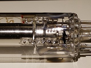
Photomultiplier tubes (photomultipliers or PMTs for short) are extremely sensitive detectors of light in the ultraviolet, visible, and near-infrared ranges of the electromagnetic spectrum. They are members of the class of vacuum tubes, more specifically vacuum phototubes. These detectors multiply the current produced by incident light by as much as 100 million times or 108 (i.e., 160 dB), in multiple dynode stages, enabling (for example) individual photons to be detected when the incident flux of light is low.
A klystron is a specialized linear-beam vacuum tube, invented in 1937 by American electrical engineers Russell and Sigurd Varian, which is used as an amplifier for high radio frequencies, from UHF up into the microwave range. Low-power klystrons are used as oscillators in terrestrial microwave relay communications links, while high-power klystrons are used as output tubes in UHF television transmitters, satellite communication, radar transmitters, and to generate the drive power for modern particle accelerators.
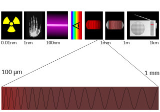
Terahertz radiation – also known as submillimeter radiation, terahertz waves, tremendously high frequency (THF), T-rays, T-waves, T-light, T-lux or THz – consists of electromagnetic waves within the ITU-designated band of frequencies from 0.3 to 3 terahertz (THz), although the upper boundary is somewhat arbitrary and is considered by some sources as 30 THz. One terahertz is 1012 Hz or 1000 GHz. Wavelengths of radiation in the terahertz band correspondingly range from 1 mm to 0.1 mm = 100 µm. Because terahertz radiation begins at a wavelength of around 1 millimeter and proceeds into shorter wavelengths, it is sometimes known as the submillimeter band, and its radiation as submillimeter waves, especially in astronomy. This band of electromagnetic radiation lies within the transition region between microwave and far infrared, and can be regarded as either.
A synchrotron light source is a source of electromagnetic radiation (EM) usually produced by a storage ring, for scientific and technical purposes. First observed in synchrotrons, synchrotron light is now produced by storage rings and other specialized particle accelerators, typically accelerating electrons. Once the high-energy electron beam has been generated, it is directed into auxiliary components such as bending magnets and insertion devices in storage rings and free electron lasers. These supply the strong magnetic fields perpendicular to the beam that are needed to stimulate the high energy electrons to emit photons.
A photocathode is a surface engineered to convert light (photons) into electrons using the photoelectric effect. Photocathodes are important in accelerator physics where they are utilised in a photoinjector to generate high brightness electron beams. Electron beams generated with photocathodes are commonly used for free electron lasers and for ultrafast electron diffraction. Photocathodes are also commonly used as the negatively charged electrode in a light detection device such as a photomultiplier, phototube and image intensifier.
An electron gun is an electrical component in some vacuum tubes that produces a narrow, collimated electron beam that has a precise kinetic energy.
Photoemission electron microscopy is a type of electron microscopy that utilizes local variations in electron emission to generate image contrast. The excitation is usually produced by ultraviolet light, synchrotron radiation or X-ray sources. PEEM measures the coefficient indirectly by collecting the emitted secondary electrons generated in the electron cascade that follows the creation of the primary core hole in the absorption process. PEEM is a surface sensitive technique because the emitted electrons originate from a shallow layer. In physics, this technique is referred to as PEEM, which goes together naturally with low-energy electron diffraction (LEED), and low-energy electron microscopy (LEEM). In biology, it is called photoelectron microscopy (PEM), which fits with photoelectron spectroscopy (PES), transmission electron microscopy (TEM), and scanning electron microscopy (SEM).
A free-electron laser (FEL) is a fourth generation light source producing extremely brilliant and short pulses of radiation. An FEL functions much as a laser but employs relativistic electrons as a gain medium instead of using stimulated emission from atomic or molecular excitations. In an FEL, a bunch of electrons passes through a magnetic structure called an undulator or wiggler to generate radiation, which re-interacts with the electrons to make them emit coherently, exponentially increasing its intensity.
The Smith–Purcell effect was the precursor of the free-electron laser (FEL). It was studied by Steve Smith, a graduate student under the guidance of Edward Purcell. In their experiment, they sent an energetic beam of electrons very closely parallel to the surface of a ruled optical diffraction grating, and thereby generated visible light. Smith showed there was negligible effect on the trajectory of the inducing electrons. Essentially, this is a form of Cherenkov radiation where the phase velocity of the light has been altered by the periodic grating. However, unlike Cherenkov radiation, there is no minimum or threshold particle velocity.
In physics, terahertz time-domain spectroscopy (THz-TDS) is a spectroscopic technique in which the properties of matter are probed with short pulses of terahertz radiation. The generation and detection scheme is sensitive to the sample's effect on both the amplitude and the phase of the terahertz radiation.
A particle beam is a stream of charged or neutral particles. In particle accelerators, these particles can move with a velocity close to the speed of light. There is a difference between the creation and control of charged particle beams and neutral particle beams, as only the first type can be manipulated to a sufficient extent by devices based on electromagnetism. The manipulation and diagnostics of charged particle beams at high kinetic energies using particle accelerators are main topics of accelerator physics.
The AWAKE facility at CERN is a proof-of-principle experiment, which investigates wakefield plasma acceleration using a proton bunch as a driver, a world-wide first. It aims to accelerate a low-energy witness bunch of electrons from 15 to 20 MeV to several GeV over a short distance by creating a high acceleration gradient of several GV/m. Particle accelerators currently in use, like CERN's LHC, use standard or superconductive RF-cavities for acceleration, but they are limited to an acceleration gradient in the order of 100 MV/m.
Extreme ultraviolet radiation or high-energy ultraviolet radiation is electromagnetic radiation in the part of the electromagnetic spectrum spanning wavelengths shorter that the hydrogen Lyman-alpha line from 121 nm down to the X-ray band of 10 nm, and therefore having photons with energies from 10.26 eV up to 124.24 eV. EUV is naturally generated by the solar corona and artificially by plasma, high harmonic generation sources and synchrotron light sources. Since UVC extends to 100 nm, there is some overlap in the terms.
Accelerators and Lasers In Combined Experiments (ALICE), or Energy Recovery Linac Prototype (ERLP) is a 35MeV energy recovery linac test facility at Daresbury Laboratory in Cheshire, England. The project was originally conceived as a test bed for the 4th Generation Light Source (4GLS), and consists of:
ASTRID2 is a synchrotron light source at the Department of Physics and Astronomy of Aarhus University. ASTRID2 was designed, constructed and is operated by the Centre for Storage Ring Facilities in Aarhus (ISA).
The European X-Ray Free-Electron Laser Facility is an X-ray research laser facility commissioned during 2017. The first laser pulses were produced in May 2017 and the facility started user operation in September 2017. The international project with twelve participating countries; nine shareholders at the time of commissioning, later joined by three other partners, is located in the German federal states of Hamburg and Schleswig-Holstein. A free-electron laser generates high-intensity electromagnetic radiation by accelerating electrons to relativistic speeds and directing them through special magnetic structures. The European XFEL is constructed such that the electrons produce X-ray light in synchronisation, resulting in high-intensity X-ray pulses with the properties of laser light and at intensities much brighter than those produced by conventional synchrotron light sources.
A particle accelerator is a machine that uses electromagnetic fields to propel charged particles to very high speeds and energies, and to contain them in well-defined beams.
An energy recovery linac (ERL) is a type of linear particle accelerator that provides a beam of electrons used to produce x-rays by synchrotron radiation. First proposed in 1965 the idea gained interest since the early 2000s.
Ultrafast electron diffraction (UED), also known as femtosecond electron diffraction (FED), is a pump-probe experimental method based on the combination of optical pump-probe spectroscopy and electron diffraction. UED provides information on the dynamical changes of the structure of materials. It is very similar to time resolved crystallography, but instead of using X-rays as the probe, it uses electrons. In the UED technique, a femtosecond (fs) laser optical pulse excites (pumps) a sample into an excited, usually non-equilibrium, state. The pump pulse may induce chemical, electronic or structural transitions. After a finite time interval, a fs electron pulse is incident upon the sample. The electron pulse undergoes diffraction as a result of interacting with the sample. The diffraction signal is, subsequently, detected by an electron counting instrument such as a CCD camera. Specifically, after the electron pulse diffracts from the sample, the scattered electrons will form a diffraction pattern (image) on a CCD camera. This pattern contains structural information about the sample. By adjusting the time difference between the arrival of the pump and probe beams, one can obtain a series of diffraction patterns as a function of the various time differences. The diffraction data series can be concatenated in order to produce a motion picture of the changes that occurred in the data. UED can provide a wealth of dynamics on charge carriers, atoms, and molecules.
In accelerator physics, the mean transverse energy (MTE) is a quantity that describes the variance of the transverse momentum of a beam. While the quantity has a defined value for any particle beam, it is generally used in the context of photoinjectors for electron beams.
This page is based on this Wikipedia article Text is available under the CC BY-SA 4.0 license; additional terms may apply. Images, videos and audio are available under their respective licenses.