Synchrotron light source

Last updated
Synchrotron radiation reflecting from a terbium crystal at the Daresbury Synchrotron Radiation Source, 1990 SynchrotronLight.jpg
Synchrotron radiation reflecting from a terbium crystal at the Daresbury Synchrotron Radiation Source, 1990

A synchrotron light source is a source of electromagnetic radiation (EM) usually produced by a storage ring, [1] for scientific and technical purposes. First observed in synchrotrons, synchrotron light is now produced by storage rings and other specialized particle accelerators, typically accelerating electrons. Once the high-energy electron beam has been generated, it is directed into auxiliary components such as bending magnets and insertion devices (undulators or wigglers) in storage rings and free electron lasers. These supply the strong magnetic fields perpendicular to the beam that are needed to stimulate the high energy electrons to emit photons.

Contents

The major applications of synchrotron light are in condensed matter physics, materials science, biology and medicine. A large fraction of experiments using synchrotron light involve probing the structure of matter from the sub-nanometer level of electronic structure to the micrometer and millimeter levels important in medical imaging. An example of a practical industrial application is the manufacturing of microstructures by the LIGA process.

Synchrotron is one of the most expensive kinds of light source known, but it is practically the only viable luminous source of wide-band radiation in far infrared wavelength range for some applications, such as far-infrared absorption spectrometry.

Spectral brightness

The primary figure of merit used to compare different sources of synchrotron radiation has been referred to as the "brightness", the "brilliance", and the "spectral brightness", with the latter term being recommended as the best choice by the Working Group on Synchrotron Nomenclature. [2] Regardless of the name chosen, the term is a measure of the total flux of photons in a given six-dimensional phase space per unit bandwidth (BW). [3]

The spectral brightness is given by

where is the number of photons per second in the beam, and are the root mean square values for the size of the beam in the axes perpendicular to the beam direction, and are the RMS values for the beam solid angle in the x and y dimensions, and is the relative bandwidth, or spread in beam frequency around the central frequency. [4] The customary value for bandwidth is 0.1%. [2]

Spectral brightness has units of time−1distance−2angle−2(% bandwidth)−1.

Properties of sources

Especially when artificially produced, synchrotron radiation is notable for its:

Synchrotron radiation from accelerators

Synchrotron radiation may occur in accelerators either as a nuisance, causing undesired energy loss in particle physics contexts, or as a deliberately produced radiation source for numerous laboratory applications. Electrons are accelerated to high speeds in several stages to achieve a final energy that is typically in the gigaelectronvolt range. The electrons are forced to travel in a closed path by strong magnetic fields. This is similar to a radio antenna, but with the difference that the relativistic speed changes the observed frequency due to the Doppler effect by a factor . Relativistic Lorentz contraction bumps the frequency by another factor of , thus multiplying the gigahertz frequency of the resonant cavity that accelerates the electrons into the X-ray range. Another dramatic effect of relativity is that the radiation pattern is distorted from the isotropic dipole pattern expected from non-relativistic theory into an extremely forward-pointing cone of radiation. This makes synchrotron radiation sources the most brilliant known sources of X-rays. The planar acceleration geometry makes the radiation linearly polarized when observed in the orbital plane, and circularly polarized when observed at a small angle to that plane.[ citation needed ]

The advantages of using synchrotron radiation for spectroscopy and diffraction have been realized by an ever-growing scientific community, beginning in the 1960s and 1970s. In the beginning, accelerators were built for particle physics, and synchrotron radiation was used in "parasitic mode" when bending magnet radiation had to be extracted by drilling extra holes in the beam pipes. The first storage ring commissioned as a synchrotron light source was Tantalus, at the Synchrotron Radiation Center, first operational in 1968. [5] As accelerator synchrotron radiation became more intense and its applications more promising, devices that enhanced the intensity of synchrotron radiation were built into existing rings. Third-generation synchrotron radiation sources were conceived and optimized from the outset to produce brilliant X-rays. Fourth-generation sources that will include different concepts for producing ultrabrilliant, pulsed time-structured X-rays for extremely demanding and also probably yet-to-be-conceived experiments are under consideration.[ citation needed ]

Bending electromagnets in accelerators were first used to generate this radiation, but to generate stronger radiation, other specialized devices – insertion devices – are sometimes employed. Current (third-generation) synchrotron radiation sources are typically reliant upon these insertion devices, where straight sections of the storage ring incorporate periodic magnetic structures (comprising many magnets in a pattern of alternating N and S poles – see diagram above) which force the electrons into a sinusoidal or helical path. Thus, instead of a single bend, many tens or hundreds of "wiggles" at precisely calculated positions add up or multiply the total intensity of the beam.[ citation needed ]

These devices are called wigglers or undulators. The main difference between an undulator and a wiggler is the intensity of their magnetic field and the amplitude of the deviation from the straight line path of the electrons.[ citation needed ]

There are openings in the storage ring to let the radiation exit and follow a beam line into the experimenters' vacuum chamber. A great number of such beamlines can emerge from modern third-generation synchrotron radiation sources.[ citation needed ]

Storage rings

The electrons may be extracted from the accelerator proper and stored in an ultrahigh vacuum auxiliary magnetic storage ring where they may circle a large number of times. The magnets in the ring also need to repeatedly recompress the beam against Coulomb (space charge) forces tending to disrupt the electron bunches. The change of direction is a form of acceleration and thus the electrons emit radiation at GeV energies.[ citation needed ]

Applications of synchrotron radiation

Beamlines

Beamlines of Soleil Schema de principe du synchrotron.jpg
Beamlines of Soleil

At a synchrotron facility, electrons are usually accelerated by a synchrotron, and then injected into a storage ring, in which they circulate, producing synchrotron radiation, but without gaining further energy. The radiation is projected at a tangent to the electron storage ring and captured by beamlines. These beamlines may originate at bending magnets, which mark the corners of the storage ring; or insertion devices, which are located in the straight sections of the storage ring. The spectrum and energy of X-rays differ between the two types. The beamline includes X-ray optical devices which control the bandwidth, photon flux, beam dimensions, focus, and collimation of the rays. The optical devices include slits, attenuators, crystal monochromators, and mirrors. The mirrors may be bent into curves or toroidal shapes to focus the beam. A high photon flux in a small area is the most common requirement of a beamline. The design of the beamline will vary with the application. At the end of the beamline is the experimental end station, where samples are placed in the line of the radiation, and detectors are positioned to measure the resulting diffraction, scattering or secondary radiation.

Experimental techniques and usage

Synchrotron light is an ideal tool for many types of research in materials science, physics, and chemistry and is used by researchers from academic, industrial, and government laboratories. Several methods take advantage of the high intensity, tunable wavelength, collimation, and polarization of synchrotron radiation at beamlines which are designed for specific kinds of experiments. The high intensity and penetrating power of synchrotron X-rays enables experiments to be performed inside sample cells designed for specific environments. Samples may be heated, cooled, or exposed to gas, liquid, or high pressure environments. Experiments which use these environments are called in situ and allow the characterization of atomic- to nano-scale phenomena which are inaccessible to most other characterization tools. In operando measurements are designed to mimic the real working conditions of a material as closely as possible. [8]

Diffraction and scattering

X-ray diffraction (XRD) and scattering experiments are performed at synchrotrons for the structural analysis of crystalline and amorphous materials. These measurements may be performed on powders, single crystals, or thin films. The high resolution and intensity of the synchrotron beam enables the measurement of scattering from dilute phases or the analysis of residual stress. Materials can be studied at high pressure using diamond anvil cells to simulate extreme geologic environments or to create exotic forms of matter.[ citation needed ]

Structure of a ribosome subunit solved at high resolution using synchrotron X-ray crystallography. 010 large subunit-1FFK.gif
Structure of a ribosome subunit solved at high resolution using synchrotron X-ray crystallography.

X-ray crystallography of proteins and other macromolecules (PX or MX) are routinely performed. Synchrotron-based crystallography experiments were integral to solving the structure of the ribosome; [9] [10] this work earned the Nobel Prize in Chemistry in 2009.

The size and shape of nanoparticles are characterized using small angle X-ray scattering (SAXS). Nano-sized features on surfaces are measured with a similar technique, grazing-incidence small angle X-ray scattering (GISAXS). [11] In this and other methods, surface sensitivity is achieved by placing the crystal surface at a small angle relative to the incident beam, which achieves total external reflection and minimizes the X-ray penetration into the material.[ citation needed ]

The atomic- to nano-scale details of surfaces, interfaces, and thin films can be characterized using techniques such as X-ray reflectivity (XRR) and crystal truncation rod (CTR) analysis. [12] X-ray standing wave (XSW) measurements can also be used to measure the position of atoms at or near surfaces; these measurements require high-resolution optics capable of resolving dynamical diffraction phenomena. [13]

Amorphous materials, including liquids and melts, as well as crystalline materials with local disorder, can be examined using X-ray pair distribution function analysis, which requires high energy X-ray scattering data. [14]

By tuning the beam energy through the absorption edge of a particular element of interest, the scattering from atoms of that element will be modified. These so-called resonant anomalous X-ray scattering methods can help to resolve scattering contributions from specific elements in the sample.[ citation needed ]

Other scattering techniques include energy dispersive X-ray diffraction, resonant inelastic X-ray scattering, and magnetic scattering.[ citation needed ]

Spectroscopy

X-ray absorption spectroscopy (XAS) is used to study the coordination structure of atoms in materials and molecules. The synchrotron beam energy is tuned through the absorption edge of an element of interest, and modulations in the absorption are measured. Photoelectron transitions cause modulations near the absorption edge, and analysis of these modulations (called the X-ray absorption near-edge structure (XANES) or near-edge X-ray absorption fine structure (NEXAFS)) reveals information about the chemical state and local symmetry of that element. At incident beam energies which are much higher than the absorption edge, photoelectron scattering causes "ringing" modulations called the extended X-ray absorption fine structure (EXAFS). Fourier transformation of the EXAFS regime yields the bond lengths and number of the surrounding the absorbing atom; it is therefore useful for studying liquids and amorphous materials [15] as well as sparse species such as impurities. A related technique, X-ray magnetic circular dichroism (XMCD), uses circularly polarized X-rays to measure the magnetic properties of an element.[ citation needed ]

X-ray photoelectron spectroscopy (XPS) can be performed at beamlines equipped with a photoelectron analyzer. Traditional XPS is typically limited to probing the top few nanometers of a material under vacuum. However, the high intensity of synchrotron light enables XPS measurements of surfaces at near-ambient pressures of gas. Ambient pressure XPS (AP-XPS) can be used to measure chemical phenomena under simulated catalytic or liquid conditions. [16] Using high-energy photons yields high kinetic energy photoelectrons which have a much longer inelastic mean free path than those generated on a laboratory XPS instrument. The probing depth of synchrotron XPS can therefore be lengthened to several nanometers, allowing the study of buried interfaces. This method is referred to as high-energy X-ray photoemission spectroscopy (HAXPES). [17] Furthermore, the tunable nature of the synchrotron X-ray photon energies presents a wide range of depth sensitivity in the order of 2-50 nm. [18] This allows for probing of samples at greater depths and for non destructive depth-profiling experiments.

Material composition can be quantitatively analyzed using X-ray fluorescence (XRF). XRF detection is also used in several other techniques, such as XAS and XSW, in which it is necessary to measure the change in absorption of a particular element.[ citation needed ]

Other spectroscopy techniques include angle resolved photoemission spectroscopy (ARPES), soft X-ray emission spectroscopy, and nuclear resonance vibrational spectroscopy, which is related to Mössbauer spectroscopy.[ citation needed ]

Imaging

X-ray nanoprobe beamline at the Advanced Photon Source APS - Nanoprobe.jpg
X-ray nanoprobe beamline at the Advanced Photon Source

Synchrotron X-rays can be used for traditional X-ray imaging, phase-contrast X-ray imaging, and tomography. The Ångström-scale wavelength of X-rays enables imaging well below the diffraction limit of visible light, but practically the smallest resolution so far achieved is about 30 nm. [19] Such nanoprobe sources are used for scanning transmission X-ray microscopy (STXM). Imaging can be combined with spectroscopy such as X-ray fluorescence or X-ray absorption spectroscopy in order to map a sample's chemical composition or oxidation state with sub-micron resolution. [20]

Other imaging techniques include coherent diffraction imaging.[ citation needed ]

Similar optics can be employed for photolithography for MEMS structures can use a synchrotron beam as part of the LIGA process.[ citation needed ]

Compact synchrotron light sources

Because of the usefulness of tuneable collimated coherent X-ray radiation, efforts have been made to make smaller more economical sources of the light produced by synchrotrons. The aim is to make such sources available within a research laboratory for cost and convenience reasons; at present, researchers have to travel to a facility to perform experiments. One method of making a compact light source is to use the energy shift from Compton scattering near-visible laser photons from electrons stored at relatively low energies of tens of megaelectronvolts (see for example the Compact Light Source (CLS) [21] ). However, a relatively low cross-section of collision can be obtained in this manner, and the repetition rate of the lasers is limited to a few hertz rather than the megahertz repetition rates naturally arising in normal storage ring emission. Another method is to use plasma acceleration to reduce the distance required to accelerate electrons from rest to the energies required for UV or X-ray emission within magnetic devices.[ citation needed ]

See also

Related Research Articles

<span class="mw-page-title-main">X-ray photoelectron spectroscopy</span> Spectroscopic technique

X-ray photoelectron spectroscopy (XPS) is a surface-sensitive quantitative spectroscopic technique based on the photoelectric effect that can identify the elements that exist within a material or are covering its surface, as well as their chemical state, and the overall electronic structure and density of the electronic states in the material. XPS is a powerful measurement technique because it not only shows what elements are present, but also what other elements they are bonded to. The technique can be used in line profiling of the elemental composition across the surface, or in depth profiling when paired with ion-beam etching. It is often applied to study chemical processes in the materials in their as-received state or after cleavage, scraping, exposure to heat, reactive gasses or solutions, ultraviolet light, or during ion implantation.

<span class="mw-page-title-main">Beamline</span> Trajectory of a beam of accelerated particles

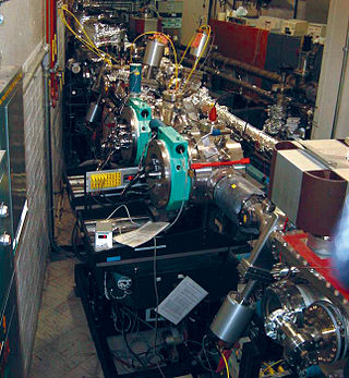
In accelerator physics, a beamline refers to the trajectory of the beam of particles, including the overall construction of the path segment along a specific path of an accelerator facility. This part is either

<span class="mw-page-title-main">Synchrotron</span> Type of cyclic particle accelerator

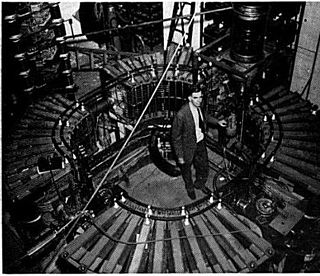
A synchrotron is a particular type of cyclic particle accelerator, descended from the cyclotron, in which the accelerating particle beam travels around a fixed closed-loop path. The magnetic field which bends the particle beam into its closed path increases with time during the accelerating process, being synchronized to the increasing kinetic energy of the particles. The synchrotron is one of the first accelerator concepts to enable the construction of large-scale facilities, since bending, beam focusing and acceleration can be separated into different components. The most powerful modern particle accelerators use versions of the synchrotron design. The largest synchrotron-type accelerator, also the largest particle accelerator in the world, is the 27-kilometre-circumference (17 mi) Large Hadron Collider (LHC) near Geneva, Switzerland, built in 2008 by the European Organization for Nuclear Research (CERN). It can accelerate beams of protons to an energy of 7 tera electronvolts (TeV or 1012 eV).

<span class="mw-page-title-main">Extended X-ray absorption fine structure</span> Measurement of X-ray absorption of a material as a function of energy

Extended X-ray absorption fine structure (EXAFS), along with X-ray absorption near edge structure (XANES), is a subset of X-ray absorption spectroscopy (XAS). Like other absorption spectroscopies, XAS techniques follow Beer's law. The X-ray absorption coefficient of a material as a function of energy is obtained by directing X-rays of a narrow energy range at a sample, while recording the incident and transmitted x-ray intensity, as the incident x-ray energy is incremented.

<span class="mw-page-title-main">Stanford Synchrotron Radiation Lightsource</span> Research center at Stanford University

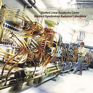
The Stanford Synchrotron Radiation Lightsource, a division of SLAC National Accelerator Laboratory, is operated by Stanford University for the Department of Energy. SSRL is a National User Facility which provides synchrotron radiation, a name given to electromagnetic radiation in the x-ray, ultraviolet, visible and infrared realms produced by electrons circulating in a storage ring at nearly the speed of light. The extremely bright light that is produced can be used to investigate various forms of matter ranging from objects of atomic and molecular size to man-made materials with unusual properties. The obtained information and knowledge is of great value to society, with impact in areas such as the environment, future technologies, health, biology, basic research, and education.

<span class="mw-page-title-main">Electron scattering</span> Deviation of electrons from their original trajectories

Electron scattering occurs when electrons are displaced from their original trajectory. This is due to the electrostatic forces within matter interaction or, if an external magnetic field is present, the electron may be deflected by the Lorentz force. This scattering typically happens with solids such as metals, semiconductors and insulators; and is a limiting factor in integrated circuits and transistors.

The National Synchrotron Light Source (NSLS) at Brookhaven National Laboratory (BNL) in Upton, New York was a national user research facility funded by the U.S. Department of Energy (DOE). Built from 1978 through 1984, and officially shut down on September 30, 2014, the NSLS was considered a second-generation synchrotron.

High-energy X-rays or HEX-rays are very hard X-rays, with typical energies of 80–1000 keV (1 MeV), about one order of magnitude higher than conventional X-rays used for X-ray crystallography. They are produced at modern synchrotron radiation sources such as the Cornell High Energy Synchrotron Source, SPring-8, and the beamlines ID15 and BM18 at the European Synchrotron Radiation Facility (ESRF). The main benefit is the deep penetration into matter which makes them a probe for thick samples in physics and materials science and permits an in-air sample environment and operation. Scattering angles are small and diffraction directed forward allows for simple detector setups.

<span class="mw-page-title-main">Advanced Light Source</span> Synchrotron radiation facility

The Advanced Light Source (ALS) is a research facility at Lawrence Berkeley National Laboratory in Berkeley, California. One of the world's brightest sources of ultraviolet and soft x-ray light, the ALS is the first "third-generation" synchrotron light source in its energy range, providing multiple extremely bright sources of intense and coherent short-wavelength light for use in scientific experiments by researchers from around the world. It is funded by the US Department of Energy (DOE) and operated by the University of California. The current director is Dimitri Argyriou.

<span class="mw-page-title-main">ALBA (synchrotron)</span>

ALBA is a 3 GeV, third-generation synchrotron light source facility located in the Barcelona Synchrotron Park in Cerdanyola del Vallès near Barcelona, in Catalonia (Spain). It was constructed and is operated by CELLS, and co-financed by the Spanish central administration and regional Catalan Government.

The Australian Synchrotron is a 3 GeV national synchrotron radiation facility located in Clayton, in the south-eastern suburbs of Melbourne, Victoria. The facility opened in 2007, and is operated by the Australian Nuclear Science and Technology Organisation.

X-ray absorption near edge structure (XANES), also known as near edge X-ray absorption fine structure (NEXAFS), is a type of absorption spectroscopy that indicates the features in the X-ray absorption spectra (XAS) of condensed matter due to the photoabsorption cross section for electronic transitions from an atomic core level to final states in the energy region of 50–100 eV above the selected atomic core level ionization energy, where the wavelength of the photoelectron is larger than the interatomic distance between the absorbing atom and its first neighbour atoms.

<span class="mw-page-title-main">Synchrotron Radiation Center</span>

The Synchrotron Radiation Center (SRC), located in Stoughton, Wisconsin and operated by the University of Wisconsin–Madison, was a national synchrotron light source research facility, operating the Aladdin storage ring. From 1968 to 1987 SRC was the home of Tantalus, the first storage ring dedicated to the production of synchrotron radiation.

<span class="mw-page-title-main">European XFEL</span>

The European X-Ray Free-Electron Laser Facility is an X-ray research laser facility commissioned during 2017. The first laser pulses were produced in May 2017 and the facility started user operation in September 2017. The international project with twelve participating countries; nine shareholders at the time of commissioning, later joined by three other partners, is located in the German federal states of Hamburg and Schleswig-Holstein. A free-electron laser generates high-intensity electromagnetic radiation by accelerating electrons to relativistic speeds and directing them through special magnetic structures. The European XFEL is constructed such that the electrons produce X-ray light in synchronisation, resulting in high-intensity X-ray pulses with the properties of laser light and at intensities much brighter than those produced by conventional synchrotron light sources.

Laser-based angle-resolved photoemission spectroscopy is a form of angle-resolved photoemission spectroscopy that uses a laser as the light source. Photoemission spectroscopy is a powerful and sensitive experimental technique to study surface physics. It is based on the photoelectric effect originally observed by Heinrich Hertz in 1887 and later explained by Albert Einstein in 1905 that when a material is shone by light, the electrons can absorb photons and escape from the material with the kinetic energy: , where is the incident photon energy, the work function of the material. Since the kinetic energy of ejected electrons are highly associated with the internal electronic structure, by analyzing the photoelectron spectroscopy one can realize the fundamental physical and chemical properties of the material, such as the type and arrangement of local bonding, electronic structure and chemical composition.

ANKA is a synchrotron light source facility at the Karlsruhe Institute of Technology (KIT). The KIT runs ANKA as a national synchrotron light source and as a large scale user facility for the international science community. Being a large scale machine of the performance category LK II of the Helmholtz Association, ANKA is part of a national and European infrastructure offering research services to scientific and commercial users for their purposes in research and development. The facility was opened to external users in 2003.

<span class="mw-page-title-main">National Synchrotron Light Source II</span> Research and Development Facility in New York, United States

The National Synchrotron Light Source II (NSLS-II) at Brookhaven National Laboratory (BNL) in Upton, New York is a national user research facility funded primarily by the U.S. Department of Energy's (DOE) Office of Science. NSLS-II is one of the world's most advanced synchrotron light sources, designed to produce x-rays 10,000 times brighter than BNL's original light source, the National Synchrotron Light Source (NSLS). NSLS-II supports basic and applied research in energy security, advanced materials synthesis and manufacturing, environment, and human health.

<span class="mw-page-title-main">Solaris (synchrotron)</span>

SOLARIS is the only synchrotron in Central-Eastern Europe. Built in Poland in 2015, under the auspices of the Jagiellonian University, it is located on the Campus of the 600th Anniversary of the Jagiellonian University Revival, in the southern part of Kraków. It is the central facility of the National Synchrotron Radiation Centre SOLARIS.

<span class="mw-page-title-main">X-ray emission spectroscopy</span>

X-ray emission spectroscopy (XES) is a form of X-ray spectroscopy in which a core electron is excited by an incident x-ray photon and then this excited state decays by emitting an x-ray photon to fill the core hole. The energy of the emitted photon is the energy difference between the involved electronic levels. The analysis of the energy dependence of the emitted photons is the aim of the X-ray emission spectroscopy.

References

  1. Handbook on Synchrotron Radiation, Volume 1a, Ernst-Eckhard Koch, Ed., North Holland, 1983, reprinted at "Synchrotron Radiation Turns the Big Five-O Archived September 16, 2008, at the Wayback Machine
  2. 1 2 Mills, D. M.; Helliwell, J. R.; Kvick, Å.; Ohta, T.; Robinson, I. A.; Authier, A. (1 May 2005). "Report of the Working Group on Synchrotron Radiation Nomenclature – brightness, spectral brightness or brilliance?". Journal of Synchrotron Radiation. 12 (3): 385. Bibcode:2005JSynR..12..385M. doi: 10.1107/S090904950500796X . PMID   15840926 . Retrieved 8 April 2022.
  3. Nielsen, Jens (2011). Elements of modern X-ray physics. Chichester, West Sussex: John Wiley. ISBN   9781119970156.
  4. Wiedemann, Helmut (2007). Particle accelerator physics (3rd ed.). Berlin: Springer. p. 782. ISBN   9783540490432.
  5. E. M. Rowe and F. E. Mills, Tantalus I: A Dedicated Storage Ring Synchrotron Radiation Source, Particle Accelerators, Vol. 4 (1973); pages 211-227.
  6. A. A. Sokolov; I. M. Ternov (1986). C. W. Kilmister (ed.). Radiation from Relativistic Electrons. Translation Series. New York: American Institute of Physics. ISBN   978-0-88318-507-0.
  7. The Physics of Electron Storage Rings: An Introduction by Matt Sands Archived 2015-05-11 at the Wayback Machine
  8. Nelson, Johanna; Misra, Sumohan; Yang, Yuan; Jackson, Ariel; Liu, Yijin; et al. (2012-03-30). "In Operando X-ray Diffraction and Transmission X-ray Microscopy of Lithium Sulfur Batteries". Journal of the American Chemical Society. 134 (14): 6337–6343. doi:10.1021/ja2121926. PMID   22432568.
  9. 1 2 Ban, N.; Nissen, P.; Hansen, J.; Moore, P.; Steitz, T. (2000-08-11). "The Complete Atomic Structure of the Large Ribosomal Subunit at 2.4 Å Resolution". Science. 289 (5481): 905–920. Bibcode:2000Sci...289..905B. doi:10.1126/science.289.5481.905. PMID   10937989.
  10. The Royal Swedish Academy of Sciences, "The Nobel Prize in Chemistry 2009: Information for the Public", accessed 2016-06-20
  11. Renaud, Gilles; Lazzari, Rémi; Leroy, Frédéric (2009). "Probing surface and interface morphology with Grazing Incidence Small Angle X-Ray Scattering". Surface Science Reports. 64 (8): 255–380. Bibcode:2009SurSR..64..255R. doi:10.1016/j.surfrep.2009.07.002.
  12. Robinson, I K; Tweet, D J (1992-05-01). "Surface X-ray diffraction". Reports on Progress in Physics. 55 (5): 599–651. Bibcode:1992RPPh...55..599R. doi:10.1088/0034-4885/55/5/002. S2CID   250899816.
  13. Golovchenko, J. A.; Patel, J. R.; Kaplan, D. R.; Cowan, P. L.; Bedzyk, M. J. (1982-08-23). "Solution to the Surface Registration Problem Using X-Ray Standing Waves" (PDF). Physical Review Letters. 49 (8): 560–563. Bibcode:1982PhRvL..49..560G. doi:10.1103/physrevlett.49.560.
  14. T. Egami, S.J.L. Billinge, "Underneath the Bragg Peaks: Structural Analysis of Complex Materials", Pergamon (2003)
  15. Sayers, Dale E.; Stern, Edward A.; Lytle, Farrel W. (1971-11-01). "New Technique for Investigating Noncrystalline Structures: Fourier Analysis of the Extended X-Ray—Absorption Fine Structure". Physical Review Letters. 27 (18): 1204–1207. Bibcode:1971PhRvL..27.1204S. doi:10.1103/physrevlett.27.1204.
  16. Bluhm, Hendrik; Hävecker, Michael; Knop-Gericke, Axel; Kiskinova, Maya; Schlögl, Robert; Salmeron, Miquel (2017). "In Situ X-Ray Photoelectron Spectroscopy Studies of Gas-Solid Interfaces at Near-Ambient Conditions". MRS Bulletin. 32 (12): 1022–1030. doi:10.1557/mrs2007.211. OSTI   927255. S2CID   55577979.
  17. Sing, M.; Berner, G.; Goß, K.; Müller, A.; Ruff, A.; Wetscherek, A.; Thiel, S.; Mannhart, J.; Pauli, S. A.; Schneider, C. W.; Willmott, P. R.; Gorgoi, M.; Schäfers, F.; Claessen, R. (2009-04-30). "Profiling the Interface Electron Gas of LaAlO3/SrTiO3 Heterostructures with Hard X-Ray Photoelectron Spectroscopy". Physical Review Letters. 102 (17): 176805. arXiv: 0809.1917 . Bibcode:2009PhRvL.102q6805S. doi:10.1103/physrevlett.102.176805. PMID   19518810. S2CID   43739895.
  18. Gong, Zhengliang; Yang, Yong (2018). "The application of synchrotron X-ray techniques to the study of rechargeable batteries". Journal of Energy Chemistry. 27 (6): 1566–1583. doi:10.1016/j.jechem.2018.03.020. S2CID   104038441.
  19. Argonne National Laboratory Center for Nanoscale Materials, "X-Ray Microscopy Capabilities", accessed 2016-06-20
  20. Beale, Andrew M.; Jacques, Simon D. M.; Weckhuysen, Bert M. (2010). "Chemical imaging of catalytic solids with synchrotron radiation". Chemical Society Reviews. 39 (12): 4656–4672. doi:10.1039/c0cs00089b. hdl: 1874/290865 . PMID   20978688.
  21. "Miniature synchrotron produces first light". Eurekalert.org. Retrieved 2009-10-19.