Resonant inelastic X-ray scattering

Last updated
Cartoon of the RIXS experiment. A photon with energy
E
i
n
{\displaystyle E_{in}}
and momentum
k
i
n
{\displaystyle {\textbf {k}}_{in}}
impinges on the sample and another photon with energy
E
o
u
t
{\displaystyle E_{out}}
and momentum
k
o
u
t
{\displaystyle {\textbf {k}}_{out}}
leaves it. The conservation laws of energy and momentum are also highlighted, being ho and q respectively the energy and momentum transferred to the sample. Rixs cartoon.png
Cartoon of the RIXS experiment. A photon with energy and momentum impinges on the sample and another photon with energy and momentum leaves it. The conservation laws of energy and momentum are also highlighted, being ħω and q respectively the energy and momentum transferred to the sample.

Resonant inelastic X-ray scattering (RIXS) is an advanced X-ray spectroscopy technique. [1] [2]

Contents

In the last two decades RIXS has been widely exploited to study the electronic, magnetic and structural properties of quantum materials and molecules. It is a resonant X-rays photon-in photon-out energy loss and momentum resolved spectroscopy, capable of measuring the energy and momentum transferred to specific excitations proper of the sample under study. [1] [2]

The use of X-rays guarantees bulk sensitivity, as opposed to electron spectroscopies, and the tuning of the incoming X-rays to a specific absorption edge allows for element and chemical specificity. [1] [2] [3]

Due to the intrinsic inefficiency of the RIXS process, extremely brilliant sources of X-rays are crucial. In addition to that, the possibility to tune the energy of the incoming X-rays is compelling to match a chosen resonance. These two strict conditions make RIXS to be necessarily performed at synchrotrons or nowadays at X-ray free electron lasers (XFELs) and set the advent of third generation synchrotrons (1994, ESRF [4] ) as a turning point for the success of the technique. [1] [2]

Exploiting different experimental setups, RIXS can be performed using both soft and hard X-rays, spanning a vast range of absorption edges and thus samples to be studied. [1]

RIXS process

RIXS is a two steps process. First an electron is resonantly excited from a core level, defined by the absorption edge, to an empty state, leaving a core hole. The intermediate state with the core hole has a lifetime of few femtoseconds, then the system radiatively decays into the final state with the filling of the core hole and the emission of another photon. Since the probability of a radiative core hole relaxation is low, the RIXS cross section is very small and a high brilliance X-ray source is needed. Being a second order process, the RIXS cross section is described by the Kramers-Heisenberg formula. [1] [5]

The scattering geometry (incidence and scattering angles) determines the momentum transfer . In order to explore the space the spectrometer angle with respect to the incoming beam can be changed, as well as the incident angle to the sample. [1] [5]

The RIXS process can be classified as either direct or indirect. This distinction is useful because the cross-sections for each are quite different. When direct scattering is allowed, it will be the dominant scattering channel, with indirect processes contributing only in higher order. In contrast, for the large class of experiments for which direct scattering is forbidden, RIXS relies exclusively on indirect scattering channels. [1] [5]

Direct RIXS

Direct RIXS process. The incoming X-rays excite an electron from a deep-lying core level into the empty valence. The empty core state is subsequently filled by an electron from the occupied states under the emission of an X-ray. This RIXS process creates a valence excitation with momentum
k
'
-
k
{\displaystyle k'-k}
and energy

o
-

o
'
{\displaystyle \hbar \omega -\hbar \omega '}
. Direct RIXS process.jpg
Direct RIXS process. The incoming X-rays excite an electron from a deep-lying core level into the empty valence. The empty core state is subsequently filled by an electron from the occupied states under the emission of an X-ray. This RIXS process creates a valence excitation with momentum and energy .

In direct RIXS, the incoming photon promotes a core-electron to an empty valence band state. Subsequently, an electron from a different state decays and annihilates the core-hole. The hole in the final state may either be in a core level at lower binding energy than in the intermediate state or in the filled valence shell. Some authors refer to this technique as resonant X-ray emission spectroscopy (RXES). The distinction between RIXS, resonant X-ray Raman and RXES in the literature is not strict. [2]

The net result is a final state with an electron-hole excitation, as an electron was created in an empty valence band state and a hole in a filled shell. If the hole is in the filled valence shell, the electron-hole excitation can propagate through the material, carrying away momentum and energy. Momentum and energy conservation require that these are equal to the momentum and energy loss of the scattered photon. [1]

For direct RIXS to occur, both photoelectric transitions—the initial one from core to valence state and succeeding one to fill the core hole—must be possible. These transitions can for instance be an initial dipolar transition of 1s → 2p followed by the decay of another electron in the 2p band from 2p → 1s. This happens at the K-edge of oxygen, carbon and silicon. Very efficient sequence often used in 3d transition metals are a 1s → 3d excitation followed by a 2p → 1s decay. [6]

Indirect RIXS

Indirect RIXS process. An electron is excited from a deep-lying core level into the valence shell. Excitations are created through the Coulomb interaction
U
c
{\displaystyle U_{c}}
between the core hole (and in some cases the excited electron) and the valence electrons. Indirect RIXS process.jpg
Indirect RIXS process. An electron is excited from a deep-lying core level into the valence shell. Excitations are created through the Coulomb interaction between the core hole (and in some cases the excited electron) and the valence electrons.

Indirect RIXS is slightly more complicated. Here, the incoming photon promotes a core-electron to an itinerant state far above the electronic chemical potential. Subsequently, the electron in this same state decays again, filling the core-hole. Scattering of the X-rays occurs via the core-hole potential that is present in the intermediate state. It shakes up the electronic system, creating excitations to which the X-ray photon loses energy and momentum. [7] [8] [9] The number of electrons in the valence sub-system is constant throughout the process. [5] [10] [11]

Experimental details

In general the natural linewidth of a spectral feature is determined by the life-times of initial and final states. Indeed, as for X-ray absorption and non-resonant X-ray emission spectroscopy the energy resolution is often limited by the relatively short life-time of the final state core-hole. As in RIXS a high energy core-hole is absent in the final state, this leads to intrinsically sharp spectra with energy and momentum resolution determined by the instrumentation. [3] [2] [1] [12]

A convolution of the incident X-ray bandpass, defined by the beamline monochromator, and the bandpass of the RIXS spectrometer for the analysis of the scattered photons energy gives the total (combined) energy resolution. Since RIXS exploits high energy photons in the X-ray range, a very large combined resolving power (103-105 depending on the goal of the experiment) is needed to detail the different spectral features. Therefore, in the last two decades efforts have been made to improve RIXS spectrometers performances, gaining orders of magnitude in terms of resolving power. [13] State of the art soft X-rays RIXS beamlines in use at the ESRF, at DLS and at NSLS II, have reached approximately 40000 of combined resolving power, leading to a record energy resolution of 25 meV at Cu L3 edge. [14] [15] [16]

As for hard X-rays, the optical design is different and requires the use of Bragg reflection crystal analyzers. Thus, the resolving power is mostly determined by the crystal analyzers in use. [17] [18]

Soft X-ray spectrometers

Schematic layout of a varied line spacing (VLS) spherical grating RIXS spectrometer. Optical layout RIXS spectrometer.png
Schematic layout of a varied line spacing (VLS) spherical grating RIXS spectrometer.
The ESRF ID32 soft X-rays RIXS spectrometer. ESRF soft RIXS spectrometer.png
The ESRF ID32 soft X-rays RIXS spectrometer.
The Diamond Light Source I21 RIXS spectrometer DLS RIXS spectrometer.jpg
The Diamond Light Source I21 RIXS spectrometer

State of the art soft X-ray RIXS spectrometers are based on grazing incidence diffraction gratings, to disperse the X-rays scattered from the sample, and on position sensitive detectors, mostly CCDs. The two-dimensional image shows a vertical dispersive direction and a non-dispersive one. Integrating along the non-dispersive direction one can obtain a spectrum. [13] [14] [15] [16] [19]

RIXS 2D image on a CCD and the corresponding spectrum in energy loss. The red arrow indicates the dispersive direction. Ccd and RIXS spectrum NiO.png
RIXS 2D image on a CCD and the corresponding spectrum in energy loss. The red arrow indicates the dispersive direction.

The whole optical path from the source to the CCD must be kept in UHV to minimize the absorption of X-rays by air. [20] The number of optical elements is typically minimized, which is important for a number of reasons. Indeed, the low reflectivity of optical elements for X-rays reduces the throughput. In addition to that, a non-negligible contribution to the combined resolving power is due to the imperfections on the surface of mirrors and gratings (slope error). Finally, the lower the number of optical elements to be aligned, the better in terms of setup time. [13] [14] [15] [16] [19]

The monochromatized X-rays impinge on the sample with a defined geometry and are scattered and collected by the spectrometer. Collection mirrors are often placed after the sample, the distance (1 cm to 1 m) depends on the optical design. This is useful to increase the acceptance angle of the spectrometer and thus the efficiency. [13] [14] [15] [16]

After the collecting optics X-rays are dispersed by the varied line spacing (VLS) grating that can be either plane or spherical. In the former case, a vertical focusing mirror is added to the optical path to focus the X-rays on the detector, in the latter the grating itself also focuses the dispersed X-rays on the CCD detector. Depending on the absorption edge chosen for the experiment, the respective positions between the grating and the detector, and the incidence angle of the grating can be tuned to optimize the spectrometer in a large energy window, without changing any optical element. [13] [14] [15] [16]

Since the spectral analysis of the scattered X-rays is done through a dispersive grating, longer spectrometers offer higher resolving power. State of the art spectrometers are more than ten meters long, more than five times the dimensions of the pioneering ones. Two examples from ESRF and DLS are in the figures. [14] [15] [16]

Hard X-ray spectrometers

Rowland circle geometry for hard X-rays RIXS experiments. Rowland circle geometry.png
Rowland circle geometry for hard X-rays RIXS experiments.
The hard X-rays RIXS spectrometer of the ID20 beamline of the ESRF. ID20 ESRF RIXS spectrometer.jpg
The hard X-rays RIXS spectrometer of the ID20 beamline of the ESRF.

The optical layout for hard X-rays RIXS spectrometers is different. The spectrometers are based on spherical crystal analyzers (typically more than one to increase the solid angle of the spectrometer) exploiting Bragg reflections and on a position sensitive detector, typically in the so called Rowland geometry. This means that the source (X-rays spot on the sample), the analyzers and the detector must sit on the Rowland circle. By scanning the positions of the analyzers and of the detector (the source is fixed for convenience) the Bragg condition is changed and thus the energy of the scattered X-rays can be analyzed. By increasing the radius of the Rowland circle, the energy resolution can be increased, loosing in terms of efficiency. Nevertheless, as opposed to soft X-rays spectrometers, the resolving power of the spectrometer is limited by the crystal analyzers. Thus, increasing too much the dimensions of the spectrometer does not pay off. [18] [22] [23]

Depending on the chosen absorption edge (and thus incidence energy), different crystal analyzers are used both on the monochromator side and on the spectrometer side. Thanks to the high penetration depth of hard X-rays, there is no need of UHV. Therefore, the exchange of optical elements, such as crystal analyzers, is less disruptive than for soft X-rays. [17] [18] [22] [23]

One of the major technical challenges in these RIXS experiments is selecting the monochromator and energy analyzer which produce, at the desired energy, the desired resolution. Some of the feasible crystal monochromator reflections and energy analyzer reflections have been tabulated. [24] [25]

RIXS properties

Compared to other inelastic scattering techniques as INS, IXS, EELS or Raman scattering that present shortcomings, RIXS has a number of unique features: it covers a large scattering phase-space thanks to the high energy photons, it is polarization dependent, element specific, bulk sensitive and requires only small sample volumes enabling studies on thin films as well as diluted solutions. RIXS is a resonant technique because the energy of the incident photon is chosen such that it coincides with, and hence resonates with, one of the atomic X-ray absorption edges of the system. The resonance greatly enhances the valence contribution to the inelastic scattering cross section, sometimes by many orders of magnitude. [3] [2] [1] [26]

Comparing the energy of a neutron, electron or photon with a wavelength of the order of the relevant length scale in a solid - as given by the de Broglie equation considering the interatomic lattice spacing is in the order of Ångströms - it derives from the relativistic energy–momentum relation that an X-ray photon has more energy than a neutron or electron. The scattering phase space (the range of energies and momenta that can be transferred in a scattering event) of X-rays is therefore without equal. In particular, high-energy X-rays carry a momentum that is comparable to the inverse lattice spacing of typical condensed matter systems so that, unlike Raman scattering experiments with visible or infrared light, RIXS can probe the full dispersion of low energy excitations in solids. [1] [2] [3]

RIXS can utilize the polarization of the photon: the nature of the excitations created in the material can be disentangled by a polarization analysis of the incident and scattered photons, which allow one, through the use of various selection rules, to characterize the symmetry and nature of the excitations. [1] [2] [3]

RIXS is element specific: chemical sensitivity arises by tuning to the absorption edges of the different types of elements in a material. RIXS can even differentiate between the same chemical element at sites with different valencies or at inequivalent crystallographic positions as long as the X-ray absorption edges in these cases are distinguishable. In addition, the type of information on the electronic excitations of a system being probed can be varied by tuning to different X-ray edges (e.g., K, L or M) of the same chemical element, where the photon excites core-electrons into different valence orbitals. [1] [2] [3]

RIXS is bulk sensitive: the penetration depth of resonant X-ray photons depends on the material and on the scattering geometry, but typically is of the order of a few micrometers in the hard X-rays regime (for example at transition metal K-edges) and on the order of 0.1 micrometers in the soft X-ray regime (e.g. transition metal L-edges). [1] [2] [3]

RIXS needs only small sample volumes: the photon-matter interaction is relatively strong, compared to for instance to the neutron-matter interaction strength. This makes RIXS feasible on very small volume samples, thin films, surfaces and nano-objects, in addition to bulk single crystal, powder samples or diluted solutions. [1] [2] [3]

RIXS spectral features

Elementary excitations that can be measured by RIXS. The indicated energy scales are the ones relevant for transition metal oxides. RIXS excitations.jpg
Elementary excitations that can be measured by RIXS. The indicated energy scales are the ones relevant for transition metal oxides.

In principle RIXS can probe a very broad class of intrinsic excitations of the system under study, as long as the excitations are overall charge neutral. This constraint arises from the fact that in RIXS the scattered photons do not add or remove charge from the sample. [1]

Starting from the low energy loss part of the spectrum, RIXS has a purely elastic response, which hosts both a diffused elastic signal, but also any kind of order proper of the system, as charge density waves. [1] [27] [28] [29] [30]

In the low-energy window, the signal is dominated by phonons and vibrational modes that are present in a RIXS spectrum through the electron-phonon coupling. Only a portion of phonons modes that characterize the sample are visible through RIXS. [1] [31] [32] [33]

Electron-hole continuum and excitons in band metals, doped systems and semiconductors are visible through RIXS, thanks to the enhancement of valence charge excitations guaranteed by the resonance character of the technique. [1] [34]

In the charge channel, also plasmons and their dispersion can be measured by RIXS, [1] [35] [36] [37] as well as orbital and crystal field excitations [38] [39] and charge transfer excitations. [1]

Spin excitations are symmetry-allowed in RIXS as well. In particular, RIXS at L and M edges, thanks to the resonant character, also spin flip excitations (magnons) can be accessed with RIXS, exploiting the spin-orbit coupling of the core level involved in the RIXS process. This makes RIXS as the paramount technique to study magnon dispersions, thanks to the higher cross-section with respect to INS. Besides magnons, RIXS can probe bi-magnons and spinons. [1] [40] [39] [41]

Moreover, it has been theoretically shown that RIXS can probe Bogoliubov quasiparticles in high-temperature superconductors, [42] and shed light on the nature and symmetry of the electron-electron pairing of the superconducting state. [43]

Pump-probe RIXS with X-ray free electron lasers (XFELs)

Cartoon of the pump-probe spectroscopy approach. The sample is first prepared in an excited state by a laser pulse and then probed by an X-ray pulse. Pump probe x ray spectroscopy.png
Cartoon of the pump-probe spectroscopy approach. The sample is first prepared in an excited state by a laser pulse and then probed by an X-ray pulse.

With the advent of XFELs, sources that can provide extremely brilliant (more than five orders of magnitude larger than synchrotron sources) and extremely short X-ray pulses, X-ray spectroscopies performed in a pump and probe fashion are nowadays available. [44] [45]

The power of pump-probe spectroscopies lies in the possibility to study how a system evolves after an external stimulus. The most straightforward example is the study of photoactivated biological process, such as the photosynthesis: the sample is illuminated by an optical laser tuned at the proper wavelength and then its evolution is observed taking snapshots as a function of time. [44] [45]

The development of high-resolution RIXS spectrometers at XFELs is opening a new field, exploiting the power of RIXS to study the photo-induced transient states in quantum materials and photoactivated processes in molecules. [46] [47] [48] [49] [50]

Applications

See also

Related Research Articles

<span class="mw-page-title-main">Spectroscopy</span> Study involving matter and electromagnetic radiation

Spectroscopy is the field of study that measures and interprets electromagnetic spectra. In narrower contexts, spectroscopy is the precise study of color as generalized from visible light to all bands of the electromagnetic spectrum.

<span class="mw-page-title-main">Photoluminescence</span> Light emission from substances after they absorb photons

Photoluminescence is light emission from any form of matter after the absorption of photons. It is one of many forms of luminescence and is initiated by photoexcitation, hence the prefix photo-. Following excitation, various relaxation processes typically occur in which other photons are re-radiated. Time periods between absorption and emission may vary: ranging from short femtosecond-regime for emission involving free-carrier plasma in inorganic semiconductors up to milliseconds for phosphoresence processes in molecular systems; and under special circumstances delay of emission may even span to minutes or hours.

<span class="mw-page-title-main">Electron energy loss spectroscopy</span> Form of microscopy using an electron beam

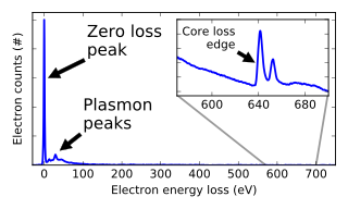
Electron energy loss spectroscopy (EELS) is a form of electron microscopy in which a material is exposed to a beam of electrons with a known, narrow range of kinetic energies. Some of the electrons will undergo inelastic scattering, which means that they lose energy and have their paths slightly and randomly deflected. The amount of energy loss can be measured via an electron spectrometer and interpreted in terms of what caused the energy loss. Inelastic interactions include phonon excitations, inter- and intra-band transitions, plasmon excitations, inner shell ionizations, and Cherenkov radiation. The inner-shell ionizations are particularly useful for detecting the elemental components of a material. For example, one might find that a larger-than-expected number of electrons comes through the material with 285 eV less energy than they had when they entered the material. This is approximately the amount of energy needed to remove an inner-shell electron from a carbon atom, which can be taken as evidence that there is a significant amount of carbon present in the sample. With some care, and looking at a wide range of energy losses, one can determine the types of atoms, and the numbers of atoms of each type, being struck by the beam. The scattering angle can also be measured, giving information about the dispersion relation of whatever material excitation caused the inelastic scattering.

<span class="mw-page-title-main">High-temperature superconductivity</span> Superconductive behavior at temperatures much higher than absolute zero

High-temperature superconductors are defined as materials with critical temperature above 77 K, the boiling point of liquid nitrogen. They are only "high-temperature" relative to previously known superconductors, which function at even colder temperatures, close to absolute zero. The "high temperatures" are still far below ambient, and therefore require cooling. The first break through of high-temperature superconductor was discovered in 1986 by IBM researchers Georg Bednorz and K. Alex Müller. Although the critical temperature is around 35.1 K, this new type of superconductor was readily modified by Ching-Wu Chu to make the first high-temperature superconductor with critical temperature 93 K. Bednorz and Müller were awarded the Nobel Prize in Physics in 1987 "for their important break-through in the discovery of superconductivity in ceramic materials". Most high-Tc materials are type-II superconductors.

<span class="mw-page-title-main">Fermi liquid theory</span> Theoretical model in physics

Fermi liquid theory is a theoretical model of interacting fermions that describes the normal state of the conduction electrons in most metals at sufficiently low temperatures. The theory describes the behavior of many-body systems of particles in which the interactions between particles may be strong. The phenomenological theory of Fermi liquids was introduced by the Soviet physicist Lev Davidovich Landau in 1956, and later developed by Alexei Abrikosov and Isaak Khalatnikov using diagrammatic perturbation theory. The theory explains why some of the properties of an interacting fermion system are very similar to those of the ideal Fermi gas, and why other properties differ.

<span class="mw-page-title-main">Raman scattering</span> Inelastic scattering of photons by matter

In physics, Raman scattering or the Raman effect is the inelastic scattering of photons by matter, meaning that there is both an exchange of energy and a change in the light's direction. Typically this effect involves vibrational energy being gained by a molecule as incident photons from a visible laser are shifted to lower energy. This is called normal Stokes-Raman scattering.

<span class="mw-page-title-main">Two-photon physics</span> Branch of particle physics concerning interactions between two photons

Two-photon physics, also called gamma–gamma physics, is a branch of particle physics that describes the interactions between two photons. Normally, beams of light pass through each other unperturbed. Inside an optical material, and if the intensity of the beams is high enough, the beams may affect each other through a variety of non-linear effects. In pure vacuum, some weak scattering of light by light exists as well. Also, above some threshold of this center-of-mass energy of the system of the two photons, matter can be created.

<span class="mw-page-title-main">Fano resonance</span> Type of scattering resonance

In physics, a Fano resonance is a type of resonant scattering phenomenon that gives rise to an asymmetric line-shape. Interference between a background and a resonant scattering process produces the asymmetric line-shape. It is named after Italian-American physicist Ugo Fano, who in 1961 gave a theoretical explanation for the scattering line-shape of inelastic scattering of electrons from helium; however, Ettore Majorana was the first to discover this phenomenon. Fano resonance is a weak coupling effect meaning that the decay rate is so high, that no hybridization occurs. The coupling modifies the resonance properties such as spectral position and width and its line-shape takes on the distinctive asymmetric Fano profile. Because it is a general wave phenomenon, examples can be found across many areas of physics and engineering.

<span class="mw-page-title-main">X-ray absorption spectroscopy</span> Panel of different types of X-ray absorption spectroscopy requiring a synchrotron radiation facility

X-ray absorption spectroscopy (XAS) is a widely used technique for determining the local geometric and/or electronic structure of matter. The experiment is usually performed at synchrotron radiation facilities, which provide intense and tunable X-ray beams. Samples can be in the gas phase, solutions, or solids.

Strongly correlated materials are a wide class of compounds that include insulators and electronic materials, and show unusual electronic and magnetic properties, such as metal-insulator transitions, heavy fermion behavior, half-metallicity, and spin-charge separation. The essential feature that defines these materials is that the behavior of their electrons or spinons cannot be described effectively in terms of non-interacting entities. Theoretical models of the electronic (fermionic) structure of strongly correlated materials must include electronic (fermionic) correlation to be accurate. As of recently, the label quantum materials is also used to refer to strongly correlated materials, among others.

X-ray absorption near edge structure (XANES), also known as near edge X-ray absorption fine structure (NEXAFS), is a type of absorption spectroscopy that indicates the features in the X-ray absorption spectra (XAS) of condensed matter due to the photoabsorption cross section for electronic transitions from an atomic core level to final states in the energy region of 50–100 eV above the selected atomic core level ionization energy, where the wavelength of the photoelectron is larger than the interatomic distance between the absorbing atom and its first neighbour atoms.

<span class="mw-page-title-main">Angle-resolved photoemission spectroscopy</span> Experimental technique to determine the distribution of electrons in solids

Angle-resolved photoemission spectroscopy (ARPES) is an experimental technique used in condensed matter physics to probe the allowed energies and momenta of the electrons in a material, usually a crystalline solid. It is based on the photoelectric effect, in which an incoming photon of sufficient energy ejects an electron from the surface of a material. By directly measuring the kinetic energy and emission angle distributions of the emitted photoelectrons, the technique can map the electronic band structure and Fermi surfaces. ARPES is best suited for the study of one- or two-dimensional materials. It has been used by physicists to investigate high-temperature superconductors, graphene, topological materials, quantum well states, and materials exhibiting charge density waves.

Inelastic electron tunneling spectroscopy (IETS) is an experimental tool for studying the vibrations of molecular adsorbates on metal oxides. It yields vibrational spectra of the adsorbates with high resolution (< 0.5 meV) and high sensitivity (< 1013 molecules are required to provide a spectrum). An additional advantage is the fact that optically forbidden transitions may be observed as well. Within IETS, an oxide layer with molecules adsorbed on it is put between two metal plates. A bias voltage is applied between the two contacts. An energy diagram of the metal-oxide-metal device under bias is shown in the top figure. The metal contacts are characterized by a constant density of states, filled up to the Fermi energy. The metals are assumed to be equal. The adsorbates are situated on the oxide material. They are represented by a single bridge electronic level, which is the upper dashed line. If the insulator is thin enough, there is a finite probability that the incident electron tunnels through the barrier. Since the energy of the electron is not changed by this process, it is an elastic process. This is shown in the left figure.

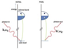
X-ray Raman scattering (XRS) is non-resonant inelastic scattering of X-rays from core electrons. It is analogous to vibrational Raman scattering, which is a widely used tool in optical spectroscopy, with the difference being that the wavelengths of the exciting photons fall in the X-ray regime and the corresponding excitations are from deep core electrons.

Laser-based angle-resolved photoemission spectroscopy is a form of angle-resolved photoemission spectroscopy that uses a laser as the light source. Photoemission spectroscopy is a powerful and sensitive experimental technique to study surface physics. It is based on the photoelectric effect originally observed by Heinrich Hertz in 1887 and later explained by Albert Einstein in 1905 that when a material is shone by light, the electrons can absorb photons and escape from the material with the kinetic energy: , where is the incident photon energy, the work function of the material. Since the kinetic energy of ejected electrons are highly associated with the internal electronic structure, by analyzing the photoelectron spectroscopy one can realize the fundamental physical and chemical properties of the material, such as the type and arrangement of local bonding, electronic structure and chemical composition.

John F. Mitchell is an American chemist and researcher. He is the deputy director of the materials science division at the U.S. Department of Energy's (DOE) Argonne National Laboratory and leads Argonne's Emerging Materials Group.

Electron orbital imaging is an X-ray synchrotron technique used to produce images of electron orbitals in real space. It utilizes the technique of X-ray Raman scattering (XRS), also known as Non-resonant Inelastic X-Ray Scattering (NIXS) to inelastically scatter electrons off a single crystal. It is an element specific spectroscopic technique for studying the valence electrons of transition metals.

<span class="mw-page-title-main">Fabrizio Carbone</span> Italian and Swiss physicist

Fabrizio Carbone is an Italian and Swiss physicist and currently an Associate Professor at École Polytechnique Fédérale de Lausanne (EPFL). His research focuses on the study of matter in out of equilibrium conditions using ultrafast spectroscopy, diffraction and imaging techniques. In 2015, he attracted international attention by publishing a photography of light displaying both its quantum and classical nature.

<span class="mw-page-title-main">Electron-on-helium qubit</span> Quantum bit

An electron-on-helium qubit is a quantum bit for which the orthonormal basis states |0⟩ and |1⟩ are defined by quantized motional states or alternatively the spin states of an electron trapped above the surface of liquid helium. The electron-on-helium qubit was proposed as the basic element for building quantum computers with electrons on helium by Platzman and Dykman in 1999. 

Aron Pinczuk was an Argentine-American experimental condensed matter physicist who was professor of physics and professor of applied physics at Columbia University. He was known for his work on correlated electronic states in two dimensional systems using photoluminescence and resonant inelastic light scattering methods. He was a fellow of the American Physical Society, the American Association for the Advancement of Science and the American Academy of Arts and Sciences.

References

  1. 1 2 3 4 5 6 7 8 9 10 11 12 13 14 15 16 17 18 19 20 21 22 23 24 25 26 Ament, Luuk J. P.; van Veenendaal, Michel; Devereaux, Thomas P.; Hill, John P.; van den Brink, Jeroen (2011-06-24). "Resonant inelastic x-ray scattering studies of elementary excitations". Reviews of Modern Physics. 83 (2): 705–767. arXiv: 1009.3630 . doi:10.1103/RevModPhys.83.705.
  2. 1 2 3 4 5 6 7 8 9 10 11 12 de Groot, Frank; Kotani, Akio (2008-03-10). Core Level Spectroscopy of Solids. CRC Press. pp. 335–435. doi:10.1201/9781420008425. ISBN   978-0-429-19579-2.
  3. 1 2 3 4 5 6 7 8 Schülke, W. (2007). Electron dynamics by inelastic X-ray scattering (1st ed.). Oxford university press. pp. 377–479. ISBN   978-0-19-851017-8.
  4. Raimondi, Pantaleo (2016). "ESRF-EBS: The Extremely Brilliant Source Project". Synchrotron Radiation News. 29 (6): 8–15. doi:10.1080/08940886.2016.1244462. ISSN   0894-0886.
  5. 1 2 3 4 Brink, J. van den; Veenendaal, M. van (2006). "Correlation functions measured by indirect resonant inelastic X-ray scattering". Europhysics Letters (EPL). IOP Publishing. 73 (1): 121–127. Bibcode:2006EL.....73..121V. doi:10.1209/epl/i2005-10366-9. ISSN   0295-5075. S2CID   250849541.
  6. 1 2 Glatzel, Pieter; Bergmann, Uwe; Yano, Junko; Visser, Hendrik; Robblee, John H.; et al. (2004). "The Electronic Structure of Mn in Oxides, Coordination Complexes, and the Oxygen-Evolving Complex of Photosystem II Studied by Resonant Inelastic X-ray Scattering". Journal of the American Chemical Society. American Chemical Society (ACS). 126 (32): 9946–9959. doi:10.1021/ja038579z. ISSN   0002-7863. PMC   3960404 . PMID   15303869.
  7. 1 2 Hasan, M. Z.; Isaacs, E. D.; Shen, Z.-X.; Miller, L. L.; Tsutsui, K.; Tohyama, T.; Maekawa, S. (2000-06-09). "Electronic Structure of Mott Insulators Studied by Inelastic X-ray Scattering". Science. 288 (5472): 1811–1814. arXiv: cond-mat/0102489 . Bibcode:2000Sci...288.1811H. doi:10.1126/science.288.5472.1811. ISSN   0036-8075. PMID   10846160. S2CID   2581764.
  8. 1 2 Hasan, M. Z.; Isaacs, E. D.; Shen, Z. -X.; Miller, L. L. (2001-03-01). "Inelastic X-ray scattering as a novel tool to study electronic excitations in complex insulators". Journal of Electron Spectroscopy and Related Phenomena. Proceeding of the Eight International Conference on Electronic Spectroscopy and Structure. 114–116: 705–709. doi:10.1016/S0368-2048(00)00401-1. ISSN   0368-2048.
  9. 1 2 Hasan, M. Z.; Isaacs, E. D.; Shen, Z-X.; Miller, L. L. (2000-11-01). "Particle-hole excitations in insulating antiferromagnet Ca2CuO2Cl2". Physica C: Superconductivity. 341–348: 781–782. Bibcode:2000PhyC..341..781H. doi:10.1016/S0921-4534(00)00690-0. ISSN   0921-4534.
  10. Hancock, J N; Chabot-Couture, G; Greven, M (2010-03-03). "Lattice coupling and Franck–Condon effects in K-edge resonant inelastic x-ray scattering". New Journal of Physics. IOP Publishing. 12 (3): 033001. arXiv: 1004.0859 . Bibcode:2010NJPh...12c3001H. doi:10.1088/1367-2630/12/3/033001. ISSN   1367-2630. S2CID   118673022.
  11. Vernay, F.; Moritz, B.; Elfimov, I. S.; Geck, J.; Hawthorn, D.; Devereaux, T. P.; Sawatzky, G. A. (2008-03-18). "CuK-edge resonant inelastic x-ray scattering in edge-sharing cuprates". Physical Review B. 77 (10): 104519. arXiv: cond-mat/0702026 . Bibcode:2008PhRvB..77j4519V. doi:10.1103/physrevb.77.104519. ISSN   1098-0121. S2CID   119045219.
  12. Glatzel, P.; Sikora, M.; Fernández-García, M. (2009). "Resonant X-ray spectroscopy to study K absorption pre-edges in 3d transition metal compounds". The European Physical Journal Special Topics. Springer Science and Business Media LLC. 169 (1): 207–214. Bibcode:2009EPJST.169..207G. doi:10.1140/epjst/e2009-00994-7. ISSN   1951-6355. S2CID   121110109.
  13. 1 2 3 4 5 6 Ghiringhelli, G.; Piazzalunga, A.; Dallera, C.; Trezzi, G.; Braicovich, L.; Schmitt, T.; Strocov, V. N.; Betemps, R.; Patthey, L.; Wang, X.; Grioni, M. (2006-11-01). "SAXES, a high resolution spectrometer for resonant x-ray emission in the 400–1600eV energy range". Review of Scientific Instruments. 77 (11). doi:10.1063/1.2372731. ISSN   0034-6748.
  14. 1 2 3 4 5 6 Brookes, N. B.; Yakhou-Harris, F.; Kummer, K.; Fondacaro, A.; Cezar, J. C.; Betto, D.; Velez-Fort, E.; Amorese, A.; Ghiringhelli, G.; Braicovich, L.; Barrett, R.; Berruyer, G.; Cianciosi, F.; Eybert, L.; Marion, P. (2018-09-21). "The beamline ID32 at the ESRF for soft X-ray high energy resolution resonant inelastic X-ray scattering and polarisation dependent X-ray absorption spectroscopy". Nuclear Instruments and Methods in Physics Research Section A: Accelerators, Spectrometers, Detectors and Associated Equipment. 903: 175–192. doi:10.1016/j.nima.2018.07.001. hdl: 11311/1128322 . ISSN   0168-9002.
  15. 1 2 3 4 5 6 Zhou, K.-J.; Walters, A.; Garcia-Fernandez, M.; Rice, T.; Hand, M.; Nag, A.; Li, J.; Agrestini, S.; Garland, P.; Wang, H.; Alcock, S.; Nistea, I.; Nutter, B.; Rubies, N.; Knap, G. (2022-03-01). "I21: an advanced high-resolution resonant inelastic X-ray scattering beamline at Diamond Light Source". Journal of Synchrotron Radiation. 29 (2): 563–580. doi:10.1107/S1600577522000601. ISSN   1600-5775. PMC   8900866 .
  16. 1 2 3 4 5 6 Dvorak, Joseph; Jarrige, Ignace; Bisogni, Valentina; Coburn, Scott; Leonhardt, William (2016-11-01). "Towards 10 meV resolution: The design of an ultrahigh resolution soft X-ray RIXS spectrometer". Review of Scientific Instruments. 87 (11). doi:10.1063/1.4964847. ISSN   0034-6748.
  17. 1 2 Gog, T.; Casa, D. M.; Knopp, J.; Kim, J.; Upton, M. H.; Krakora, R.; Jaski, A.; Said, A.; Yavaş, H.; Gretarsson, H.; Huang, X. R. (2018-07-01). "Performance of quartz- and sapphire-based double-crystal high-resolution (∼10 meV) RIXS monochromators under varying power loads". Journal of Synchrotron Radiation. 25 (4): 1030–1035. doi:10.1107/S1600577518005945. ISSN   1600-5775.
  18. 1 2 3 Moretti Sala, M.; Martel, K.; Henriquet, C.; Al Zein, A.; Simonelli, L.; Sahle, C.; Gonzalez, H.; Lagier, M.-C.; Ponchut, C.; Huotari, S.; Verbeni, R.; Krisch, M.; Monaco, G. (2018-03-01). "A high-energy-resolution resonant inelastic X-ray scattering spectrometer at ID20 of the European Synchrotron Radiation Facility". Journal of Synchrotron Radiation. 25 (2): 580–591. doi: 10.1107/S1600577518001200 . hdl: 11311/1049071 . ISSN   1600-5775.
  19. 1 2 Dinardo, M. E.; Piazzalunga, A.; Braicovich, L.; Bisogni, V.; Dallera, C.; Giarda, K.; Marcon, M.; Tagliaferri, A.; Ghiringhelli, G. (2007-01-01). "Gaining efficiency and resolution in soft X-ray emission spectrometers thanks to directly illuminated CCD detectors". Nuclear Instruments and Methods in Physics Research Section A: Accelerators, Spectrometers, Detectors and Associated Equipment. 570 (1): 176–181. doi:10.1016/j.nima.2006.10.024. ISSN   0168-9002.
  20. "Physics.nist.gov". Physics.nist.gov. Retrieved 2023-07-06.
  21. Gao, Xing; Gu, Songqi; Gao, Qian; Zou, Yang; Jiang, Zheng; Zhang, Shuo; Wei, Xiangjun; Yu, Haisheng; Sheng, Guodong; Duan, Peiquan; Huang, Yuying (2013). "A high-resolution X-ray fluorescence spectrometer and its application at SSRF: A high-resolution X-ray fluorescence spectrometer and its application". X-Ray Spectrometry. 42 (6): 502–507. doi:10.1002/xrs.2511.
  22. 1 2 Huotari, S.; Albergamo, F.; Vankò, Gy.; Verbeni, R.; Monaco, G. (2006). "Resonant inelastic hard x-ray scattering with diced analyzer crystals and position-sensitive detectors". Review of Scientific Instruments. 77 (5). doi:10.1063/1.2198805.
  23. 1 2 Moretti Sala, M.; Henriquet, C.; Simonelli, L.; Verbeni, R.; Monaco, G. (2013-06-01). "High energy-resolution set-up for Ir L3 edge RIXS experiments". Journal of Electron Spectroscopy and Related Phenomena. Progress in Resonant Inelastic X-Ray Scattering. 188: 150–154. doi:10.1016/j.elspec.2012.08.002. ISSN   0368-2048.
  24. [ "Archived copy". Archived from the original on 2013-02-09. Retrieved 2012-06-06.{{cite web}}: CS1 maint: archived copy as title (link)
  25. "Archived copy". Archived from the original on 2013-02-09. Retrieved 2012-06-06.{{cite web}}: CS1 maint: archived copy as title (link)
  26. Barbiellini, Bernardo; Hancock, Jason N.; Monney, Claude; Joly, Yves; Ghiringhelli, Giacomo; Braicovich, Lucio; Schmitt, Thorsten (2014-06-30). "Inelastic x-ray scattering from valence electrons near absorption edges of FeTe and TiSe2". Physical Review B. 89 (23): 235138. arXiv: 1009.3630 . Bibcode:2014PhRvB..89w5138B. doi:10.1103/PhysRevB.89.235138. S2CID   119231739.
  27. Ghiringhelli, G.; Le Tacon, M.; Minola, M.; Blanco-Canosa, S.; Mazzoli, C.; Brookes, N. B.; De Luca, G. M.; Frano, A.; Hawthorn, D. G.; He, F.; Loew, T.; Sala, M. Moretti; Peets, D. C.; Salluzzo, M.; Schierle, E. (2012-08-17). "Long-Range Incommensurate Charge Fluctuations in (Y,Nd)Ba 2 Cu 3 O 6+ x". Science. 337 (6096): 821–825. arXiv: 1207.0915 . doi:10.1126/science.1223532. ISSN   0036-8075.
  28. Arpaia, R.; Caprara, S.; Fumagalli, R.; De Vecchi, G.; Peng, Y. Y.; Andersson, E.; Betto, D.; De Luca, G. M.; Brookes, N. B.; Lombardi, F.; Salluzzo, M.; Braicovich, L.; Di Castro, C.; Grilli, M.; Ghiringhelli, G. (2019-08-30). "Dynamical charge density fluctuations pervading the phase diagram of a Cu-based high-Tc superconductor". Science. 365 (6456): 906–910. arXiv: 1809.04949 . doi:10.1126/science.aav1315. ISSN   0036-8075.
  29. Comin, Riccardo; Damascelli, Andrea (2016-03-10). "Resonant X-Ray Scattering Studies of Charge Order in Cuprates". Annual Review of Condensed Matter Physics. 7 (1): 369–405. arXiv: 1509.03313 . doi:10.1146/annurev-conmatphys-031115-011401. ISSN   1947-5454.
  30. Arpaia, Riccardo; Ghiringhelli, Giacomo (2021-11-15). "Charge Order at High Temperature in Cuprate Superconductors". Journal of the Physical Society of Japan. 90 (11): 111005. arXiv: 2106.00731 . doi:10.7566/jpsj.90.111005. ISSN   0031-9015.
  31. Devereaux, T. P.; Shvaika, A. M.; Wu, K.; Wohlfeld, K.; Jia, C. J.; Wang, Y.; Moritz, B.; Chaix, L.; Lee, W.-S.; Shen, Z.-X.; Ghiringhelli, G.; Braicovich, L. (2016-10-25). "Directly Characterizing the Relative Strength and Momentum Dependence of Electron-Phonon Coupling Using Resonant Inelastic X-Ray Scattering". Physical Review X. 6 (4). arXiv: 1605.03129 . doi:10.1103/physrevx.6.041019. ISSN   2160-3308.
  32. Rossi, Matteo; Arpaia, Riccardo; Fumagalli, Roberto; Moretti Sala, Marco; Betto, Davide; Kummer, Kurt; De Luca, Gabriella M.; van den Brink, Jeroen; Salluzzo, Marco; Brookes, Nicholas B.; Braicovich, Lucio; Ghiringhelli, Giacomo (2019-07-08). "Experimental Determination of Momentum-Resolved Electron-Phonon Coupling". Physical Review Letters. 123 (2). arXiv: 1902.09163 . doi:10.1103/physrevlett.123.027001. ISSN   0031-9007.
  33. Braicovich, Lucio (2016), "High Resolution Resonant Inelastic X-Ray Scattering from Solids in the Soft Range", Synchrotron Light Sources and Free-Electron Lasers, Cham: Springer International Publishing, pp. 1797–1822, doi:10.1007/978-3-319-14394-1_42 , retrieved 2023-07-06
  34. Jia, Chunjing; Wohlfeld, Krzysztof; Wang, Yao; Moritz, Brian; Devereaux, Thomas P. (2016-05-13). "Using RIXS to Uncover Elementary Charge and Spin Excitations". Physical Review X. 6 (2). arXiv: 1510.05068 . doi:10.1103/physrevx.6.021020. ISSN   2160-3308.
  35. Nag, Abhishek; Zhu, M.; Bejas, Matías; Li, J.; Robarts, H. C.; Yamase, Hiroyuki; Petsch, A. N.; Song, D.; Eisaki, H.; Walters, A. C.; García-Fernández, M.; Greco, Andrés; Hayden, S. M.; Zhou, Ke-Jin (2020-12-14). "Detection of Acoustic Plasmons in Hole-Doped Lanthanum and Bismuth Cuprate Superconductors Using Resonant Inelastic X-Ray Scattering". Physical Review Letters. 125 (25). doi:10.1103/physrevlett.125.257002. hdl: 1983/4135a219-e672-4666-a98d-94d300a4287b . ISSN   0031-9007.
  36. Hepting, M.; Bejas, M.; Nag, A.; Yamase, H.; Coppola, N.; Betto, D.; Falter, C.; Garcia-Fernandez, M.; Agrestini, S.; Zhou, Ke-Jin; Minola, M.; Sacco, C.; Maritato, L.; Orgiani, P.; Wei, H. I. (2022-07-19). "Gapped Collective Charge Excitations and Interlayer Hopping in Cuprate Superconductors". Physical Review Letters. 129 (4). arXiv: 2206.14083 . doi:10.1103/physrevlett.129.047001. ISSN   0031-9007.
  37. Vojta, Matthias; Vojta, Thomas; Kaul, Ribhu K. (2006-08-28). "Spin Excitations in Fluctuating Stripe Phases of Doped Cuprate Superconductors". Physical Review Letters. 97 (9). arXiv: cond-mat/0510448 . doi:10.1103/physrevlett.97.097001. ISSN   0031-9007.
  38. Moretti Sala, M; Bisogni, V; Aruta, C; Balestrino, G; Berger, H; Brookes, N B; Luca, G M de; Di Castro, D; Grioni, M; Guarise, M; Medaglia, P G; Miletto Granozio, F; Minola, M; Perna, P; Radovic, M (2011-04-19). "Energy and symmetry of dd excitations in undoped layered cuprates measured by CuL3resonant inelastic x-ray scattering". New Journal of Physics. 13 (4): 043026. arXiv: 1009.4882 . doi: 10.1088/1367-2630/13/4/043026 . ISSN   1367-2630.
  39. 1 2 3 Schlappa, J.; Wohlfeld, K.; Zhou, K. J.; Mourigal, M.; Haverkort, M. W.; et al. (2012-04-18). "Spin–orbital separation in the quasi-one-dimensional Mott insulator Sr2CuO3". Nature. Springer Science and Business Media LLC. 485 (7396): 82–85. arXiv: 1205.1954 . Bibcode:2012Natur.485...82S. doi:10.1038/nature10974. ISSN   0028-0836. PMID   22522933. S2CID   43990784.
  40. Braicovich, L.; van den Brink, J.; Bisogni, V.; Sala, M. Moretti; Ament, L. J. P.; Brookes, N. B.; De Luca, G. M.; Salluzzo, M.; Schmitt, T.; Strocov, V. N.; Ghiringhelli, G. (2010-02-19). "Magnetic Excitations and Phase Separation in the Underdoped La 2− x Sr x CuO 4 Superconductor Measured by Resonant Inelastic X-Ray Scattering". Physical Review Letters. 104 (7). doi:10.1103/physrevlett.104.077002. ISSN   0031-9007.
  41. Martinelli, Leonardo; Betto, Davide; Kummer, Kurt; Arpaia, Riccardo; Braicovich, Lucio; Di Castro, Daniele; Brookes, Nicholas B.; Moretti Sala, Marco; Ghiringhelli, Giacomo (2022-05-19). "Fractional Spin Excitations in the Infinite-Layer Cuprate CaCuO2". Physical Review X. 12 (2). arXiv: 2110.06666 . doi: 10.1103/physrevx.12.021041 . ISSN   2160-3308.
  42. Marra, Pasquale; Sykora, Steffen; Wohlfeld, Krzysztof; van den Brink, Jeroen (2013). "Resonant Inelastic X-Ray Scattering as a Probe of the Phase and Excitations of the Order Parameter of Superconductors". Physical Review Letters. 110 (11): 117005. arXiv: 1212.0112 . Bibcode:2013PhRvL.110k7005M. doi:10.1103/PhysRevLett.110.117005. ISSN   0031-9007. PMID   25166567. S2CID   21978160.
  43. Marra, Pasquale; van den Brink, Jeroen; Sykora, Steffen (2016-05-06). "Theoretical approach to resonant inelastic x-ray scattering in iron-based superconductors at the energy scale of the superconducting gap". Scientific Reports. 6 (1): 25386. arXiv: 1405.5556 . Bibcode:2016NatSR...625386M. doi: 10.1038/srep25386 . ISSN   2045-2322. PMC   4858731 . PMID   27151253.
  44. 1 2 Willmott, Philip (2019). An Introduction to Synchrotron Radiation. Wiley. pp. 107–126. doi:10.1002/9781119280453. ISBN   978-1-119-28039-2.
  45. 1 2 Decking, W.; Abeghyan, S.; Abramian, P.; Abramsky, A.; Aguirre, A.; Albrecht, C.; Alou, P.; Altarelli, M.; Altmann, P.; Amyan, K.; Anashin, V.; Apostolov, E.; Appel, K.; Auguste, D.; Ayvazyan, V. (2020). "A MHz-repetition-rate hard X-ray free-electron laser driven by a superconducting linear accelerator". Nature Photonics. 14 (6): 391–397. doi:10.1038/s41566-020-0607-z. ISSN   1749-4893.
  46. Dean, M. P. M.; Cao, Y.; Liu, X.; Wall, S.; Zhu, D.; Mankowsky, R.; Thampy, V.; Chen, X. M.; Vale, J. G.; Casa, D.; Kim, Jungho; Said, A. H.; Juhas, P.; Alonso-Mori, R.; Glownia, J. M.; Robert, A.; Robinson, J.; Sikorski, M.; Song, S.; Kozina, M.; Lemke, H.; Patthey, L.; Owada, S.; Katayama, T.; Yabashi, M.; Tanaka, Yoshikazu; Togashi, T.; Liu, J.; Rayan Serrao, C.; Kim, B. J.; Huber, L.; Chang, C.-L.; McMorrow, D. F.; Först, M.; Hill, J. P. (June 2016). "Ultrafast energy- and momentum-resolved dynamics of magnetic correlations in the photo-doped Mott insulator Sr2IrO4". Nature Materials. 15 (6): 601–605. arXiv: 1604.02439 . doi:10.1038/nmat4641.
  47. Mazzone, Daniel G.; Meyers, Derek; Cao, Yue; Vale, James G.; Dashwood, Cameron D.; Shi, Youguo; James, Andrew J. A.; Robinson, Neil J.; Lin, Jiaqi; Thampy, Vivek; Tanaka, Yoshikazu; Johnson, Allan S.; Miao, Hu; Wang, Ruitang; Assefa, Tadesse A.; Kim, Jungho; Casa, Diego; Mankowsky, Roman; Zhu, Diling; Alonso-Mori, Roberto; Song, Sanghoon; Yavas, Hasan; Katayama, Tetsuo; Yabashi, Makina; Kubota, Yuya; Owada, Shigeki; Liu, Jian; Yang, Junji; Konik, Robert M.; Robinson, Ian K.; Hill, John P.; McMorrow, Desmond F.; Först, Michael; Wall, Simon; Liu, Xuerong; Dean, Mark P. M. (June 2021). "Laser-induced transient magnons in Sr 3 Ir 2 O 7 throughout the Brillouin zone". Proceedings of the National Academy of Sciences. 118 (22). doi: 10.1073/pnas.2103696118 . PMC   8179144 .
  48. Chen, Yuan; Wang, Yao; Jia, Chunjing; Moritz, Brian; Shvaika, Andrij M.; Freericks, James K.; Devereaux, Thomas P. (2019-03-22). "Theory for time-resolved resonant inelastic x-ray scattering". Physical Review B. 99 (10). arXiv: 1901.11255 . doi:10.1103/physrevb.99.104306. ISSN   2469-9950.
  49. Mitrano, Matteo; Wang, Yao (2020-10-19). "Probing light-driven quantum materials with ultrafast resonant inelastic X-ray scattering". Communications Physics. 3 (1). arXiv: 2009.11315 . doi:10.1038/s42005-020-00447-6. ISSN   2399-3650.
  50. Mitrano, Matteo; Lee, Sangjun; Husain, Ali A.; Delacretaz, Luca; Zhu, Minhui; de la Peña Munoz, Gilberto; Sun, Stella X.-L.; Joe, Young Il; Reid, Alexander H.; Wandel, Scott F.; Coslovich, Giacomo; Schlotter, William; van Driel, Tim; Schneeloch, John; Gu, G. D. (2019-08-02). "Ultrafast time-resolved x-ray scattering reveals diffusive charge order dynamics in La2-xBaxCuO4". Science Advances. 5 (8). doi:10.1126/sciadv.aax3346. ISSN   2375-2548. PMC   6697434 .
  51. Stewart, Theodora J. (2017). "Chapter 5. Lead Speciation in Microorganisms". In Astrid, S.; Helmut, S.; Sigel, R. K. O. (eds.). Lead: Its Effects on Environment and Health. Metal Ions in Life Sciences. Vol. 17. de Gruyter. pp. 79–98. doi:10.1515/9783110434330-005. PMID   28731298.
  52. Hasan, M. Z.; Montano, P. A.; Isaacs, E. D.; Shen, Z.-X.; Eisaki, H.; Sinha, S. K.; Islam, Z.; Motoyama, N.; Uchida, S. (2002-04-16). "Momentum-Resolved Charge Excitations in a Prototype One-Dimensional Mott Insulator". Physical Review Letters. 88 (17): 177403. arXiv: cond-mat/0102485 . Bibcode:2002PhRvL..88q7403H. doi:10.1103/PhysRevLett.88.177403. PMID   12005784. S2CID   30809135.
  53. Hasan, M. Z.; Chuang, Y.-D.; Li, Y.; Montano, P.; Beno, M.; Hussain, Z.; Eisaki, H.; Uchida, S.; Gog, T.; Casa, D. M. (2003-08-10). "Direct Spectroscopic Evidence of Holons in a Quantum Antiferromagnetic Spin-1/2 Chain". International Journal of Modern Physics B. 17 (18n20): 3479–3483. Bibcode:2003IJMPB..17.3479H. doi:10.1142/S0217979203021241. ISSN   0217-9792.
  54. Wray, L.; Qian, D.; Hsieh, D.; Xia, Y.; Eisaki, H.; Hasan, M. Z. (2007-09-19). "Dispersive collective charge modes in an incommensurately modulated cuprate Mott insulator". Physical Review B. 76 (10): 100507. arXiv: cond-mat/0612207 . Bibcode:2007PhRvB..76j0507W. doi:10.1103/PhysRevB.76.100507. S2CID   119333385.
  55. 1 2 3 Markiewicz, R. S.; Hasan, M. Z.; Bansil, A. (2008-03-25). "Acoustic plasmons and doping evolution of Mott physics in resonant inelastic x-ray scattering from cuprate superconductors". Physical Review B. 77 (9): 094518. Bibcode:2008PhRvB..77i4518M. doi:10.1103/PhysRevB.77.094518.
  56. Kotani, A.; Okada, K.; Vankó, György; Dhalenne, G.; Revcolevschi, A.; Giura, P.; Shukla, Abhay (2008-05-20). "Cu Kαresonant x-ray emission spectroscopy of high-Tc-related cuprates". Physical Review B. American Physical Society (APS). 77 (20): 205116. Bibcode:2008PhRvB..77t5116K. doi:10.1103/physrevb.77.205116. ISSN   1098-0121.
  57. Braicovich, L.; Ament, L. J. P.; Bisogni, V.; Forte, F.; Aruta, C.; et al. (2009-04-20). "Dispersion of Magnetic Excitations in the Cuprate La2CuO4 and CaCuO2 Compounds Measured Using Resonant X-Ray Scattering". Physical Review Letters. American Physical Society (APS). 102 (16): 167401. Bibcode:2009PhRvL.102p7401B. doi:10.1103/physrevlett.102.167401. hdl: 2066/75508 . ISSN   0031-9007. PMID   19518752. S2CID   2543028.
  58. Le Tacon, M.; Ghiringhelli, G.; Chaloupka, J.; Sala, M. Moretti; Hinkov, V.; et al. (2011-07-10). "Intense paramagnon excitations in a large family of high-temperature superconductors". Nature Physics. 7 (9): 725–730. arXiv: 1106.2641 . Bibcode:2011NatPh...7..725L. doi:10.1038/nphys2041. ISSN   1745-2473. S2CID   8992693.
  59. Dean, M. P. M.; Springell, R. S.; Monney, C.; Zhou, K. J.; Pereiro, J.; et al. (2012-09-02). "Spin excitations in a single La2CuO4 layer". Nature Materials. 11 (10): 850–854. arXiv: 1208.0018 . Bibcode:2012NatMa..11..850D. doi:10.1038/nmat3409. ISSN   1476-1122. PMID   22941330. S2CID   6600719.
  60. Dean, M. P. M.; Dellea, G.; Springell, R. S.; Yakhou-Harris, F.; Kummer, K.; et al. (2013-08-04). "Persistence of magnetic excitations in La2−xSrxCuO4 from the undoped insulator to the heavily overdoped non-superconducting metal". Nature Materials. 12 (11): 1019–1023. arXiv: 1303.5359 . Bibcode:2013NatMa..12.1019D. doi:10.1038/nmat3723. ISSN   1476-1122. PMID   23913170. S2CID   10231086.
  61. Hancock, J. N.; Viennois, R.; van der Marel, D.; Rønnow, H. M.; Guarise, M.; et al. (2010-07-23). "Evidence for core-hole-mediated inelastic x-ray scattering from metallic Fe1.087Te". Physical Review B. American Physical Society (APS). 82 (2): 020513(R). arXiv: 1004.3759 . Bibcode:2010PhRvB..82b0513H. doi:10.1103/physrevb.82.020513. ISSN   1098-0121. S2CID   119225498.
  62. Magnuson, M.; Schmitt, T.; Strocov, V. N.; Schlappa, J.; Kalabukhov, A. S.; Duda, L.-C. (2014-11-12). "Self-doping processes between planes and chains in the metal-to-superconductor transition of YBa2Cu3O6.9". Scientific Reports. 4 (1): 7017. arXiv: 1411.3301 . Bibcode:2014NatSR...4E7017M. doi: 10.1038/srep07017 . ISSN   2045-2322. PMC   4228345 . PMID   25388860.
  63. Guarise, M.; Piazza, B. Dalla; Berger, H.; Giannini, E.; Schmitt, T.; et al. (2014). "Anisotropic softening of magnetic excitations along the nodal direction in superconducting cuprates". Nature Communications. Springer Science and Business Media LLC. 5 (1): 5760. Bibcode:2014NatCo...5.5760G. doi: 10.1038/ncomms6760 . ISSN   2041-1723. PMID   25519803.
  64. Guarise, M.; Dalla Piazza, B.; Moretti Sala, M.; Ghiringhelli, G.; Braicovich, L.; et al. (2010-10-08). "Measurement of Magnetic Excitations in the Two-Dimensional Antiferromagnetic Sr2CuO2Cl2 Insulator Using Resonant X-Ray Scattering: Evidence for Extended Interactions". Physical Review Letters. 105 (15): 157006. arXiv: 1004.2441 . Bibcode:2010PhRvL.105o7006G. doi:10.1103/physrevlett.105.157006. ISSN   0031-9007. PMID   21230933. S2CID   22995067.
  65. Zhou, Ke-Jin; Huang, Yao-Bo; Monney, Claude; Dai, Xi; Strocov, Vladimir N.; et al. (2013-02-12). "Persistent high-energy spin excitations in iron-pnictide superconductors". Nature Communications. Springer Science and Business Media LLC. 4 (1): 1470. arXiv: 1301.1289 . Bibcode:2013NatCo...4.1470Z. doi: 10.1038/ncomms2428 . ISSN   2041-1723. PMID   23403571.
  66. Kim, Young-June; Hill, J. P.; Yamaguchi, H.; Gog, T.; Casa, D. (2010-05-04). "Resonant inelastic x-ray scattering study of the electronic structure of Cu2O". Physical Review B. 81 (19): 195202. arXiv: 0904.3937 . Bibcode:2010PhRvB..81s5202K. doi:10.1103/physrevb.81.195202. ISSN   1098-0121. S2CID   117289462.
  67. Grenier, S.; Hill, J. P.; Kiryukhin, V.; Ku, W.; Kim, Y.-J.; et al. (2005-02-03). "d−d Excitations in Manganites Probed by Resonant Inelastic X-Ray Scattering". Physical Review Letters. American Physical Society (APS). 94 (4): 047203. arXiv: cond-mat/0407326 . Bibcode:2005PhRvL..94d7203G. doi:10.1103/physrevlett.94.047203. ISSN   0031-9007. PMID   15783591. S2CID   2908407.
  68. Harada, Yoshihisa; Taguchi, Munetaka; Miyajima, Yoshiharu; Tokushima, Takashi; Horikawa, Yuka; et al. (2009-04-15). "Ligand Energy Controls the Heme-Fe Valence in Aqueous Myoglobins". Journal of the Physical Society of Japan. Physical Society of Japan. 78 (4): 044802. Bibcode:2009JPSJ...78d4802H. doi: 10.1143/jpsj.78.044802 . ISSN   0031-9015.
  69. Glatzel, Pieter; Singh, Jagdeep; Kvashnina, Kristina O.; van Bokhoven, Jeroen A. (2010-03-03). "In Situ Characterization of the 5d Density of States of Pt Nanoparticles upon Adsorption of CO". Journal of the American Chemical Society. American Chemical Society (ACS). 132 (8): 2555–2557. doi:10.1021/ja907760p. ISSN   0002-7863. PMID   20121279.
  70. Fuchs, O.; Zharnikov, M.; Weinhardt, L.; Blum, M.; Weigand, M.; et al. (2008-01-16). "Isotope and Temperature Effects in Liquid Water Probed by X-Ray Absorption and Resonant X-Ray Emission Spectroscopy". Physical Review Letters. American Physical Society (APS). 100 (2): 027801. Bibcode:2008PhRvL.100b7801F. doi:10.1103/physrevlett.100.027801. ISSN   0031-9007. PMID   18232928.
  71. Tokushima, T.; Harada, Y.; Takahashi, O.; Senba, Y.; Ohashi, H.; Pettersson, L.G.M.; Nilsson, A.; Shin, S. (2008). "High resolution X-ray emission spectroscopy of liquid water: The observation of two structural motifs". Chemical Physics Letters. Elsevier BV. 460 (4–6): 387–400. Bibcode:2008CPL...460..387T. doi:10.1016/j.cplett.2008.04.077. ISSN   0009-2614.
  72. Forsberg, Johan; Gråsjö, Johan; Brena, Barbara; Nordgren, Joseph; Duda, Laurent-C.; Rubensson, Jan-Erik (2009-04-13). "Angular anisotropy of resonant inelastic soft x-ray scattering from liquid water". Physical Review B. American Physical Society (APS). 79 (13): 132203. Bibcode:2009PhRvB..79m2203F. doi:10.1103/physrevb.79.132203. ISSN   1098-0121.
  73. Yin, Zhong; Rajkovic, Ivan; Kubicek, Katharina; Quevedo, Wilson; Pietzsch, Annette; et al. (2014-07-28). "Probing the Hofmeister Effect with Ultrafast Core–Hole Spectroscopy". The Journal of Physical Chemistry B. American Chemical Society (ACS). 118 (31): 9398–9403. doi:10.1021/jp504577a. hdl: 11858/00-001M-0000-0023-C074-7 . ISSN   1520-6106. PMID   25029209.
  74. Yin, Zhong; Rajkovic, Ivan; Thekku Veedu, Sreevidya; Deinert, Sascha; Raiser, Dirk; et al. (2015-01-28). "Ionic Solutions Probed by Resonant Inelastic X-ray Scattering". Zeitschrift für Physikalische Chemie. Walter de Gruyter GmbH. 229 (10–12): 1855. doi:10.1515/zpch-2015-0610. hdl: 11858/00-001M-0000-0028-2D54-A . ISSN   0942-9352. S2CID   42044741.
  75. Horikawa, Yuka; Tokushima, Takashi; Harada, Yoshihisa; Takahashi, Osamu; Chainani, Ashish; et al. (2009). "Identification of valence electronic states of aqueous acetic acid in acid–base equilibrium using site-selective X-ray emission spectroscopy". Physical Chemistry Chemical Physics. Royal Society of Chemistry (RSC). 11 (39): 8676–8679. Bibcode:2009PCCP...11.8676H. doi:10.1039/b910039c. ISSN   1463-9076. PMID   20449008.
  76. Gråsjö, Johan; Andersson, Egil; Forsberg, Johan; Duda, Laurent; Henke, Ev; et al. (2009-12-10). "Local Electronic Structure of Functional Groups in Glycine As Anion, Zwitterion, and Cation in Aqueous Solution". The Journal of Physical Chemistry B. American Chemical Society (ACS). 113 (49): 16002–16006. doi:10.1021/jp905998x. ISSN   1520-6106. PMID   19916538.
  77. Rueff, Jean-Pascal; Shukla, Abhay (2010-03-18). "Inelastic x-ray scattering by electronic excitations under high pressure". Reviews of Modern Physics. 82 (1): 847–896. arXiv: 0812.0538 . Bibcode:2010RvMP...82..847R. doi:10.1103/revmodphys.82.847. ISSN   0034-6861. S2CID   118507544.
  78. Rossi, Matteo; Henriquet, Christian; Jacobs, Jeroen; Donnerer, Christian; Boseggia, Stefano; Al-Zein, Ali; Fumagalli, Roberto; Yao, Yi; Vale, James G.; Hunter, Emily C.; Perry, Robin S.; Kantor, Innokenty; Garbarino, Gaston; Crichton, Wilson; Monaco, Giulio (2019-08-15). "Resonant inelastic X-ray scattering of magnetic excitations under pressure". Journal of Synchrotron Radiation. 26 (5): 1725–1732. arXiv: 1907.09519 . doi:10.1107/s1600577519008877. ISSN   1600-5775.

Further reading