Related Research Articles
Crystallography is the experimental science of determining the arrangement of atoms in crystalline solids. Crystallography is a fundamental subject in the fields of materials science and solid-state physics. The word crystallography is derived from the Ancient Greek word κρύσταλλος, with its meaning extending to all solids with some degree of transparency, and γράφειν. In July 2012, the United Nations recognised the importance of the science of crystallography by proclaiming that 2014 would be the International Year of Crystallography.

X-ray crystallography is the experimental science determining the atomic and molecular structure of a crystal, in which the crystalline structure causes a beam of incident X-rays to diffract into many specific directions. By measuring the angles and intensities of these diffracted beams, a crystallographer can produce a three-dimensional picture of the density of electrons within the crystal. From this electron density, the positions of the atoms in the crystal can be determined, as well as their chemical bonds, crystallographic disorder, and various other information.
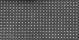
In X-ray crystallography, wide-angle X-ray scattering (WAXS) or wide-angle X-ray diffraction (WAXD) is the analysis of Bragg peaks scattered to wide angles, which are caused by sub-nanometer-sized structures. It is an X-ray-diffraction method and commonly used to determine a range of information about crystalline materials. The term WAXS is commonly used in polymer sciences to differentiate it from SAXS but many scientists doing "WAXS" would describe the measurements as Bragg/X-ray/powder diffraction or crystallography.

Small-angle neutron scattering (SANS) is an experimental technique that uses elastic neutron scattering at small scattering angles to investigate the structure of various substances at a mesoscopic scale of about 1–100 nm.
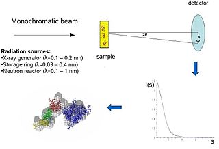
X-ray scattering techniques are a family of non-destructive analytical techniques which reveal information about the crystal structure, chemical composition, and physical properties of materials and thin films. These techniques are based on observing the scattered intensity of an X-ray beam hitting a sample as a function of incident and scattered angle, polarization, and wavelength or energy.

Neutron diffraction or elastic neutron scattering is the application of neutron scattering to the determination of the atomic and/or magnetic structure of a material. A sample to be examined is placed in a beam of thermal or cold neutrons to obtain a diffraction pattern that provides information of the structure of the material. The technique is similar to X-ray diffraction but due to their different scattering properties, neutrons and X-rays provide complementary information: X-Rays are suited for superficial analysis, strong x-rays from synchrotron radiation are suited for shallow depths or thin specimens, while neutrons having high penetration depth are suited for bulk samples.

A synchrotron light source is a source of electromagnetic radiation (EM) usually produced by a storage ring, for scientific and technical purposes. First observed in synchrotrons, synchrotron light is now produced by storage rings and other specialized particle accelerators, typically accelerating electrons. Once the high-energy electron beam has been generated, it is directed into auxiliary components such as bending magnets and insertion devices in storage rings and free electron lasers. These supply the strong magnetic fields perpendicular to the beam that are needed to stimulate the high energy electrons to emit photons.

In polymer chemistry, a copolymer is a polymer derived from more than one species of monomer. The polymerization of monomers into copolymers is called copolymerization. Copolymers obtained from the copolymerization of two monomer species are sometimes called bipolymers. Those obtained from three and four monomers are called terpolymers and quaterpolymers, respectively. Copolymers can be characterized by a variety of techniques such as NMR spectroscopy and size-exclusion chromatography to determine the molecular size, weight, properties, and composition of the material.
Biological small-angle scattering is a small-angle scattering method for structure analysis of biological materials. Small-angle scattering is used to study the structure of a variety of objects such as solutions of biological macromolecules, nanocomposites, alloys, and synthetic polymers. Small-angle X-ray scattering (SAXS) and small-angle neutron scattering (SANS) are the two complementary techniques known jointly as small-angle scattering (SAS). SAS is an analogous method to X-ray and neutron diffraction, wide angle X-ray scattering, as well as to static light scattering. In contrast to other X-ray and neutron scattering methods, SAS yields information on the sizes and shapes of both crystalline and non-crystalline particles. When used to study biological materials, which are very often in aqueous solution, the scattering pattern is orientation averaged.
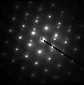
Selected area (electron) diffraction is a crystallographic experimental technique typically performed using a transmission electron microscope (TEM). It is a specific case of electron diffraction used primarily in material science and solid state physics as one of the most common experimental techniques. Especially with appropriate analytical software, SAD patterns (SADP) can be used to determine crystal orientation, measure lattice constants or examine its defects.

Dynamic light scattering (DLS) is a technique in physics that can be used to determine the size distribution profile of small particles in suspension or polymers in solution. In the scope of DLS, temporal fluctuations are usually analyzed using the intensity or photon autocorrelation function. In the time domain analysis, the autocorrelation function (ACF) usually decays starting from zero delay time, and faster dynamics due to smaller particles lead to faster decorrelation of scattered intensity trace. It has been shown that the intensity ACF is the Fourier transform of the power spectrum, and therefore the DLS measurements can be equally well performed in the spectral domain. DLS can also be used to probe the behavior of complex fluids such as concentrated polymer solutions.
Small-angle scattering (SAS) is a scattering technique based on deflection of collimated radiation away from the straight trajectory after it interacts with structures that are much larger than the wavelength of the radiation. The deflection is small (0.1-10°) hence the name small-angle. SAS techniques can give information about the size, shape and orientation of structures in a sample.
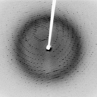
Diffraction topography is a imaging technique based on Bragg diffraction. Diffraction topographic images ("topographies") record the intensity profile of a beam of X-rays diffracted by a crystal. A topography thus represents a two-dimensional spatial intensity mapping of reflected X-rays, i.e. the spatial fine structure of a Laue reflection. This intensity mapping reflects the distribution of scattering power inside the crystal; topographs therefore reveal the irregularities in a non-ideal crystal lattice. X-ray diffraction topography is one variant of X-ray imaging, making use of diffraction contrast rather than absorption contrast which is usually used in radiography and computed tomography (CT). Topography is exploited to a lesser extends with neutrons, and has similarities to dark field imaging in the electron microscope community.
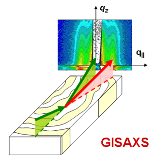
Grazing-incidence small-angle scattering (GISAS) is a scattering technique used to study nanostructured surfaces and thin films. The scattered probe is either photons or neutrons. GISAS combines the accessible length scales of small-angle scattering and the surface sensitivity of grazing incidence diffraction (GID).

Asymmetrical flow field-flow fractionation (AF4) is most versatile and most widely used sub-technique within the family of field flow fractionation (FFF) methods. AF4 can be used in aqueous and organic solvents and is able to characterize nanoparticles, polymers and proteins. The theory for AF4 was conceived in 1986 and was established in 1987 and first published by Wahlund and Giddings. AF4 is distinct from symmetrical Flow FFF because it contains only one permeable wall so the cross-flow is caused only by the carrier liquid. The cross-flow is induced by the carrier liquid constantly exiting by way of the semi-permeable wall on the bottom of the channel.

In computational chemistry, conformational ensembles, also known as structural ensembles, are experimentally constrained computational models describing the structure of intrinsically unstructured proteins. Such proteins are flexible in nature, lacking a stable tertiary structure, and therefore cannot be described with a single structural representation. The techniques of ensemble calculation are relatively new on the field of structural biology, and are still facing certain limitations that need to be addressed before it will become comparable to classical structural description methods such as biological macromolecular crystallography.
A neutron may pass by a nucleus with a probability determined by the nuclear interaction distance, or be absorbed, or undergo scattering that may be either coherent or incoherent. The interference effects in coherent scattering can be computed via the coherent scattering length of neutrons, being proportional to the amplitude of the spherical scattered waves according to Huygens–Fresnel theory. This scattering length varies by isotope in a way that appears random, whereas the X-ray scattering length is just the product of atomic number and Thomson scattering length, thus monotonically increasing with atomic number.
Frank Steven Bates is an American chemical engineer and materials scientist. Bates is a Regent's Professor (2007–present), a Distinguished McKnight University Professor (1996–present), and department head (1999-2014) in the department of chemical engineering and materials science at the University of Minnesota, where he has been a faculty member since 1989. Prior to his appointment at the University of Minnesota, Bates was a member of the technical staff at AT&T Bell Laboratories from 1982-1989.
Spin echo small angle neutron scattering (SESANS) measures structures from around 20 to 2000 nm in size. The information is presented as a real-space as opposed to a reciprocal space mapping. This can simplify the interpretation for some systems. SESANS is useful for studying processes that occur over relatively long time scales, as data collection is often slow, but large length scales. Aggregation of colloids, block copolymer micelles, Stöber silica particles being a prime examples.

Polysulfobetaines are zwitterionic polymers that contain a positively charged quaternary ammonium and a negatively charged sulfonate group within one constitutional repeat unit. In recent years, polysulfobetaines have received increasing attention owing to their good biotolerance and ultralow-fouling behavior towards surfaces. These properties are mainly referred to a tightly bound hydration layer around each zwitterionic group, which effectively suppresses protein adsorption and thus, improves anti-fouling behavior. Therefore, polysulfobetaines have been typically employed as ultrafiltration membranes, blood-contacting devices, and drug delivery materials.
References
- ↑ Hamley, I.W. "Small-Angle Scattering: Theory, Instrumentation, Data, and Applications" – Wiley, 2022. ISBN 978-1-119-76830-2.
- ↑ Glatter O; Kratky O, eds. (1982). Small Angle X-ray Scattering. Academic Press. ISBN 0-12-286280-5. Archived from the original on April 21, 2008.
- ↑ Sztucki, M; Narayanan, T (2007). "Development of an ultra-small-angle X-ray scattering instrument for probing the microstructure and the dynamics of soft matter". Journal of Applied Crystallography. 40: s459–s462. doi: 10.1107/S0021889806045833 . ISSN 1600-5767.
- ↑ Narayanan, T; Sztucki, M; Van Vaerenbergh, P; Léonardon, J; Gorini, J; Claustre, L; Sever, F; Morse, J; Boesecke, P (2018). "A multipurpose instrument for time-resolved ultra-small-angle and coherent X-ray scattering". Journal of Applied Crystallography. 51 (6): 1511–1524. doi:10.1107/S1600576718012748. ISSN 1600-5767. PMC 6276275 . PMID 30546286.
- ↑ Patil, N; Narayanan, T; Michels, L; Skjønsfjell, ETB; Guizar-Sicairos, M; Van den Brande, N; Claessens, R; Van Mele, B; Breiby, DW (May 2019). "Probing Organic Thin Films by Coherent X-ray Imaging and X-ray Scattering". ACS Applied Polymer Materials. 1 (7): 1787–1797. doi:10.1021/acsapm.9b00324. ISSN 2637-6105. S2CID 189992231.
- ↑ Burger, Virginia M., Daniel J. Arenas, and Collin M. Stultz. "A structure-free method for quantifying conformational flexibility in proteins." Scientific reports 6 (2016): 29040. DOI: 10.1038/srep29040 (2016).| http://hdl.handle.net/1721.1/108809
- ↑ Pedersen, JS (July 1997). "Analysis of small-angle scattering data from colloids and polymer solutions: modeling and least-squares fitting". Advances in Colloid and Interface Science. 70: 171–210. doi:10.1016/S0001-8686(97)00312-6. ISSN 0001-8686.
- ↑ Pedersen, JS (2000). "Form factors of block copolymer micelles with spherical, ellipsoidal and cylindrical cores". Journal of Applied Crystallography. 33 (3): 637–640. doi:10.1107/S0021889899012248. ISSN 1600-5767.
- ↑ Pedersen, JS (1994). "Determination of size distribution from small-angle scattering data for systems with effective hard-sphere interactions". Journal of Applied Crystallography. 27 (4): 595–608. doi:10.1107/S0021889893013810. ISSN 1600-5767.
- ↑ Gommes, CJ; Jaksch, S; Frielinghaus, H (2021). "Small-Angle Scattering for Beginners". Journal of Applied Crystallography. 54 (6): 1832–1843. doi: 10.1107/S1600576721010293 . PMC 8662971 . PMID 34963770.
- ↑ Hollamby, Martin J.; Aratsu, Keisuke; Pauw, Brian R.; Rogers, Sarah E.; Smith, Andrew J.; Yamauchi, Mitsuaki; Lin, Xu; Yagai, Shiki (2016-08-16). "Simultaneous SAXS and SANS Analysis for the Detection of Toroidal Supramolecular Polymers Composed of Noncovalent Supermacrocycles in Solution". Angewandte Chemie. 128 (34): 10044–10047. Bibcode:2016AngCh.12810044H. doi:10.1002/ange.201603370.
- ↑ Fanova, Anastasiia; Janata, Miroslav; Filippov, Sergey K.; Šlouf, Miroslav; Netopilík, Miloš; Mariani, Alessandro; Štěpánek, Miroslav (2019-08-27). "Evolution of Structure in a Comb Copolymer–Surfactant Coacervate". Macromolecules. 52 (16): 6303–6310. Bibcode:2019MaMol..52.6303F. doi:10.1021/acs.macromol.9b00332. ISSN 0024-9297. S2CID 202079335.
- ↑ Zhang, Xiaohan; Niebuur, Bart-Jan; Chytil, Petr; Etrych, Tomas; Filippov, Sergey K.; Kikhney, Alexey; Wieland, D. C. Florian; Svergun, Dmitri I.; Papadakis, Christine M. (2018-02-12). "Macromolecular p HPMA-Based Nanoparticles with Cholesterol for Solid Tumor Targeting: Behavior in HSA Protein Environment". Biomacromolecules. 19 (2): 470–480. doi:10.1021/acs.biomac.7b01579. ISSN 1525-7797. PMID 29381335.
- ↑ Fanova, Anastasiia; Šindelka, Karel; Uchman, Mariusz; Limpouchová, Zuzana; Filippov, Sergey K.; Pispas, Stergios; Procházka, Karel; Štěpánek, Miroslav (2018-09-25). "Coassembly of Poly( N -isopropylacrylamide) with Dodecyl and Carboxyl Terminal Groups with Cationic Surfactant: Critical Comparison of Experimental and Simulation Data". Macromolecules. 51 (18): 7295–7308. Bibcode:2018MaMol..51.7295F. doi:10.1021/acs.macromol.8b01161. ISSN 0024-9297. S2CID 105195163.
- ↑ Leisner, Dietrich; Imae, Toyoko (2003-08-01). "Interpolyelectrolyte Complex and Coacervate Formation of Poly(glutamic acid) with a Dendrimer Studied by Light Scattering and SAXS". The Journal of Physical Chemistry B. 107 (32): 8078–8087. doi:10.1021/jp027365l. ISSN 1520-6106.
- ↑ Murmiliuk, Anastasiia; Matějíček, Pavel; Filippov, Sergey K.; Janata, Miroslav; Šlouf, Miroslav; Pispas, Stergios; Štěpánek, Miroslav (2018). "Formation of core/corona nanoparticles with interpolyelectrolyte complex cores in aqueous solution: insight into chain dynamics in the complex from fluorescence quenching". Soft Matter. 14 (37): 7578–7585. Bibcode:2018SMat...14.7578M. doi:10.1039/C8SM01174E. ISSN 1744-683X. PMID 30140809.
- ↑ Dähling, Claudia; Lotze, Gudrun; Drechsler, Markus; Mori, Hideharu; Pergushov, Dmitry V.; Plamper, Felix A. (2016). "Temperature-induced structure switch in thermo-responsive micellar interpolyelectrolyte complexes: toward core–shell–corona and worm-like morphologies". Soft Matter. 12 (23): 5127–5137. Bibcode:2016SMat...12.5127D. doi:10.1039/C6SM00757K. ISSN 1744-683X. PMID 27194585.
- ↑ Sommer, Cornelia; Pedersen, Jan Skov; Garamus, Vasil M. (2005-03-01). "Structure and Interactions of Block Copolymer Micelles of Brij 700 Studied by Combining Small-Angle X-ray and Neutron Scattering". Langmuir. 21 (6): 2137–2149. doi:10.1021/la047489k. ISSN 0743-7463. PMID 15752000.
- ↑ Filippov, Sergey K.; Chytil, Petr; Konarev, Petr V.; Dyakonova, Margarita; Papadakis, ChristineM.; Zhigunov, Alexander; Plestil, Josef; Stepanek, Petr; Etrych, Tomas; Ulbrich, Karel; Svergun, Dmitri I. (2012-08-13). "Macromolecular HPMA-Based Nanoparticles with Cholesterol for Solid-Tumor Targeting: Detailed Study of the Inner Structure of a Highly Efficient Drug Delivery System". Biomacromolecules. 13 (8): 2594–2604. doi:10.1021/bm3008555. ISSN 1525-7797. PMID 22793269.
- ↑ Filippov, Sergey K.; Franklin, John M.; Konarev, Petr V.; Chytil, Petr; Etrych, Tomas; Bogomolova, Anna; Dyakonova, Margarita; Papadakis, Christine M.; Radulescu, Aurel; Ulbrich, Karel; Stepanek, Petr (2013-11-11). "Hydrolytically Degradable Polymer Micelles for Drug Delivery: A SAXS/SANS Kinetic Study". Biomacromolecules. 14 (11): 4061–4070. doi:10.1021/bm401186z. ISSN 1525-7797. PMID 24083567. S2CID 36632159.
- ↑ Riabtseva, Anna; Kaberov, Leonid I.; Noirez, Laurence; Ryukhtin, Vasyl; Nardin, Corinne; Verbraeken, Bart; Hoogenboom, Richard; Stepanek, Petr; Filippov, Sergey K. (February 2018). "Structural characterization of nanoparticles formed by fluorinated poly(2-oxazoline)-based polyphiles". European Polymer Journal. 99: 518–527. doi:10.1016/j.eurpolymj.2018.01.007. hdl: 1854/LU-8561215 . S2CID 102663271.
- ↑ Filippov, Sergey K.; Verbraeken, Bart; Konarev, Petr V.; Svergun, Dmitri I.; Angelov, Borislav; Vishnevetskaya, Natalya S.; Papadakis, Christine M.; Rogers, Sarah; Radulescu, Aurel; Courtin, Tim; Martins, José C. (2017-08-17). "Block and Gradient Copoly(2-oxazoline) Micelles: Strikingly Different on the Inside". The Journal of Physical Chemistry Letters. 8 (16): 3800–3804. doi: 10.1021/acs.jpclett.7b01588 . ISSN 1948-7185. PMID 28759235. S2CID 206664063.
- ↑ Suzuki, Daisuke; Nagase, Yasuhisa; Kureha, Takuma; Sato, Takaaki (2014-02-27). "Internal Structures of Thermosensitive Hybrid Microgels Investigated by Means of Small-Angle X-ray Scattering". The Journal of Physical Chemistry B. 118 (8): 2194–2204. doi:10.1021/jp410983x. ISSN 1520-6106. PMID 24517119.
- ↑ Chaves, Matheus Andrade; Oseliero Filho, Pedro Leonidas; Jange, Camila Garcia; Sinigaglia-Coimbra, Rita; Oliveira, Cristiano Luis Pinto; Pinho, Samantha Cristina (July 2018). "Structural characterization of multilamellar liposomes coencapsulating curcumin and vitamin D3". Colloids and Surfaces A: Physicochemical and Engineering Aspects. 549: 112–121. doi:10.1016/j.colsurfa.2018.04.018. S2CID 103002028.
- ↑ Di Cola, Emanuela; Grillo, Isabelle; Ristori, Sandra (2016-03-28). "Small Angle X-ray and Neutron Scattering: Powerful Tools for Studying the Structure of Drug-Loaded Liposomes". Pharmaceutics. 8 (2): 10. doi: 10.3390/pharmaceutics8020010 . ISSN 1999-4923. PMC 4932473 . PMID 27043614.
- ↑ Zaborova, Olga V.; Filippov, Sergey K.; Chytil, Petr; Kováčik, Lubomir; Ulbrich, Karel; Yaroslavov, Alexander A.; Etrych, Tomaš (April 2018). "A Novel Approach to Increase the Stability of Liposomal Containers via In Prep Coating by Poly[ N -(2-Hydroxypropyl)Methacrylamide] with Covalently Attached Cholesterol Groups". Macromolecular Chemistry and Physics. 219 (7): 1700508. doi:10.1002/macp.201700508.
- ↑ Bressel, Katharina; Muthig, Michael; Prevost, Sylvain; Gummel, Jeremie; Narayanan, Theyencheri; Gradzielski, Michael (2012-07-24). "Shaping Vesicles–Controlling Size and Stability by Admixture of Amphiphilic Copolymer". ACS Nano. 6 (7): 5858–5865. doi:10.1021/nn300359q. ISSN 1936-0851. PMID 22713309.
- ↑ Mable, Charlotte J.; Derry, Matthew J.; Thompson, Kate L.; Fielding, Lee A.; Mykhaylyk, Oleksandr O.; Armes, Steven P. (2017-06-13). "Time-Resolved SAXS Studies of the Kinetics of Thermally Triggered Release of Encapsulated Silica Nanoparticles from Block Copolymer Vesicles". Macromolecules. 50 (11): 4465–4473. Bibcode:2017MaMol..50.4465M. doi:10.1021/acs.macromol.7b00475. ISSN 0024-9297. PMC 5472368 . PMID 28626247.
- ↑ Filippov, Sergey K.; Bogomolova, Anna; Kaberov, Leonid; Velychkivska, Nadiia; Starovoytova, Larisa; Cernochova, Zulfiya; Rogers, Sarah E.; Lau, Wing Man; Khutoryanskiy, Vitaliy V.; Cook, Michael T. (2016-05-31). "Internal Nanoparticle Structure of Temperature-Responsive Self-Assembled PNIPAM- b -PEG- b -PNIPAM Triblock Copolymers in Aqueous Solutions: NMR, SANS, and Light Scattering Studies". Langmuir. 32 (21): 5314–5323. doi: 10.1021/acs.langmuir.6b00284 . ISSN 0743-7463. PMID 27159129.
- ↑ Cook, Michael T.; Filippov, Sergey K.; Khutoryanskiy, Vitaliy V. (August 2017). "Synthesis and solution properties of a temperature-responsive PNIPAM–b-PDMS–b-PNIPAM triblock copolymer". Colloid and Polymer Science. 295 (8): 1351–1358. doi:10.1007/s00396-017-4084-y. hdl: 2299/19649 . ISSN 0303-402X. S2CID 100587522.
- ↑ Sergeeva, Olga; Vlasov, Petr S.; Domnina, Nina S.; Bogomolova, Anna; Konarev, Petr V.; Svergun, Dmitri I.; Walterova, Zuzana; Horsky, Jiri; Stepanek, Petr; Filippov, Sergey K. (2014). "Novel thermosensitive telechelic PEGs with antioxidant activity: synthesis, molecular properties and conformational behaviour". RSC Adv. 4 (79): 41763–41771. Bibcode:2014RSCAd...441763S. doi:10.1039/C4RA06978A. ISSN 2046-2069.
- ↑ Kaberov, Leonid I.; Kaberova, Zhansaya; Murmiliuk, Anastasiia; Trousil, Jiří; Sedláček, Ondřej; Konefal, Rafal; Zhigunov, Alexander; Pavlova, Ewa; Vít, Martin; Jirák, Daniel; Hoogenboom, Richard (2021-06-28). "Fluorine-Containing Block and Gradient Copoly(2-oxazoline)s Based on 2-(3,3,3-Trifluoropropyl)-2-oxazoline: A Quest for the Optimal Self-Assembled Structure for 19F Imaging". Biomacromolecules. 22 (7): 2963–2975. doi:10.1021/acs.biomac.1c00367. ISSN 1525-7797. PMID 34180669. S2CID 235659596.
- ↑ Bernadó, Pau; Svergun, Dmitri I. (2012). "Structural analysis of intrinsically disordered proteins by small-angle X-ray scattering". Mol. BioSyst. 8 (1): 151–167. doi:10.1039/C1MB05275F. ISSN 1742-206X. PMID 21947276.
- ↑ Janisova, Larisa; Gruzinov, Andrey; Zaborova, Olga V.; Velychkivska, Nadiia; Vaněk, Ondřej; Chytil, Petr; Etrych, Tomáš; Janoušková, Olga; Zhang, Xiaohan; Blanchet, Clement; Papadakis, Christine M. (2020-01-28). "Molecular Mechanisms of the Interactions of N-(2-Hydroxypropyl)methacrylamide Copolymers Designed for Cancer Therapy with Blood Plasma Proteins". Pharmaceutics. 12 (2): 106. doi: 10.3390/pharmaceutics12020106 . ISSN 1999-4923. PMC 7076460 . PMID 32013056.
- ↑ Schuelke, Winfried (21 June 2007). Electron Dynamics by Inelastic X-Ray Scattering. Oxford University Press. ISBN 9780191523281.