Retinal ganglion cell

Last updated
Retinal ganglion cell
Gray882.png
Diagram showing cross-section of retinal layers. The area labeled "Ganglionic layer" contains retinal ganglion cells
Identifiers
MeSH D012165
NeuroLex ID nifext_17
Anatomical terms of neuroanatomy

A retinal ganglion cell (RGC) is a type of neuron located near the inner surface (the ganglion cell layer) of the retina of the eye. It receives visual information from photoreceptors via two intermediate neuron types: bipolar cells and retina amacrine cells. Retina amacrine cells, particularly narrow field cells, are important for creating functional subunits within the ganglion cell layer and making it so that ganglion cells can observe a small dot moving a small distance. [1] Retinal ganglion cells collectively transmit image-forming and non-image forming visual information from the retina in the form of action potential to several regions in the thalamus, hypothalamus, and mesencephalon, or midbrain.

Contents

Retinal ganglion cells vary significantly in terms of their size, connections, and responses to visual stimulation but they all share the defining property of having a long axon that extends into the brain. These axons form the optic nerve, optic chiasm, and optic tract.

A small percentage of retinal ganglion cells contribute little or nothing to vision, but are themselves photosensitive; their axons form the retinohypothalamic tract and contribute to circadian rhythms and pupillary light reflex, the resizing of the pupil.

Function

There are about 0.7 to 1.5 million retinal ganglion cells in the human retina. [2] With about 4.6 million cone cells and 92 million rod cells, or 96.6 million photoreceptors per retina, [3] on average each retinal ganglion cell receives inputs from about 100 rods and cones. However, these numbers vary greatly among individuals and as a function of retinal location. In the fovea (center of the retina), a single ganglion cell will communicate with as few as five photoreceptors. In the extreme periphery (edge of the retina), a single ganglion cell will receive information from many thousands of photoreceptors.[ citation needed ]

Retinal ganglion cells spontaneously fire action potentials at a base rate while at rest. Excitation of retinal ganglion cells results in an increased firing rate while inhibition results in a depressed rate of firing.

A false-color image of a flat-mounted rat retina viewed through a fluorescence microscope at 50x magnification. The optic nerve was injected with a fluorophore, causing retinal ganglion cells to fluoresce. 50x RGC axotomy 1 day.png
A false-color image of a flat-mounted rat retina viewed through a fluorescence microscope at 50x magnification. The optic nerve was injected with a fluorophore, causing retinal ganglion cells to fluoresce.

Types

There is wide variability in ganglion cell types across species. In primates, including humans, there are generally three classes of RGCs:

Based on their projections and functions, there are at least five main classes of retinal ganglion cells:

P-type

P-type retinal ganglion cells project to the parvocellular layers of the lateral geniculate nucleus. These cells are known as midget retinal ganglion cells, based on the small sizes of their dendritic trees and cell bodies. About 80% of all retinal ganglion cells are midget cells in the parvocellular pathway. They receive inputs from relatively few rods and cones. They have slow conduction velocity, and respond to changes in color but respond only weakly to changes in contrast unless the change is great. They have simple center-surround receptive fields, where the center may be either ON or OFF while the surround is the opposite.

Simulated array of parvocellular +M-L (green on) responses (right) to a natural video (left). Notice the relatively high spatial acuity, and sustained temporal responses in this pathway. [5]

M-type

M-type retinal ganglion cells project to the magnocellular layers of the lateral geniculate nucleus. These cells are known as parasol retinal ganglion cells, based on the large sizes of their dendritic trees and cell bodies. About 10% of all retinal ganglion cells are parasol cells, and these cells are part of the magnocellular pathway. They receive inputs from relatively many rods and cones. They have fast conduction velocity, and can respond to low-contrast stimuli, but are not very sensitive to changes in color. They have much larger receptive fields which are nonetheless also center-surround.

Simulated array of magnocellular OFF responses (right) to a natural video (left). Notice more transient temporal responses in this pathway, compared to the P-type. This retinal pathway is largely color blind. [5]

K-type

BiK-type retinal ganglion cells project to the koniocellular layers of the lateral geniculate nucleus. K-type retinal ganglion cells have been identified only relatively recently. Koniocellular means "cells as small as dust"; their small size made them hard to find. About 10% of all retinal ganglion cells are bistratified cells, and these cells go through the koniocellular pathway. They receive inputs from intermediate numbers of rods and cones. They may be involved in color vision. They have very large receptive fields that only have centers (no surrounds) and are always ON to the blue cone and OFF to both the red and green cone.

Simulated array of koniocellular +S (blue on) responses (right) to a natural video (left). Notice the low spatial acuity, reflecting the very large receptive fields. [5]

Photosensitive ganglion cell

Photosensitive ganglion cells, including but not limited to the giant retinal ganglion cells, contain their own photopigment, melanopsin, which makes them respond directly to light even in the absence of rods and cones. They project to, among other areas, the suprachiasmatic nucleus (SCN) via the retinohypothalamic tract for setting and maintaining circadian rhythms. Other retinal ganglion cells projecting to the lateral geniculate nucleus (LGN) include cells making connections with the Edinger-Westphal nucleus (EW), for control of the pupillary light reflex, and giant retinal ganglion cells.

Physiology

Most mature ganglion cells are able to fire action potentials at a high frequency because of their expression of Kv3 potassium channels. [6] [7] [8]

Pathology

Degeneration of axons of the retinal ganglion cells (the optic nerve) is a hallmark of glaucoma. [9]

Developmental biology

Retinal growth: the beginning

Retinal ganglion cells (RGCs) are born between embryonic day 11 and post-natal day zero in the mouse and between week 5 and week 18 in utero in human development. [10] [11] [12] In mammals, RGCs are typically added at the beginning in the dorsal central aspect of the optic cup, or eye primordium. Then RC growth sweeps out ventrally and peripherally from there in a wave-like pattern. [13] This process depends on a host of factors, ranging from signaling factors like FGF3 and FGF8 to proper inhibition of the Notch signaling pathway. Most importantly, the bHLH (basic helix-loop-helix)-domain containing transcription factor Atoh7 and its downstream effectors, such as Brn3b and Isl-1, work to promote RGC survival and differentiation. [10] The "differentiation wave" that drives RGC development across the retina is also regulated in particular of the bHLH factors Neurog2 and Ascl1 and FGF/Shh signaling, deriving from the periphery. [10] [13] [14]

Growth within the retinal ganglion cell (optic fiber) layer

Early progenitor RGCs will typically extend processes connecting to the inner and outer limiting membranes of the retina with the outer layer adjacent to the retinal pigment epithelium and inner adjacent to the future vitreous humor. The cell soma will pull towards the pigment epithelium, undergo a terminal cell division and differentiation, and then migrate backwards towards the inner limiting membrane in a process called somal translocation. The kinetics of RGC somal translocation and underlying mechanisms are best understood in the zebrafish. [15] The RGC will then extend an axon in the retinal ganglion cell layer, which is directed by laminin contact. [16] The retraction of the apical process of the RGC is likely mediated by Slit–Robo signaling. [10]

RGCs will grow along glial end feet positioned on the inner surface (side closest to the future vitreous humor). Neural cell adhesion molecule (N-CAM) will mediate this attachment via homophilic interactions between molecules of like isoforms (A or B). Slit signaling also plays a role, preventing RGCs from growing into layers beyond the optic fiber layer. [17]

Axons from the RGCs will grow and extend towards the optic disc, where they exit the eye. Once differentiated, they are bordered by an inhibitory peripheral region and a central attractive region, thus promoting extension of the axon towards the optic disc. CSPGs exist along the retinal neuroepithelium (surface over which the RGCs lie) in a peripheral high–central low gradient. [10] Slit is also expressed in a similar pattern, secreted from the cells in the lens. [17] Adhesion molecules, like N-CAM and L1, will promote growth centrally and will also help to properly fasciculate (bundle) the RGC axons together. Shh is expressed in a high central, low peripheral gradient, promoting central-projecting RGC axons extension via Patched-1, the principal receptor for Shh, mediated signaling. [18]

Growth into and through the optic nerve

RGCs exit the retinal ganglion cell layer through the optic disc, which requires a 45° turn. [10] This requires complex interactions with optic disc glial cells which will express local gradients of Netrin-1, a morphogen that will interact with the Deleted in Colorectal Cancer (DCC) receptor on growth cones of the RGC axon. This morphogen initially attracts RGC axons, but then, through an internal change in the growth cone of the RGC, netrin-1 becomes repulsive, pushing the axon away from the optic disc. [19] This is mediated through a cAMP-dependent mechanism. Additionally, CSPGs and Eph–ephrin signaling may also be involved.

RGCs will grow along glial cell end feet in the optic nerve. These glia will secrete repulsive semaphorin 5a and Slit in a surround fashion, covering the optic nerve which ensures that they remain in the optic nerve. Vax1, a transcription factor, is expressed by the ventral diencephalon and glial cells in the region where the chiasm is formed, and it may also be secreted to control chiasm formation. [20]

Growth at the optic chiasm

When RGCs approach the optic chiasm, the point at which the two optic nerves meet, at the ventral diencephalon around embryonic days 10–11 in the mouse, they have to make the decision to cross to the contralateral optic tract or remain in the ipsilateral optic tract. In the mouse, about 5% of RGCs, mostly those coming from the ventral-temporal crescent (VTc) region of the retina, will remain ipsilateral, while the remaining 95% of RGCs will cross. [10] This is largely controlled by the degree of binocular overlap between the two fields of sight in both eyes. Mice do not have a significant overlap, whereas, humans, who do, will have about 50% of RGCs cross and 50% will remain ipsilateral.

Building the repulsive outline of the chiasm

Once RGCs reach the chiasm, the glial cells supporting them will change from an intrafascicular to radial morphology. A group of diencephalic cells that express the cell surface antigen stage-specific embryonic antigen (SSEA)-1 and CD44 will form an inverted V-shape. [21] They will establish the posterior aspect of the optic chiasm border. Additionally, Slit signaling is important here: Heparin sulfate proteoglycans, proteins in the ECM, will anchor the Slit morphogen at specific points in the posterior chiasm border. [22] RGCs will begin to express Robo, the receptor for Slit, at this point, thus facilitating the repulsion.

Contralateral projecting RGCs

RGC axons traveling to the contralateral optic tract need to cross. Shh, expressed along the midline in the ventral diencephalon, provides a repulsive cue to prevent RGCs from crossing the midline ectopically. However, a hole is generated in this gradient, thus allowing RGCs to cross.

Molecules mediating attraction include NrCAM, which is expressed by growing RGCs and the midline glia and acts along with Sema6D, mediated via the plexin-A1 receptor. [10] VEGF-A is released from the midline directs RGCs to take a contralateral path, mediated by the neuropilin-1 (NRP1) receptor. [23] cAMP seems to be very important in regulating the production of NRP1 protein, thus regulating the growth cones response to the VEGF-A gradient in the chiasm. [24]

Ipsilateral projecting RGCs

The only component in mice projecting ipsilaterally are RGCs from the ventral-temporal crescent in the retina, and only because they express the Zic2 transcription factor. Zic2 will promote the expression of the tyrosine kinase receptor EphB1, which, through forward signaling (see review by Xu et al. [25] ) will bind to ligand ephrin B2 expressed by midline glia and be repelled to turn away from the chiasm. Some VTc RGCs will project contralaterally because they express the transcription factor Islet-2, which is a negative regulator of Zic2 production. [26]

Shh plays a key role in keeping RGC axons ipsilateral as well. Shh is expressed by the contralaterally projecting RGCs and midline glial cells. Boc, or Brother of CDO (CAM-related/downregulated by oncogenes), a co-receptor for Shh that influences Shh signaling through Ptch1, [27] seems to mediate this repulsion, as it is only on growth cones coming from the ipsilaterally projecting RGCs. [18]

Other factors influencing ipsilateral RGC growth include the Teneurin family, which are transmembrane adhesion proteins that use homophilic interactions to control guidance, and Nogo, which is expressed by midline radial glia. [28] [29] The Nogo receptor is only expressed by VTc RGCs. [10]

Finally, other transcription factors seem to play a significant role in altering. For example, Foxg1, also called Brain-Factor 1, and Foxd1, also called Brain Factor 2, are winged-helix transcription factors that are expressed in the nasal and temporal optic cups and the optic vesicles begin to evaginate from the neural tube. These factors are also expressed in the ventral diencephalon, with Foxd1 expressed near the chiasm, while Foxg1 is expressed more rostrally. They appear to play a role in defining the ipsilateral projection by altering expression of Zic2 and EphB1 receptor production. [10] [30]

Growth in the optic tract

Once out of the optic chiasm, RGCs will extend dorsocaudally along the ventral diencephalic surface making the optic tract, which will guide them to the superior colliculus and lateral geniculate nucleus in the mammals, or the tectum in lower vertebrates. [10] Sema3d seems to be promote growth, at least in the proximal optic tract, and cytoskeletal re-arrangements at the level of the growth cone appear to be significant. [31]

Myelination

In most mammals, the axons of retinal ganglion cells are not myelinated where they pass through the retina. However, the parts of axons that are beyond the retina, are myelinated. This myelination pattern is functionally explained by the relatively high opacity of myelin—myelinated axons passing over the retina would absorb some of the light before it reaches the photoreceptor layer, reducing the quality of vision. There are human eye diseases where this does, in fact, happen. In some vertebrates, such as the chicken, the ganglion cell axons are myelinated inside the retina. [32]

See also

Related Research Articles

<span class="mw-page-title-main">Retina</span> Part of the eye

The retina is the innermost, light-sensitive layer of tissue of the eye of most vertebrates and some molluscs. The optics of the eye create a focused two-dimensional image of the visual world on the retina, which then processes that image within the retina and sends nerve impulses along the optic nerve to the visual cortex to create visual perception. The retina serves a function which is in many ways analogous to that of the film or image sensor in a camera.

<span class="mw-page-title-main">Optic chiasm</span> Part of the brain where the optic nerves cross

In neuroanatomy, the optic chiasm, or optic chiasma, is the part of the brain where the optic nerves cross. It is located at the bottom of the brain immediately inferior to the hypothalamus. The optic chiasm is found in all vertebrates, although in cyclostomes, it is located within the brain.

The development of the nervous system, or neural development (neurodevelopment), refers to the processes that generate, shape, and reshape the nervous system of animals, from the earliest stages of embryonic development to adulthood. The field of neural development draws on both neuroscience and developmental biology to describe and provide insight into the cellular and molecular mechanisms by which complex nervous systems develop, from nematodes and fruit flies to mammals.

<span class="mw-page-title-main">Lateral geniculate nucleus</span> Component of the visual system in the brains thalamus

In neuroanatomy, the lateral geniculate nucleus is a structure in the thalamus and a key component of the mammalian visual pathway. It is a small, ovoid, ventral projection of the thalamus where the thalamus connects with the optic nerve. There are two LGNs, one on the left and another on the right side of the thalamus. In humans, both LGNs have six layers of neurons alternating with optic fibers.

<span class="mw-page-title-main">Photoreceptor cell</span> Type of neuroepithelial cell

A photoreceptor cell is a specialized type of neuroepithelial cell found in the retina that is capable of visual phototransduction. The great biological importance of photoreceptors is that they convert light into signals that can stimulate biological processes. To be more specific, photoreceptor proteins in the cell absorb photons, triggering a change in the cell's membrane potential.

<span class="mw-page-title-main">Retina bipolar cell</span> Type of neuron

As a part of the retina, bipolar cells exist between photoreceptors and ganglion cells. They act, directly or indirectly, to transmit signals from the photoreceptors to the ganglion cells.

<span class="mw-page-title-main">Melanopsin</span> Mammalian protein found in Homo sapiens

Melanopsin is a type of photopigment belonging to a larger family of light-sensitive retinal proteins called opsins and encoded by the gene Opn4. In the mammalian retina, there are two additional categories of opsins, both involved in the formation of visual images: rhodopsin and photopsin in the rod and cone photoreceptor cells, respectively.

Axon guidance is a subfield of neural development concerning the process by which neurons send out axons to reach their correct targets. Axons often follow very precise paths in the nervous system, and how they manage to find their way so accurately is an area of ongoing research.

Intrinsically photosensitive retinal ganglion cells (ipRGCs), also called photosensitive retinal ganglion cells (pRGC), or melanopsin-containing retinal ganglion cells (mRGCs), are a type of neuron in the retina of the mammalian eye. The presence of ipRGCs was first suspected in 1927 when rodless, coneless mice still responded to a light stimulus through pupil constriction, This implied that rods and cones are not the only light-sensitive neurons in the retina. Yet research on these cells did not advance until the 1980s. Recent research has shown that these retinal ganglion cells, unlike other retinal ganglion cells, are intrinsically photosensitive due to the presence of melanopsin, a light-sensitive protein. Therefore, they constitute a third class of photoreceptors, in addition to rod and cone cells.

<span class="mw-page-title-main">Floor plate</span> Embryonic structure

The floor plate is a structure integral to the developing nervous system of vertebrate organisms. Located on the ventral midline of the embryonic neural tube, the floor plate is a specialized glial structure that spans the anteroposterior axis from the midbrain to the tail regions. It has been shown that the floor plate is conserved among vertebrates, such as zebrafish and mice, with homologous structures in invertebrates such as the fruit fly Drosophila and the nematode C. elegans. Functionally, the structure serves as an organizer to ventralize tissues in the embryo as well as to guide neuronal positioning and differentiation along the dorsoventral axis of the neural tube.

<span class="mw-page-title-main">Retinohypothalamic tract</span> Neural pathway involved with circadian rhythms

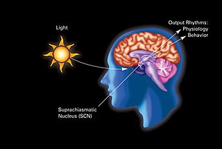
In neuroanatomy, the retinohypothalamic tract (RHT) is a photic neural input pathway involved in the circadian rhythms of mammals. The origin of the retinohypothalamic tract is the intrinsically photosensitive retinal ganglion cells (ipRGC), which contain the photopigment melanopsin. The axons of the ipRGCs belonging to the retinohypothalamic tract project directly, monosynaptically, to the suprachiasmatic nuclei (SCN) via the optic nerve and the optic chiasm. The suprachiasmatic nuclei receive and interpret information on environmental light, dark and day length, important in the entrainment of the "body clock". They can coordinate peripheral "clocks" and direct the pineal gland to secrete the hormone melatonin.

<span class="mw-page-title-main">Ephrin</span>

Ephrins are a family of proteins that serve as the ligands of the Eph receptor. Eph receptors in turn compose the largest known subfamily of receptor protein-tyrosine kinases (RTKs).

<span class="mw-page-title-main">Roundabout family</span>

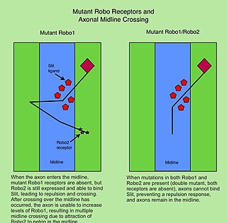
The Roundabout (Robo) family of proteins are single-pass transmembrane receptors that are highly conserved across many branches of the animal kingdom, from C. elegans to humans. They were first discovered in Drosophila, through a mutant screen for genes involved in axon guidance. The Drosophila roundabout mutant was named after its phenotype, which resembled the circular traffic junctions. The Robo receptors are most well known for their role in the development of the nervous system, where they have been shown to respond to secreted Slit ligands. One well-studied example is the requirement for Slit-Robo signaling in regulation of axonal midline crossing. Slit-Robo signaling is also critical for many neurodevelopmental processes including formation of the olfactory tract, the optic nerve, and motor axon fasciculation. In addition, Slit-Robo signaling contributes to cell migration and the development of other tissues such as the lung, kidney, liver, muscle and breast. Mutations in Robo genes have been linked to multiple neurodevelopmental disorders in humans.

Slit is a family of secreted extracellular matrix proteins which play an important signalling role in the neural development of most bilaterians. While lower animal species, including insects and nematode worms, possess a single Slit gene, humans, mice and other vertebrates possess three Slit homologs: Slit1, Slit2 and Slit3. Human Slits have been shown to be involved in certain pathological conditions, such as cancer and inflammation.

<span class="mw-page-title-main">Midget cell</span>

A midget cell is one type of retinal ganglion cell (RGC). Midget cells originate in the ganglion cell layer of the retina, and project to the parvocellular layers of the lateral geniculate nucleus (LGN). The axons of midget cells travel through the optic nerve and optic tract, ultimately synapsing with parvocellular cells in the LGN. These cells are known as midget retinal ganglion cells due to the small sizes of their dendritic trees and cell bodies. About 80% of RGCs are midget cells. They receive inputs from relatively few rods and cones. In many cases, they are connected to midget bipolar cells, which are linked to one cone each.

Slit-Robo is the name of a cell signaling protein complex with many diverse functions including axon guidance and angiogenesis.

<span class="mw-page-title-main">Ephrin A5</span> Protein-coding gene in the species Homo sapiens

Ephrin A5 is a protein that in humans is encoded by the EFNA5 gene.

<span class="mw-page-title-main">Tropic cues involved in growth cone guidance</span>

The growth cone is a highly dynamic structure of the developing neuron, changing directionality in response to different secreted and contact-dependent guidance cues; it navigates through the developing nervous system in search of its target. The migration of the growth cone is mediated through the interaction of numerous trophic and tropic factors; netrins, slits, ephrins and semaphorins are four well-studied tropic cues (Fig.1). The growth cone is capable of modifying its sensitivity to these guidance molecules as it migrates to its target; this sensitivity regulation is an important theme seen throughout development.

<span class="mw-page-title-main">Retinal precursor cells</span> Type of cell in the human eye

Retinal precursor cells are biological cells that differentiate into the various cell types of the retina during development. In the vertebrate, these retinal cells differentiate into seven cell types, including retinal ganglion cells, amacrine cells, bipolar cells, horizontal cells, rod photoreceptors, cone photoreceptors, and Müller glia cells. During embryogenesis, retinal cells originate from the anterior portion of the neural plate termed the eye field. Eye field cells with a retinal fate express several transcription factor markers including Rx1, Pax6, and Lhx2. The eye field gives rise to the optic vesicle and then to the optic cup. The retina is generated from the precursor cells within the inner layer of the optic cup, as opposed to the retinal pigment epithelium that originate from the outer layer of the optic cup. In general, the developing retina is organized so that the least-committed precursor cells are located in the periphery of the retina, while the committed cells are located in the center of the retina. The differentiation of retinal precursor cells into the mature cell types found in the retina is coordinated in time and space by factors within the cell as well as factors in the environment of the cell. One example of an intrinsic regulator of this process is the transcription factor Ath5. Ath5 expression in retinal progenitor cells biases their differentiation into a retinal ganglion cell fate. An example of an environmental factor is the morphogen sonic hedge hog (Shh). Shh has been shown to repress the differentiation of precursor cells into retinal ganglion cells.

<span class="mw-page-title-main">Forkhead box d1</span> Human protein-coding gene

Forkhead box D1 is a protein that in humans is encoded by the FOXD1 gene. Forkhead d1 is a kidney expressed transcription factor maps at the chromosome 5 at position 5q12—q13, identified in Drosophila forkhead protein and mammalian HNF3 transcription factor. The name of was derived from two spiked head structures in the embryos of Drosophila forkhead mutant. It belong to transcription factor family that displays remarkable functional diversity and involved in a wide variety of biological processes. The most commonly used synonyms for Forkhead D1 are, FOX D1, FREAC-4 and BF2.

References

  1. Masland RH (January 2012). "The tasks of amacrine cells". Visual Neuroscience. 29 (1): 3–9. doi:10.1017/s0952523811000344. PMC   3652807 . PMID   22416289.
  2. Watson AB (June 2014). "A formula for human retinal ganglion cell receptive field density as a function of visual field location" (PDF). Journal of Vision. 14 (7): 15. doi: 10.1167/14.7.15 . PMID   24982468.
  3. Curcio CA, Sloan KR, Kalina RE, Hendrickson AE (February 1990). "Human photoreceptor topography" (PDF). The Journal of Comparative Neurology. 292 (4): 497–523. doi:10.1002/cne.902920402. PMID   2324310. S2CID   24649779.
  4. Principles of Neural Science 4th Ed. Kandel et al.
  5. 1 2 3 Schottdorf M, Lee BB (June 2021). "A quantitative description of macaque ganglion cell responses to natural scenes: the interplay of time and space". The Journal of Physiology. 599 (12): 3169–3193. doi:10.1113/JP281200. PMC   8998785 . PMID   33913164. S2CID   233448275.
  6. "Ionic conductances underlying excitability in tonically firing retinal ganglion cells of adult rat".
  7. Henne J, Pöttering S, Jeserich G (December 2000). "Voltage-gated potassium channels in retinal ganglion cells of trout: a combined biophysical, pharmacological, and single-cell RT-PCR approach". Journal of Neuroscience Research. 62 (5): 629–37. doi:10.1002/1097-4547(20001201)62:5<629::AID-JNR2>3.0.CO;2-X. PMID   11104501. S2CID   44513007.
  8. Henne J, Jeserich G (January 2004). "Maturation of spiking activity in trout retinal ganglion cells coincides with upregulation of Kv3.1- and BK-related potassium channels". Journal of Neuroscience Research. 75 (1): 44–54. doi:10.1002/jnr.10830. PMID   14689447. S2CID   38851244.
  9. Jadeja RN, Thounaojam MC, Martin PM (2020). "Implications of NAD + Metabolism in the Aging Retina and Retinal Degeneration". Oxidative Medicine and Cellular Longevity . 2020: 2692794. doi: 10.1155/2020/2692794 . PMC   7238357 . PMID   32454935.
  10. 1 2 3 4 5 6 7 8 9 10 11 Erskine L, Herrera E (2014-01-01). "Connecting the retina to the brain". ASN Neuro. 6 (6): 175909141456210. doi:10.1177/1759091414562107. PMC   4720220 . PMID   25504540.
  11. Petros TJ, Rebsam A, Mason CA (2008-01-01). "Retinal axon growth at the optic chiasm: to cross or not to cross". Annual Review of Neuroscience. 31: 295–315. doi:10.1146/annurev.neuro.31.060407.125609. PMID   18558857.
  12. Pacal M, Bremner R (May 2014). "Induction of the ganglion cell differentiation program in human retinal progenitors before cell cycle exit". Developmental Dynamics. 243 (5): 712–29. doi: 10.1002/dvdy.24103 . PMID   24339342. S2CID   4133348.
  13. 1 2 Hufnagel RB, Le TT, Riesenberg AL, Brown NL (April 2010). "Neurog2 controls the leading edge of neurogenesis in the mammalian retina". Developmental Biology. 340 (2): 490–503. doi:10.1016/j.ydbio.2010.02.002. PMC   2854206 . PMID   20144606.
  14. Lo Giudice Q, Leleu M, La Manno G, Fabre PJ (September 2019). "Single-cell transcriptional logic of cell-fate specification and axon guidance in early-born retinal neurons". Development. 146 (17): dev178103. doi: 10.1242/dev.178103 . PMID   31399471.
  15. Icha J, Kunath C, Rocha-Martins M, Norden C (October 2016). "Independent modes of ganglion cell translocation ensure correct lamination of the zebrafish retina". The Journal of Cell Biology. 215 (2): 259–275. doi:10.1083/jcb.201604095. PMC   5084647 . PMID   27810916.
  16. Randlett O, Poggi L, Zolessi FR, Harris WA (April 2011). "The oriented emergence of axons from retinal ganglion cells is directed by laminin contact in vivo". Neuron. 70 (2): 266–80. doi:10.1016/j.neuron.2011.03.013. PMC   3087191 . PMID   21521613.
  17. 1 2 Thompson H, Andrews W, Parnavelas JG, Erskine L (November 2009). "Robo2 is required for Slit-mediated intraretinal axon guidance". Developmental Biology. 335 (2): 418–26. doi:10.1016/j.ydbio.2009.09.034. PMC   2814049 . PMID   19782674.
  18. 1 2 Sánchez-Camacho C, Bovolenta P (November 2008). "Autonomous and non-autonomous Shh signalling mediate the in vivo growth and guidance of mouse retinal ganglion cell axons". Development. 135 (21): 3531–41. doi:10.1242/dev.023663. PMID   18832395.
  19. Höpker VH, Shewan D, Tessier-Lavigne M, Poo M, Holt C (September 1999). "Growth-cone attraction to netrin-1 is converted to repulsion by laminin-1". Nature. 401 (6748): 69–73. Bibcode:1999Natur.401...69H. doi:10.1038/43441. PMID   10485706. S2CID   205033254.
  20. Kim N, Min KW, Kang KH, Lee EJ, Kim HT, Moon K, et al. (September 2014). "Regulation of retinal axon growth by secreted Vax1 homeodomain protein". eLife. 3: e02671. doi: 10.7554/eLife.02671 . PMC   4178304 . PMID   25201875.
  21. Sretavan DW, Feng L, Puré E, Reichardt LF (May 1994). "Embryonic neurons of the developing optic chiasm express L1 and CD44, cell surface molecules with opposing effects on retinal axon growth". Neuron. 12 (5): 957–75. doi:10.1016/0896-6273(94)90307-7. PMC   2711898 . PMID   7514428.
  22. Wright KM, Lyon KA, Leung H, Leahy DJ, Ma L, Ginty DD (December 2012). "Dystroglycan organizes axon guidance cue localization and axonal pathfinding". Neuron. 76 (5): 931–44. doi:10.1016/j.neuron.2012.10.009. PMC   3526105 . PMID   23217742.
  23. Erskine L, Reijntjes S, Pratt T, Denti L, Schwarz Q, Vieira JM, et al. (June 2011). "VEGF signaling through neuropilin 1 guides commissural axon crossing at the optic chiasm". Neuron. 70 (5): 951–65. doi:10.1016/j.neuron.2011.02.052. PMC   3114076 . PMID   21658587.
  24. Dell AL, Fried-Cassorla E, Xu H, Raper JA (July 2013). "cAMP-induced expression of neuropilin1 promotes retinal axon crossing in the zebrafish optic chiasm". The Journal of Neuroscience. 33 (27): 11076–88. doi:10.1523/JNEUROSCI.0197-13.2013. PMC   3719991 . PMID   23825413.
  25. Xu NJ, Henkemeyer M (February 2012). "Ephrin reverse signaling in axon guidance and synaptogenesis". Seminars in Cell & Developmental Biology. 23 (1): 58–64. doi:10.1016/j.semcdb.2011.10.024. PMC   3288821 . PMID   22044884.
  26. Pak W, Hindges R, Lim YS, Pfaff SL, O'Leary DD (November 2004). "Magnitude of binocular vision controlled by islet-2 repression of a genetic program that specifies laterality of retinal axon pathfinding". Cell. 119 (4): 567–78. doi: 10.1016/j.cell.2004.10.026 . PMID   15537545. S2CID   16663526.
  27. Allen BL, Song JY, Izzi L, Althaus IW, Kang JS, Charron F, et al. (June 2011). "Overlapping roles and collective requirement for the coreceptors GAS1, CDO, and BOC in SHH pathway function". Developmental Cell. 20 (6): 775–87. doi:10.1016/j.devcel.2011.04.018. PMC   3121104 . PMID   21664576.
  28. Wang J, Chan CK, Taylor JS, Chan SO (June 2008). "Localization of Nogo and its receptor in the optic pathway of mouse embryos". Journal of Neuroscience Research. 86 (8): 1721–33. doi:10.1002/jnr.21626. PMID   18214994. S2CID   25123173.
  29. Kenzelmann D, Chiquet-Ehrismann R, Leachman NT, Tucker RP (March 2008). "Teneurin-1 is expressed in interconnected regions of the developing brain and is processed in vivo". BMC Developmental Biology. 8: 30. doi: 10.1186/1471-213X-8-30 . PMC   2289808 . PMID   18366734.
  30. Herrera E, Marcus R, Li S, Williams SE, Erskine L, Lai E, Mason C (November 2004). "Foxd1 is required for proper formation of the optic chiasm". Development. 131 (22): 5727–39. doi: 10.1242/dev.01431 . PMID   15509772.
  31. Sakai JA, Halloran MC (March 2006). "Semaphorin 3d guides laterality of retinal ganglion cell projections in zebrafish". Development. 133 (6): 1035–44. doi:10.1242/dev.02272. PMID   16467361.
  32. Villegas GM (July 1960). "Electron microscopic study of the vertebrate retina". The Journal of General Physiology. 43(6)Suppl (6): 15–43. doi:10.1085/jgp.43.6.15. PMC   2195075 . PMID   13842313.