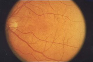
Retinopathy is any damage to the retina of the eyes, which may cause vision impairment. Retinopathy often refers to retinal vascular disease, or damage to the retina caused by abnormal blood flow. Age-related macular degeneration is technically included under the umbrella term retinopathy but is often discussed as a separate entity. Retinopathy, or retinal vascular disease, can be broadly categorized into proliferative and non-proliferative types. Frequently, retinopathy is an ocular manifestation of systemic disease as seen in diabetes or hypertension. Diabetes is the most common cause of retinopathy in the U.S. as of 2008. Diabetic retinopathy is the leading cause of blindness in working-aged people. It accounts for about 5% of blindness worldwide and is designated a priority eye disease by the World Health Organization.

Micropsia is a condition affecting human visual perception in which objects are perceived to be smaller than they actually are. Micropsia can be caused by optical factors, by distortion of images in the eye, by changes in the brain, and from psychological factors. Dissociative phenomena are linked with micropsia, which may be the result of brain-lateralization disturbance.

Amaurosis fugax is a painless temporary loss of vision in one or both eyes.

Visual snow syndrome (VSS) is a form of visual hallucination that is characterized by the perception of small, bilateral, simultaneous, diffuse, mobile, asynchronous dots usually throughout the entire visual field, but it can be partial, and it is present in all conditions of illumination. The dots remain individual and do not clump together or change in size. Visual snow exists in one of two forms: the pulse type and the broadband type.

A scotoma is an area of partial alteration in the field of vision consisting of a partially diminished or entirely degenerated visual acuity that is surrounded by a field of normal – or relatively well-preserved – vision.
The visual field is "that portion of space in which objects are visible at the same moment during steady fixation of the gaze in one direction"; in ophthalmology and neurology the emphasis is on the structure inside the visual field and it is then considered “the field of functional capacity obtained and recorded by means of perimetry”.

Diplopia is the simultaneous perception of two images of a single object that may be displaced horizontally or vertically in relation to each other. Also called double vision, it is a loss of visual focus under regular conditions, and is often voluntary. However, when occurring involuntarily, it results in impaired function of the extraocular muscles, where both eyes are still functional, but they cannot turn to target the desired object. Problems with these muscles may be due to mechanical problems, disorders of the neuromuscular junction, disorders of the cranial nerves that innervate the muscles, and occasionally disorders involving the supranuclear oculomotor pathways or ingestion of toxins.

An aura is a perceptual disturbance experienced by some with epilepsy or migraine. An epileptic aura is a seizure.
Acephalgic migraine is a neurological syndrome. It is a relatively uncommon variant of migraine in which the patient may experience some migraine symptoms such as aura, nausea, photophobia, and hemiparesis, but does not experience headache. It is generally classified as an event fulfilling the conditions of migraine with aura with no headache. It is sometimes distinguished from visual-only migraine aura without headache, also called ocular migraine.
Photopsia is the presence of perceived flashes of light in the field of vision.
Eales disease is a type of obliterative vasculopathy, also known as angiopathia retinae juvenilis, periphlebitis retinae or primary perivasculitis of the retina. It was first described by the British ophthalmologist Henry Eales (1852–1913) in 1880 and is a rare ocular disease characterized by inflammation and possible blockage of retinal blood vessels, abnormal growth of new blood vessels (neovascularization), and recurrent retinal and vitreal hemorrhages.

Progressive outer retinal necrosis (PORN) syndrome is a form of chorioretinitis, an infection in the retina, the back of the eye. The disease is most commonly caused by the varicella zoster virus and is found almost exclusively in patients with HIV/AIDS. Progressive outer retinal necrosis is the second most common opportunistic retinal infection in North America among people with AIDS. The reason this disease process is considered opportunistic is precisely because it only presents in patients with AIDS, a disease that attacks and weakens the immune system, making space for other infections, like Varicella zoster virus (VZV) and Herpes simplex virus (HSV), to attack the body.
Coats' disease is a rare congenital, nonhereditary eye disorder, causing full or partial blindness, characterized by abnormal development of blood vessels behind the retina. Coats' disease can also fall under glaucoma.

Scintillating scotoma is a common visual aura that was first described by 19th-century physician Hubert Airy (1838–1903). Originating from the brain, it may precede a migraine headache, but can also occur acephalgically, also known as visual migraine or migraine aura. It is often confused with retinal migraine, which originates in the eyeball or socket.
Optic neuropathy is damage to the optic nerve from any cause. The optic nerve is a bundle of millions of fibers in the retina that sends visual signals to the brain. [1].

Cotton wool spots are opaque fluffy white patches on the retina of the eye that are considered an abnormal finding during a funduscopic exam. Cotton wool spots are typically a sign of another disease state, most common of which is diabetic retinopathy. The irregularly shaped white patches are a result of ischemia, or reduced blood flow and oxygen, in the retinal nerve fiber layer, which is located in the distribution of the capillaries of the superficial layer of the retina. These areas with reduced blood flow reflect the obstruction of axoplasmic flow due to mechanical or vascular causes and the consequential accumulation as a result of decreased axonal transport. This reduced axonal transport can then cause swelling or bulging on the surface layer of the retina, increasing the potential for nerve fiber damage.

Blurred vision is an ocular symptom where vision becomes less precise and there is added difficulty to resolve fine details.

Intraocular hemorrhage is bleeding inside the eye. Bleeding can occur from any structure of the eye where there is vasculature or blood flow, including the anterior chamber, vitreous cavity, retina, choroid, suprachoroidal space, or optic disc.
The classification of all headaches, including migraines, is organized by the International Headache Society, and published in the International Classification of Headache Disorders (ICHD). The current version, the ICHD-3 beta, was published in 2013.
Cerebral diplopia or polyopia describes seeing two or more images arranged in ordered rows, columns, or diagonals after fixation on a stimulus. The polyopic images occur monocular bilaterally and binocularly, differentiating it from ocular diplopia or polyopia. The number of duplicated images can range from one to hundreds. Some patients report difficulty in distinguishing the replicated images from the real images, while others report that the false images differ in size, intensity, or color. Cerebral polyopia is sometimes confused with palinopsia, in which multiple images appear while watching an object. However, in cerebral polyopia, the duplicated images are of a stationary object which are perceived even after the object is removed from the visual field. Movement of the original object causes all of the duplicated images to move, or the polyopic images disappear during motion. In palinoptic polyopia, movement causes each polyopic image to leave an image in its wake, creating hundreds of persistent images (entomopia).
