In anatomy, the temporomandibular joints (TMJ) are the two joints connecting the jawbone to the skull. It is a bilateral synovial articulation between the temporal bone of the skull above and the mandible below; it is from these bones that its name is derived. The joints are unique in their bilateral function, being connected via the mandible.

The temporal bones are situated at the sides and base of the skull, and lateral to the temporal lobes of the cerebral cortex.

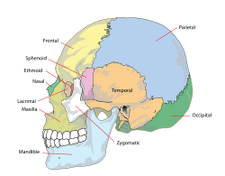
In anatomy, the zygomatic arch, or cheek bone, is a part of the skull formed by the zygomatic process of the temporal bone and the temporal process of the zygomatic bone, the two being united by an oblique suture ; the tendon of the temporal muscle passes medial to the arch, to gain insertion into the coronoid process of the mandible (jawbone).

In anatomy, the temporalis muscle, also known as the temporal muscle, is one of the muscles of mastication (chewing). It is a broad, fan-shaped convergent muscle on each side of the head that fills the temporal fossa, superior to the zygomatic arch so it covers much of the temporal bone.Temporal refers to the head's temples.

In human anatomy, the forehead is an area of the head bounded by three features, two of the skull and one of the scalp. The top of the forehead is marked by the hairline, the edge of the area where hair on the scalp grows. The bottom of the forehead is marked by the supraorbital ridge, the bone feature of the skull above the eyes. The two sides of the forehead are marked by the temporal ridge, a bone feature that links the supraorbital ridge to the coronal suture line and beyond. However, the eyebrows do not form part of the forehead.

The four classical muscles of mastication elevate the mandible and move it forward/backward and laterally, facilitating biting and chewing. Other muscles are responsible for opening the jaw, namely the geniohyoid, mylohyoid, and digastric muscles.

The middle meningeal artery is typically the third branch of the first portion of the maxillary artery. After branching off the maxillary artery in the infratemporal fossa, it runs through the foramen spinosum to supply the dura mater and the calvaria. The middle meningeal artery is the largest of the three (paired) arteries that supply the meninges, the others being the anterior meningeal artery and the posterior meningeal artery.

In anatomy, the masseter is one of the muscles of mastication. Found only in mammals, it is particularly powerful in herbivores to facilitate chewing of plant matter. The most obvious muscle of mastication is the masseter muscle, since it is the most superficial and one of the strongest.

The temporal fossa is a fossa on the side of the skull bounded by the temporal lines above, and the zygomatic arch below. Its floor is formed by the outer surfaces of four bones of the skull. The fossa is filled by the temporalis muscle.

The splenius capitis is a broad, straplike muscle in the back of the neck. It pulls on the base of the skull from the vertebrae in the neck and upper thorax. It is involved in movements such as shaking the head.

The greater wing of the sphenoid bone, or alisphenoid, is a bony process of the sphenoid bone, positioned in the skull behind each eye. There is one on each side, extending from the side of the body of the sphenoid and curving upward, laterally, and backward.

The zygomaticotemporal nerve (zygomaticotemporal branch, temporal branch) is a cutaneous (sensory) nerve of the head. It is a branch of the zygomatic nerve (itself a branch of the maxillary nerve (CN V2)). It arises in the orbit and exits the orbit through the zygomaticotemporal foramen in the zygomatic bone to enter the temporal fossa. It is distributed to the skin of the side of the forehead. It also contains a parasympathetic secretomotor component for the lacrimal gland which it confers to the lacrimal nerve (which then delivers it to the gland).

The superficial temporal vein is a vein of the side of the head which collects venous blood from the region of the temple. It arises from an anastomosing venous plexus on the side and vertex of the head. The superficial temporal vein terminates within the substance of the parotid gland by uniting with the maxillary vein to form the retromandibular vein.

The squamous part of temporal bone, or temporal squama, forms the front and upper part of the temporal bone, and is scale-like, thin, and translucent.
The mastoid part of the temporal bone is the posterior (back) part of the temporal bone, one of the bones of the skull. Its rough surface gives attachment to various muscles and it has openings for blood vessels. From its borders, the mastoid part articulates with two other bones.

The infratemporal fossa is an irregularly shaped cavity that is a part of the skull. It is situated below and medial to the zygomatic arch. It is not fully enclosed by bone in all directions. It contains superficial muscles, including the lower part of the temporalis muscle, the lateral pterygoid muscle, and the medial pterygoid muscle. It also contains important blood vessels such as the middle meningeal artery, the pterygoid plexus, and the retromandibular vein, and nerves such as the mandibular nerve (CN V3) and its branches.

The deep cervical fascia lies under cover of the platysma, and invests the muscles of the neck; it also forms sheaths for the carotid vessels, and for the structures situated in front of the vertebral column. Its attachment to the hyoid bone prevents the formation of a dewlap.

The pterion is the region where the frontal, parietal, temporal, and sphenoid bones join. It is located on the side of the skull, just behind the temple. It is also considered to be the weakest part of the skull, which makes it clinically significant, as if there is a fracture around the pterion it could be accompanied by an epidural hematoma.

The deep temporal arteries are two arteries of the head. They ascend between the temporalis muscle and the pericranium. They anastomose with the middle temporal artery, among other vessels. They supply the temporalis muscle.
Vetusodon is an extinct genus of cynodonts belonging to the clade Epicynodontia. It contains one species, Vetusodon elikhulu, which is known from four specimens found in the Late Permian Daptocephalus Assemblage Zone of South Africa. With a skull length of about 18 centimetres (7.1 in), Vetusodon is the largest known cynodont from the Permian. Through convergent evolution, it possessed several unusual features reminiscent of the contemporary therocephalian Moschorhinus, including broad, robust jaws, large incisors and canines, and small, single-cusped postcanine teeth.