Tonsil stones

Last updated

Tonsil stones
Other namesTonsillolith, tonsillolithiasis, tonsillar stones, chronic caseous tonsillitis
Tonsillolith in mouth.jpg
A tonsillolith lodged in the tonsillar crypt
Specialty Otorhinolaryngology
Symptoms Discomfort, bad breath [1]
Risk factors Recurrent throat infections [2]
Differential diagnosis Calcified granulomatous disease, mycosis, syphilis [2]
TreatmentGargling with salt water, tonsillectomy [1]
Medication Chlorhexidine or cetylpyridinium chloride [1]
FrequencyUp to 10% [1]

Tonsil stones, also known as tonsilloliths, are mineralizations of debris within the crevices of the tonsils. [1] [3] When not mineralized, the presence of debris is known as chronic caseous tonsillitis (CCT). [1] Symptoms may include bad breath, [1] foreign body sensation, sore throat, pain or discomfort with swallowing, and cough. [4] Generally there is no pain, though there may be the feeling of something present. [1] The presence of tonsil stones may be otherwise undetectable, however some people have reported seeing white material in the rear of their throat.

Contents

Risk factors may include recurrent throat infections. [2] Tonsil stones contain a biofilm composed of a number of different bacteria, calcium salts either alone or in combination with other mineral salts. [5] [1] While they most commonly occur in the palatine tonsils, they may also occur in the adenoids, lingual tonsils and tubal tonsil. [3] [6] [7] Tonsil stones have been recorded weighing from 0.3 g to 42 g, [3] and they are typically small in size. However, there are occasional reports of large tonsilloliths. They are often discovered during medical imaging for other reasons and more recently, due to the impact and influence of social media platforms such as TikTok, medical professionals have experienced an increase in patient concern and tonsillolith evaluations. [8] [9]

They are usually benign, so if tonsil stones do not bother the patient, no treatment is needed. [1] However in rare cases, tonsilloliths have presented patients with further complications necessitating surgical extraction. Tonsilloliths that exceed the average size are typically seen in older individuals as the likelihood of developing tonsil stones is linear. Otherwise, gargling salt water and manual removal may be tried. [1] Chlorhexidine or cetylpyridinium chloride may also be tried. [1] Surgical treatment may include partial or complete tonsil removal. [1] [10] Up to 10% of people have tonsil stones. [1] Biological sex does not influence the chance of having tonsil stones, [1] but older people are more commonly affected. [2] Many people opt to extract their own tonsil stones manually or with developments in dental hygiene products. Water flossers have become a more common mechanism to extract tonsilloliths and alleviate the discomfort and complications they exacerbate. Tonsil stones can become dislodged on their own while eating, drinking, gargling, and coughing. Additionally, an exhalation technique that vigorously shakes the tonsils may be performed to dislodge them. This involves loudly producing a voiceless velar fricative sound, at various pitches to shake both the palatine and lingual tonsils.

Signs and symptoms

Tonsil stones may produce no symptoms or they may be associated with bad breath. [1] In fact, many dental professionals argue that tonsil stones are the leading cause of bad breath in their patients. The smell may be that of rotting eggs. [11] Tonsil stones tend to happen most often in people with longterm inflammation in their tonsils. [12]

Occasionally there may be pain when swallowing. [13] Even when they are large, some tonsil stones are only discovered incidentally on X-rays or CAT scans. Other symptoms include a metallic taste, throat closing or tightening, coughing fits, itchy throat, and choking.

Larger tonsil stones may cause recurrent bad breath, which frequently accompanies a tonsil infection, sore throat, white debris, a bad taste in the back of the throat, difficulty swallowing, ear ache, and tonsil swelling. [12] A medical study conducted in 2007 found an association between tonsilloliths and bad breath in patients with a certain type of recurrent tonsillitis. Among those with bad breath, 75% of the subjects had tonsilloliths, while only 6% of subjects with normal halitometry values (normal breath) had tonsilloliths. A foreign body sensation may also exist in the back of the throat. The condition may also be an asymptomatic condition, with detection upon palpating a hard intratonsillar or submucosal mass.

Pathophysiology

Low-power microscope magnification of a cross-section through one of the tonsillar crypts (running diagonally) as it opens onto the surface of the throat (at the top). Stratified epithelium (e) covers the throat's surface and continues as a lining of the crypt. Beneath the surface are numerous nodules (f) of lymphoid tissue. Many lymph cells (dark-colored region) pass from the nodules toward the surface and will eventually mix with the saliva as salivary corpuscles (s). Gray1027.png
Low-power microscope magnification of a cross-section through one of the tonsillar crypts (running diagonally) as it opens onto the surface of the throat (at the top). Stratified epithelium (e) covers the throat's surface and continues as a lining of the crypt. Beneath the surface are numerous nodules (f) of lymphoid tissue. Many lymph cells (dark-colored region) pass from the nodules toward the surface and will eventually mix with the saliva as salivary corpuscles (s).

The mechanism by which these calculi form is subject to debate, [3] though they appear to result from the accumulation of material retained within the crypts, along with the growth of bacteria and fungi—sometimes in association with persistent chronic purulent tonsillitis.

In 2009, an association between biofilms and tonsilloliths was shown. Central to the biofilm concept is the assumption that bacteria form a three dimensional structure, dormant bacteria being in the center to serve as a constant nidus of infection. This impermeable structure renders the biofilm immune to antibiotic treatment. By use of confocal microscopy and microelectrodes, biofilms similar to dental biofilms were shown to be present in the tonsillolith, with oxygen respiration at the outer layer of tonsillolith, denitrification toward the middle, and acidification toward the bottom. [14]

Cause and diagnosis

CT scan of tonsilloliths CT of tonsilloliths.jpg
CT scan of tonsilloliths

Diagnosis is usually made upon inspection. Tonsilloliths are difficult to diagnose in the absence of clear manifestations, and often constitute casual findings of routine radiological studies. The cause of tonsil stones can include a multitude of sources from bacterial infections, streptococcus bacteria, viral infections, adenoviruses, influenza virus, enteroviruses and parainfluenza virus. [15]

Classification

Tonsilloliths or tonsil stones are calcifications that form in the crypts of the palatal tonsils. They are also known to form in the throat and on the roof of the mouth. Tonsils are filled with crevices where bacteria and other materials, including dead cells and mucus, can become trapped. When this occurs, the debris can become concentrated in white formations that occur in the pockets. [12] Researchers found aerobic bacteria present on the surface of tonsilloliths and anaerobic bacteria at the core of tonsilloliths. They have the potential to cause oral halitosis as they contain volatile sulfur compounds and sulfur derived gases, foul smelling compounds produced during bacterial metabolism. [16] Tonsilloliths are formed when this trapped debris accumulates and are expressed from the tonsil. They are generally soft, sometimes rubbery. This tends to occur most often in people who suffer from chronic inflammation in their tonsils or repeated bouts of tonsillitis. [12] They are often associated with post-nasal drip.

Enlarged Tonsil Stones

General size of a tonsilolith compared to a finger Comparison Finder and Tonsillolith.jpg
General size of a tonsilolith compared to a finger

Much rarer than the typical tonsil stones are giant tonsilloliths. Giant tonsilloliths may often be mistaken for other oral maladies, including peritonsillar abscess, and tumors of the tonsil. [17] On average, tonsil stones should appear within a similar range of the image on the right; however, individuals with extenuating cases have been reported. In these instances, extensive care such as extraction by a licensed medical professional may be needed.

Differential diagnosis

Imaging diagnostic techniques can identify a radiopaque mass that may be mistaken for foreign bodies, displaced teeth or calcified blood vessels. CT scan may reveal nonspecific calcified images in the tonsillar zone. The differential diagnosis must be established with acute and chronic tonsillitis, tonsillar hypertrophy, peritonsillar abscesses, foreign bodies, phlebolites, ectopic bone or cartilage, lymph nodes, granulomatous lesions or calcification of the stylohyoid ligament in the context of Eagle syndrome (elongated styloid process). [18]

Differential diagnosis of tonsilloliths includes foreign body, calcified granuloma, malignancy, an enlarged temporal styloid process or rarely, isolated bone which is usually derived from embryonic rests originating from the branchial arches. [19]

Treatment

If tonsil stones do not bother a person, no treatment is needed. [1] Otherwise gargling saltwater and manual removal may be tried. [1] Chlorhexidine or cetylpyridinium chloride may also be tried. [1] Surgical treatment may include partial or complete tonsil removal. [1]

Some people are able to remove tonsil stones using a cotton swab. Oral irrigators are also effective. Most electric oral irrigators are unsuitable for tonsil stone removal because they are too powerful and are likely to cause discomfort and rupture the tonsils, which could result in further complications such as infection. Irrigators that connect directly to the sink tap via a threaded attachment or otherwise are suitable for tonsil stone removal and everyday washing of the tonsils because they can jet water at low-pressure levels that the user can adjust by simply manipulating the sink tap, allowing for a continuous range of pressures to suit each user's requirements. [12]

There are also manually pressurized tonsil stone removers. A manual pump-type tonsil stone remover can adjust the water pressure depending on the number of pumps, effectively removing tonsil stones.

More simply still, gargling with warm, salty water may help alleviate the discomfort of tonsillitis, which often accompanies tonsil stones. Vigorous gargling each morning can also keep the tonsil crypts clear of all but the most persistent tonsilloliths. [12]

Curettage

Larger tonsil stones may require removal by curettage (scooping) or otherwise, although thorough irrigation will still be required afterward to effectively wash out smaller pieces. Larger lesions may require local excision, although these treatments may not completely help the bad breath issues that are often associated with this condition.

Laser

Another option is to decrease the surface area (crypts, crevices, etc.) of the tonsils via laser resurfacing. The procedure is called a laser cryptolysis. It can be performed using a local anesthetic. A scanned carbon dioxide laser selectively vaporizes and smooths the surface of the tonsils. This technique flattens the edges of the crypts and crevices that collect the debris, preventing trapped material from forming stones.

Surgery

Tonsillectomy may be indicated if bad breath due to tonsillar stones persists despite other measures. [20]

Epidemiology

Tonsilloliths or tonsillar concretions occur in up to 10% of the population, frequently due to episodes of tonsillitis. [21] While small concretions in the tonsils are common, true stones are less so. [3] They commonly occur in young adults and are rare in children. [3]

Related Research Articles

<span class="mw-page-title-main">Streptococcal pharyngitis</span> Medical condition

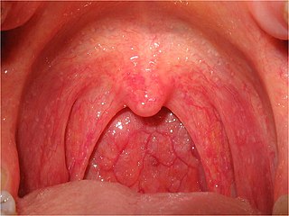
Streptococcal pharyngitis, also known as streptococcal sore throat, is pharyngitis caused by Streptococcus pyogenes, a gram-positive, group A streptococcus. Common symptoms include fever, sore throat, red tonsils, and enlarged lymph nodes in the front of the neck. A headache and nausea or vomiting may also occur. Some develop a sandpaper-like rash which is known as scarlet fever. Symptoms typically begin one to three days after exposure and last seven to ten days.

<span class="mw-page-title-main">Pharyngitis</span> Inflammation of the back of the throat

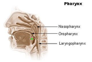
Pharyngitis is inflammation of the back of the throat, known as the pharynx. It typically results in a sore throat and fever. Other symptoms may include a runny nose, cough, headache, difficulty swallowing, swollen lymph nodes, and a hoarse voice. Symptoms usually last 3–5 days, but can be longer depending on cause. Complications can include sinusitis and acute otitis media. Pharyngitis is a type of upper respiratory tract infection.

<span class="mw-page-title-main">Necrotizing gingivitis</span> Non-contagious, painful bacterial infection of the gums

Necrotizing gingivitis (NG) is a common, non-contagious infection of the gums with sudden onset. The main features are painful, bleeding gums, and ulceration of inter-dental papillae. This disease, along with necrotizing periodontitis (NP) and necrotizing stomatitis, is classified as a necrotizing periodontal disease, one of the three general types of gum disease caused by inflammation of the gums (periodontitis).

<span class="mw-page-title-main">Sore throat</span> Medical condition

Sore throat, also known as throat pain, is pain or irritation of the throat. Usually, causes of sore throat include:

<span class="mw-page-title-main">Palatine tonsil</span> Lymphoid organs at the back of the throat on both sides

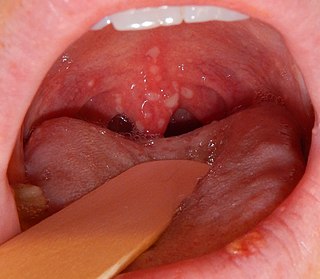
Palatine tonsils, commonly called the tonsils and occasionally called the faucial tonsils, are tonsils located on the left and right sides at the back of the throat, which can often be seen as flesh-colored, pinkish lumps. Tonsils only present as "white lumps" if they are inflamed or infected with symptoms of exudates and severe swelling.

<span class="mw-page-title-main">Tonsillectomy</span> Surgical removal of the tonsils

Tonsillectomy is a surgical procedure in which both palatine tonsils are fully removed from the back of the throat. The procedure is mainly performed for recurrent tonsillitis, throat infections and obstructive sleep apnea (OSA). For those with frequent throat infections, surgery results in 0.6 fewer sore throats in the following year, but there is no evidence of long term benefits. In children with OSA, it results in improved quality of life.

<span class="mw-page-title-main">Bad breath</span> Presence of unpleasant odors in exhaled breath

Bad breath, also known as halitosis, is a symptom in which a noticeably unpleasant breath odour is present. It can result in anxiety among those affected. It is also associated with depression and symptoms of obsessive compulsive disorder.

<span class="mw-page-title-main">Adenoidectomy</span> Surgical removal of the adenoid

Adenoidectomy is the surgical removal of the adenoid for reasons which include impaired breathing through the nose, chronic infections, or recurrent earaches. The effectiveness of removing the adenoids in children to improve recurrent nasal symptoms and/or nasal obstruction has not been well studied. The surgery is less commonly performed in adults in whom the adenoid is much smaller and less active than it is in children. It is most often done on an outpatient basis under general anesthesia. Post-operative pain is generally minimal and reduced by icy or cold foods. The procedure is often combined with tonsillectomy, for which the recovery time is an estimated 10–14 days, sometimes longer, mostly dependent on age.

<span class="mw-page-title-main">Upper respiratory tract infection</span> Medical condition

An upper respiratory tract infection (URTI) is an illness caused by an acute infection, which involves the upper respiratory tract, including the nose, sinuses, pharynx, larynx or trachea. This commonly includes nasal obstruction, sore throat, tonsillitis, pharyngitis, laryngitis, sinusitis, otitis media, and the common cold. Most infections are viral in nature, and in other instances, the cause is bacterial. URTIs can also be fungal or helminthic in origin, but these are less common.

<span class="mw-page-title-main">Tonsillitis</span> Inflammation of the tonsils

Tonsillitis is inflammation of the tonsils in the upper part of the throat. It can be acute or chronic. Acute tonsillitis typically has a rapid onset. Symptoms may include sore throat, fever, enlargement of the tonsils, trouble swallowing, and enlarged lymph nodes around the neck. Complications include peritonsillar abscess (Quinsy).

<span class="mw-page-title-main">Herpangina</span> Medical condition

Herpangina, also called mouth blisters, is a painful mouth infection caused by coxsackieviruses. Usually, herpangina is produced by one particular strain of coxsackie virus A, but it can also be caused by coxsackievirus B or echoviruses. Most cases of herpangina occur in the summer, affecting mostly children. However, it occasionally occurs in adolescents and adults. It was first characterized in 1920.

<span class="mw-page-title-main">Peritonsillar abscess</span> Pus behind the tonsil due to an infection

Peritonsillar abscess (PTA), also known as quinsy, is an accumulation of pus due to an infection behind the tonsil. Symptoms include fever, throat pain, trouble opening the mouth, and a change to the voice. Pain is usually worse on one side. Complications may include blockage of the airway or aspiration pneumonitis.

<span class="mw-page-title-main">Keratosis pharyngis</span> Medical condition

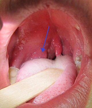
Keratosis Pharyngis is a medical condition where keratin grows on the surface of the pharynx, that is the part of the throat at the back of the mouth. Keratin is a protein that normally occurs as the main component of hair and nails. It is characterized by the presence of whitish-yellow dots on the pharyngeal wall, tonsils or lingual tonsils. They are firmly adherent and cannot be wiped off. The surrounding region does not show any sign or inflammation or any other symptoms that make affect the rest of the body.

<span class="mw-page-title-main">Sialolithiasis</span> Medical condition

Sialolithiasis is a crystallopathy where a calcified mass or sialolith forms within a salivary gland, usually in the duct of the submandibular gland. Less commonly the parotid gland or rarely the sublingual gland or a minor salivary gland may develop salivary stones.

<span class="mw-page-title-main">Tonsil</span> Lymphoid organs in the mouth and throat

The tonsils are a set of lymphoid organs facing into the aerodigestive tract, which is known as Waldeyer's tonsillar ring and consists of the adenoid tonsil, two tubal tonsils, two palatine tonsils, and the lingual tonsils. These organs play an important role in the immune system.

<span class="mw-page-title-main">Adenoiditis</span> Medical condition

Adenoiditis is the inflammation of the adenoid tissue usually caused by an infection. Adenoiditis is treated using medication or surgical intervention.

<span class="mw-page-title-main">Tonsillar crypts</span> Deep indentations in human tonsils

The human palatine tonsils (PT) are covered by stratified squamous epithelium that extends into deep and partly branched tonsillar crypts, of which there are about 10 to 30. The crypts greatly increase the contact surface between environmental influences and lymphoid tissue. In an average adult palatine tonsil the estimated epithelial surface area of the crypts is 295 cm2, in addition to the 45 cm2 of epithelium covering the oropharyngeal surface.

<i>Trichomonas tenax</i> Species of single-celled organism

Trichomonas tenax, or oral trichomonas, is a species of Trichomonas commonly found in the oral cavity of humans. Routine hygiene is generally not sufficient to eliminate the parasite, hence its Latin name, meaning "tenacious". The parasite is frequently encountered in periodontal infections, affecting more than 50% of the population in some areas, but it is usually considered insignificant. T. tenax is generally not found on the gums of healthy patients. It is known to play a pathogenic role in necrotizing ulcerative gingivitis and necrotizing ulcerative periodontitis, worsening preexisting periodontal disease. This parasite is also implicated in some chronic lung diseases; in such cases, removal of the parasite is sufficient to allow recovery.

Carcinoma of the tonsil is a type of squamous cell carcinoma. The tonsil is the most common site of squamous cell carcinoma in the oropharynx. It comprises 23.1% of all malignancies of the oropharynx. The tumors frequently present at advanced stages, and around 70% of patients present with metastasis to the cervical lymph nodes. . The most reported complaints include sore throat, otalgia or dysphagia. Some patients may complain of feeling the presence of a lump in the throat. Approximately 20% patients present with a node in the neck as the only symptom.

<span class="mw-page-title-main">Coblation tonsillectomy</span>

Coblation tonsillectomy is a surgical procedure in which the patient's tonsils are removed by destroying the surrounding tissues that attach them to the pharynx. It was first implemented in 2001. The word coblation is short for ‘controlled ablation’, which means a controlled procedure used to destroy soft tissue.

References

  1. 1 2 3 4 5 6 7 8 9 10 11 12 13 14 15 16 17 18 19 20 Ferguson M, Aydin M, Mickel J (October 2014). "Halitosis and the tonsils: a review of management". Otolaryngology–Head and Neck Surgery. 151 (4): 567–74. doi:10.1177/0194599814544881. PMID   25096359. S2CID   39801742.
  2. 1 2 3 4 White SC, Pharoah MJ (2014). Oral Radiology - E-Book: Principles and Interpretation. Elsevier Health Sciences. p. 527. ISBN   978-0-323-09634-8.
  3. 1 2 3 4 5 6 Ram S, Siar CH, Ismail SM, Prepageran N (July 2004). "Pseudo bilateral tonsilloliths: a case report and review of the literature". Oral Surg Oral Med Oral Pathol Oral Radiol Endod. 98 (1): 110–4. doi:10.1016/j.tripleo.2003.11.015. PMID   15243480.
  4. Bhasme A, Lamba P, Sajjan U (April 2023). "A Case Report of Tonsillolith Treated With Individualized Homoeopathy". Alternative Therapies in Health and Medicine. 29 (3): 88–91. ISSN   1078-6791. PMID   35839105.
  5. Silvestre-Donat FJ, Pla-Mocholi A, Estelles-Ferriol E, Martinez-Mihi V (2005). "Giant tonsillolith: report of a case". Medicina Oral, Patologia Oral y Cirugia Bucal. 10 (3): 239–242. ISSN   1698-6946. PMID   15876967.
  6. Leonard JA, Reilly BK (2 February 2022). "Adenoid stones, an unknown culprit in pediatric throat pain". Ear, Nose & Throat Journal. doi: 10.1177/01455613221074139 . PMID   35107383.
  7. Bayramov N, Usdat Ozturk A, Ercalik Yalcinkaya S (27 May 2022). "Incidental Soft Tissue Calcifications in Cone-Beam Computed Tomography Images". Turkiye Klinikleri Journal of Dental Sciences. 28 (2): 291–298. doi: 10.5336/dentalsci.2021-84346 . Retrieved 17 January 2023.
  8. Sulibhavi A, Isaacson G (26 September 2021). "TikTok Tonsils". Ear, Nose, & Throat Journal. 103 (3): NP183–NP184. doi: 10.1177/01455613211038340 . ISSN   1942-7522. PMID   34569296.
  9. Textbook of Oral Radiology. Elsevier India. 2009. p. 607. ISBN   978-81-312-1148-9.
  10. Wong Chung J, van Benthem P, Blom HM (May 2018). "Tonsillotomy versus tonsillectomy in adults suffering from tonsil-related afflictions: a systematic review". Acta Oto-Laryngologica. 138 (5): 492–501. doi: 10.1080/00016489.2017.1412500 . PMID   29241412.
  11. DeVault KR, Wallace MB, Aqel BA, Lindor KD (2016). Practical Gastroenterology and Hepatology Board Review Toolkit. John Wiley & Sons. p. 59. ISBN   978-1-118-82907-3.
  12. 1 2 3 4 5 6 "Tonsil Stones (Tonsilloliths)". WebMD.com. Retrieved 6 March 2016.
  13. Giudice M, Cristofaro MG, Fava MG, Giudice A (July 2005). "An unusual tonsillolithiasis in a patient with chronic obstructive sialoadenitis". Dentomaxillofac Radiol. 34 (4): 247–50. doi:10.1259/dmfr/19689789. PMID   15961601.
  14. Stoodley P, Debeer D, Longwell M, Nistico L, Hall-Stoodley L, Wenig B, Krespi YP (September 2009). "Tonsillolith: not just a stone but a living biofilm". Otolaryngology–Head and Neck Surgery. 141 (3): 316–21. doi:10.1016/j.otohns.2009.05.019. PMID   19716006. S2CID   209346548.
  15. "Should You Worry About Tonsil Stones?: Dr. Raminder Singh: General Dentistry". www.elitedentalanddenture.com. Retrieved 4 May 2023.
  16. Yellamma Bai K, Vinod Kumar B (July 2015). "Tonsillolith: A polymicrobial biofilm". Medical Journal Armed Forces India. 71 (Suppl 1): S95–S98. doi:10.1016/j.mjafi.2011.12.009. ISSN   0377-1237. PMC   4529540 . PMID   26265885.
  17. Padmanabhan TK, Chandra Dutt GS, Vasudevan DM, Vijayakumar (May–June 1984). "Giant tonsillolith simulating tumour of the tonsil – a case report". Indian J Cancer. 21 (2): 90–1. PMID   6530236.
  18. Silvestre-Donat F, Pla-Mocholi A, Estelles-Ferriol E, Martinez-Mihi V (2005). "Giant tonsillolith: report of a case" (PDF). Medicina Oral, Patología Oral y Cirugía Bucal. 10 (3): 239–42. PMID   15876967.
  19. "Images". Archived from the original on 3 December 2012. Retrieved 28 November 2007.
  20. Darrow DH, Siemens C (August 2002). "Indications for tonsillectomy and adenoidectomy". Laryngoscope. 112 (8 Pt 2 Suppl 100): 6–10. doi:10.1002/lary.5541121404. PMID   12172229. S2CID   24862952.
  21. S. G. Nour, Mafee, Mahmood F., Valvassori, Galdino E., Galdino E. Valbasson, Minerva Becker (2005). Imaging of the head and neck . Stuttgart: Thieme. p.  716. ISBN   1-58890-009-6.