
Histology, also known as microscopic anatomy or microanatomy, is the branch of biology that studies the microscopic anatomy of biological tissues. Histology is the microscopic counterpart to gross anatomy, which looks at larger structures visible without a microscope. Although one may divide microscopic anatomy into organology, the study of organs, histology, the study of tissues, and cytology, the study of cells, modern usage places all of these topics under the field of histology. In medicine, histopathology is the branch of histology that includes the microscopic identification and study of diseased tissue. In the field of paleontology, the term paleohistology refers to the histology of fossil organisms.

Pathology is the study of disease. The word pathology also refers to the study of disease in general, incorporating a wide range of biology research fields and medical practices. However, when used in the context of modern medical treatment, the term is often used in a narrower fashion to refer to processes and tests that fall within the contemporary medical field of "general pathology", an area that includes a number of distinct but inter-related medical specialties that diagnose disease, mostly through analysis of tissue and human cell samples. Idiomatically, "a pathology" may also refer to the predicted or actual progression of particular diseases. The suffix pathy is sometimes used to indicate a state of disease in cases of both physical ailment and psychological conditions. A physician practicing pathology is called a pathologist.

Anatomical pathology (Commonwealth) or anatomic pathology (U.S.) is a medical specialty that is concerned with the diagnosis of disease based on the macroscopic, microscopic, biochemical, immunologic and molecular examination of organs and tissues. Over the 20th century, surgical pathology has evolved tremendously: from historical examination of whole bodies (autopsy) to a more modernized practice, centered on the diagnosis and prognosis of cancer to guide treatment decision-making in oncology. Its modern founder was the Italian scientist Giovanni Battista Morgagni from Forlì.

A microscope slide is a thin flat piece of glass, typically 75 by 26 mm and about 1 mm thick, used to hold objects for examination under a microscope. Typically the object is mounted (secured) on the slide, and then both are inserted together in the microscope for viewing. This arrangement allows several slide-mounted objects to be quickly inserted and removed from the microscope, labeled, transported, and stored in appropriate slide cases or folders etc.

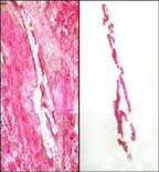
Histopathology is the microscopic examination of tissue in order to study the manifestations of disease. Specifically, in clinical medicine, histopathology refers to the examination of a biopsy or surgical specimen by a pathologist, after the specimen has been processed and histological sections have been placed onto glass slides. In contrast, cytopathology examines free cells or tissue micro-fragments.
A microtome is a cutting tool used to produce extremely thin slices of material known as sections, with the process being termed microsectioning. Important in science, microtomes are used in microscopy for the preparation of samples for observation under transmitted light or electron radiation.

Hematoxylin and eosin stain is one of the principal tissue stains used in histology. It is the most widely used stain in medical diagnosis and is often the gold standard. For example, when a pathologist looks at a biopsy of a suspected cancer, the histological section is likely to be stained with H&E.
Articles related specifically to biomedical engineering include:
Laser capture microdissection (LCM), also called microdissection, laser microdissection (LMD), or laser-assisted microdissection, is a method for isolating specific cells of interest from microscopic regions of tissue/cells/organisms.
In the fields of histology, pathology, and cell biology, fixation is the preservation of biological tissues from decay due to autolysis or putrefaction. It terminates any ongoing biochemical reactions and may also increase the treated tissues' mechanical strength or stability. Tissue fixation is a critical step in the preparation of histological sections, its broad objective being to preserve cells and tissue components and to do this in such a way as to allow for the preparation of thin, stained sections. This allows the investigation of the tissues' structure, which is determined by the shapes and sizes of such macromolecules as proteins and nucleic acids.

Virtual microscopy is a method of posting microscope images on, and transmitting them over, computer networks. This allows independent viewing of images by large numbers of people in diverse locations. It involves a synthesis of microscopy technologies and digital technologies. The use of virtual microscopes can transform traditional teaching methods by removing the reliance on physical space, equipment, and specimens to a model that is solely dependent upon computer-internet access. This increases the convenience of accessing the slide sets and making the slides available to a broader audience. Digitized slides can have a high resolution and are resistant to being damaged or broken over time.
Digital pathology is a sub-field of pathology that focuses on managing and analyzing information generated from digitized specimen slides. It utilizes computer-based technology and virtual microscopy to view, manage, share, and analyze digital slides on computer monitors. This field has applications in diagnostic medicine and aims to achieve more efficient and cost-effective diagnoses, prognoses, and disease predictions through advancements in machine learning and artificial intelligence in healthcare.

Automated tissue image analysis or histopathology image analysis (HIMA) is a process by which computer-controlled automatic test equipment is used to evaluate tissue samples, using computations to derive quantitative measurements from an image to avoid subjective errors.
Bioimage informatics is a subfield of bioinformatics and computational biology. It focuses on the use of computational techniques to analyze bioimages, especially cellular and molecular images, at large scale and high throughput. The goal is to obtain useful knowledge out of complicated and heterogeneous image and related metadata.

MBF Bioscience is a biotech company that develops microscopy software and hardware for bioscience research and education. MBF Bioscience’s primary location is Williston, Vermont, United States and has offices that market, sell, and support its line of hardware and software products throughout North America, Europe, and Asia.
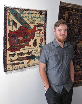
Todd Huffman is an American technology entrepreneur and prolific photographer. He was a co-founder of the biomedical imaging company 3Scan, a member of the disaster aid group Synergy Strike Force, a researcher for DARPA, and a co-founder of the unconference BIL Conference.
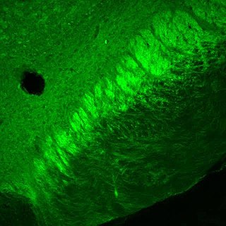
Microscopy with UV Surface Excitation (MUSE) is a novel microscopy method that utilizes the shallow penetration of UV photons excitation. Compared to conventional microscopes, which usually require sectioning to exclude blurred signals from outside of the focal plane, MUSE's low penetration depth limits the excitation volume to a thin layer, and removes the tissue sectioning requirement. The entire signal collected is the desired light, and all photons collected contribute to the image formation.
Microtechnique is an aggregate of methods used to prepare micro-objects for studying. It is currently being employed in many fields in life science. Two well-known branches of microtechnique are botanical (plant) microtechnique and zoological (animal) microtechnique.
Tissue image cytometry or tissue cytometry is a method of digital histopathology and combines classical digital pathology and computational pathology into one integrated approach with solutions for all kinds of diseases, tissue and cell types as well as molecular markers and corresponding staining methods to visualize these markers. Tissue cytometry uses virtual slides as they can be generated by multiple, commercially available slide scanners, as well as dedicated image analysis software – preferentially including machine and deep learning algorithms. Tissue cytometry enables cellular analysis within thick tissues, retaining morphological and contextual information, including spatial information on defined cellular subpopulations.

Megan Klimen is an American entrepreneur, biotechnology executive, and advocate for decentralized scientific data storage. She is best known as one of the co-founders and chief operating officer of 3Scan and currently serves as the Founding Director of the Filecoin Foundation. Megan is a founding member of Women in Hardware, a member of the disaster aid group Synergy Strike Force and a co-founder of the unconference BIL Conference. Megan was a speaker at the World Economic Forum in 2022 and 2024.