Automated tissue image analysis

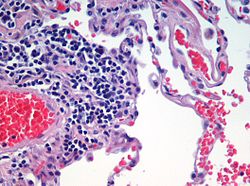
Last updated
A stained histologic specimen, sandwiched between a glass microscope slide and coverslip, mounted on the stage of a light microscope. Microscope with stained slide.jpg
A stained histologic specimen, sandwiched between a glass microscope slide and coverslip, mounted on the stage of a light microscope.
Microscopic view of a histologic specimen of human lung tissue stained with hematoxylin and eosin. Emphysema H and E.jpg
Microscopic view of a histologic specimen of human lung tissue stained with hematoxylin and eosin.

Automated tissue image analysis or histopathology image analysis (HIMA) is a process by which computer-controlled automatic test equipment is used to evaluate tissue samples, using computations to derive quantitative measurements from an image to avoid subjective errors.

Contents

In a typical application, automated tissue image analysis could be used to measure the aggregate activity of cancer cells in a biopsy of a cancerous tumor taken from a patient. In breast cancer patients, for example, automated tissue image analysis may be used to test for high levels of proteins known to be present in more aggressive forms of breast cancers.

Applications

Automated tissue imaging analysis can significantly reduce uncertainty in characterizing tumors compared to evaluations done by histologists, [1] or improve the prediction rate of recurrence of some cancers. [2] [3] As it is a digital system, suitable for networking, it also facilitates cooperative efforts between distant sites. [4] Systems for automatically analyzing tissue samples also reduce costs and save time. [1]

High-performance CCD cameras are used for acquiring the digital images. Coupled with advanced widefield microscopes and various algorithms for image restoration, this approach can provide better results than confocal techniques at comparable speeds and lower costs. [5]

Processes

The United States Food and Drug Administration classifies these systems as medical devices, under the general instrumentation category of automatic test equipment. [6]

ATIS have seven basic processes (sample preparation, image acquisition, image analysis, results reporting, data storage, network communication, and self-system diagnostics) and realization of these functions highly accurate hardware and well-integrated, complex, and expensive software. [7]

Preparation

Specimen preparation is critical for evaluating the tumor in the automated system. In the first part of the preparation process the biopsied tissue is cut to an appropriate size (typically 4 mm), fixed in buffered formalin, dehydrated in ethanol-xylene, embedded in paraffin, thin sectioned typically to 4 um slices, then mounted onto at least two barcoded slides (a control and a test). Next the paraffin is removed from the tissue, the tissue is rehydrated, then stained. Any inconsistency in these procedures from case to case may result in uncertainties in the outcome of the analysis. These potential and irreducible inconsistencies in analysis results motivated the development of Automated Tissue Image Systems.

Acquisition

Digital micrographs are acquired of the stained specimen on the glass slide. The images are taken by a set of charge-coupled devices (CCD). [8]

Analysis

Image analysis involves complex computer algorithms which identify and characterize cellular color, shape, and quantity of the tissue sample using image pattern recognition technology based on vector quantization. Vector representations of objects in the image, as opposed to bitmap representations, have superior zoom-in ability. Once the sample image has been acquired and resident in the computer's random access memory as a large array of 0's and 1's, a programmer knowledgeable in cellular architecture can develop deterministic algorithms applied to the entire memory space to detect cell patterns from previously defined cellular structures and formations known to be significant. [9]

The aggregate algorithm outcome is a set of measurements that is far superior to any human sensitivity to intensity or luminance and color hue, while at the same time improving test consistency from eyeball to eyeball.[ citation needed ]

Reporting

The systems have the capability of presenting the resulting data in text and graphically, including on high definition monitors, to the system user. Computer printers, as relatively low image resolution devices, are used mostly to present final pathology reports that could include text and graphics.[ citation needed ]

Storage

Storage of the acquired data (graphical digital slide files and text data) involves saving system information in a data storage device system having at least convenient retrieval, and file management capabilities.[ citation needed ]

Medical imaging industry standards includes the Picture Archiving and Communication Systems (PACS), of European origin, which are image and information management solutions in computer networks that allow hospitals and clinics to acquire, distribute and archive medical images and diagnostic reports across the enterprise. Another standard of European origin is the Data and Picture Archiving and Communication System (DPACS). Although medical images can be stored in various formats, a common format has been Digital Imaging and Communications in Medicine (DICOM).[ citation needed ]

See also

Related Research Articles

Computer vision is an interdisciplinary scientific field that deals with how computers can gain high-level understanding from digital images or videos. From the perspective of engineering, it seeks to understand and automate tasks that the human visual system can do.

Digital signal processing (DSP) is the use of digital processing, such as by computers or more specialized digital signal processors, to perform a wide variety of signal processing operations. The digital signals processed in this manner are a sequence of numbers that represent samples of a continuous variable in a domain such as time, space, or frequency. In digital electronics, a digital signal is represented as a pulse train, which is typically generated by the switching of a transistor.

Pathology Study of the causes and effects of disease or injury, also how they arise

Pathology is the study of the causes and effects of disease or injury. The word pathology also refers to the study of disease in general, incorporating a wide range of biology research fields and medical practices. However, when used in the context of modern medical treatment, the term is often used in a narrower fashion to refer to processes and tests which fall within the contemporary medical field of "general pathology", an area which includes a number of distinct but inter-related medical specialties that diagnose disease, mostly through analysis of tissue, cell, and body fluid samples. Idiomatically, "a pathology" may also refer to the predicted or actual progression of particular diseases, and the affix pathy is sometimes used to indicate a state of disease in cases of both physical ailment and psychological conditions. A physician practicing pathology is called a pathologist.

Radiology Branch of Medicine

Radiology is the medical discipline that uses medical imaging to diagnose and treat diseases within the bodies of animals and humans.

Image registration

Image registration is the process of transforming different sets of data into one coordinate system. Data may be multiple photographs, data from different sensors, times, depths, or viewpoints. It is used in computer vision, medical imaging, military automatic target recognition, and compiling and analyzing images and data from satellites. Registration is necessary in order to be able to compare or integrate the data obtained from these different measurements.

Medical imaging Technique and process of creating visual representations of the interior of a body

Medical imaging is the technique and process of imaging the interior of a body for clinical analysis and medical intervention, as well as visual representation of the function of some organs or tissues (physiology). Medical imaging seeks to reveal internal structures hidden by the skin and bones, as well as to diagnose and treat disease. Medical imaging also establishes a database of normal anatomy and physiology to make it possible to identify abnormalities. Although imaging of removed organs and tissues can be performed for medical reasons, such procedures are usually considered part of pathology instead of medical imaging.

Histopathology Microscopic examination of tissue in order to study and diagnose disease

Histopathology refers to the microscopic examination of tissue in order to study the manifestations of disease. Specifically, in clinical medicine, histopathology refers to the examination of a biopsy or surgical specimen by a pathologist, after the specimen has been processed and histological sections have been placed onto glass slides. In contrast, cytopathology examines free cells or tissue micro-fragments.

Computer-aided diagnosis Type of diagnosis assisted by computers

Computer-aided detection (CADe), also called computer-aided diagnosis (CADx), are systems that assist doctors in the interpretation of medical images. Imaging techniques in X-ray, MRI, and ultrasound diagnostics yield a great deal of information that the radiologist or other medical professional has to analyze and evaluate comprehensively in a short time. CAD systems process digital images for typical appearances and to highlight conspicuous sections, such as possible diseases, in order to offer input to support a decision taken by the professional.

Medical laboratory

A medical laboratory or clinical laboratory is a laboratory where tests are carried out on clinical specimens to obtain information about the health of a patient to aid in diagnosis, treatment, and prevention of disease. Clinical Medical laboratories are an example of applied science, as opposed to research laboratories that focus on basic science, such as found in some academic institutions.

Numerical relay

In utility and industrial electric power transmission and distribution systems, a numerical relay is a computer-based system with software-based protection algorithms for the detection of electrical faults. Such relays are also termed as microprocessor type protective relays. They are functional replacements for electro-mechanical protective relays and may include many protection functions in one unit, as well as providing metering, communication, and self-test functions.

3D Slicer

3D Slicer (Slicer) is a free and open source software package for image analysis and scientific visualization. Slicer is used in a variety of medical applications, including autism, multiple sclerosis, systemic lupus erythematosus, prostate cancer, lung cancer, breast cancer, schizophrenia, orthopedic biomechanics, COPD, cardiovascular disease and neurosurgery.

Digital pathology

Digital pathology is a sub-field of pathology that focuses on data management based on information generated from digitized specimen slides. Through the use of computer-based technology, digital pathology utilizes virtual microscopy. Glass slides are converted into digital slides that can be viewed, managed, shared and analyzed on a computer monitor. With the practice of Whole-Slide Imaging (WSI), which is another name for virtual microscopy, the field of digital pathology is growing and has applications in diagnostic medicine, with the goal of achieving efficient and cheaper diagnoses, prognosis, and prediction of diseases due to the success in Artificial Intelligence and Machine Learning.

Bioimage informatics is a subfield of bioinformatics and computational biology. It focuses on the use of computational techniques to analyze bioimages, especially cellular and molecular images, at large scale and high throughput. The goal is to obtain useful knowledge out of complicated and heterogeneous image and related metadata.

Medical image computing (MIC) is an interdisciplinary field at the intersection of computer science, information engineering, electrical engineering, physics, mathematics and medicine. This field develops computational and mathematical methods for solving problems pertaining to medical images and their use for biomedical research and clinical care.

Dynamic angiothermography (DATG) is a technique for the diagnosis of breast cancer. This technique, though springing from the thermography of old conception, is based on a completely different principle. DATG records the temperature variations linked to the vascular changes in the breast due to angiogenesis. The presence, change, and growth of tumors and lesions in breast tissue change the vascular network in the breast. Consequently, measuring the vascular structure over time, DATG effectively monitors the change in breast tissue due to tumors and lesions. It is currently used in combination with other techniques for diagnosis of breast cancer. This diagnostic method is a low cost one compared with other techniques.

Automated whole-breast ultrasound (AWBU) is a medical imaging technique used in radiology to obtain volumetric ultrasound data of the entire breast.

In the field of medicine, radiomics is a method that extracts a large number of features from medical images using data-characterisation algorithms. These features, termed radiomic features, have the potential to uncover tumoral patterns and characteristics that fail to be appreciated by the naked eye. The hypothesis of radiomics is that the distinctive imaging features between disease forms may be useful for predicting prognosis and therapeutic response for various cancer types, thus providing valuable information for personalized therapy. Radiomics emerged from the medical fields of radiology and oncology and is the most advanced in applications within these fields. However, the technique can be applied to any medical study where a pathological process can be imaged.

Artificial intelligence in healthcare Overview of the use of artificial intelligence in healthcare

Artificial intelligence in healthcare is an overarching term used to describe the use of machine-learning algorithms and software, or artificial intelligence (AI), to mimic human cognition in the analysis, presentation, and comprehension of complex medical and health care data. Specifically, AI is the ability of computer algorithms to approximate conclusions based solely on input data.

Applied Spectral Imaging

Applied Spectral Imaging or ASI is a multinational biomedical company that develops and manufactures microscopy imaging and digital analysis tools for hospitals, service laboratories and research centers. The company provides cytogenetic, pathology and research laboratories with brightfield, fluorescence and spectral imaging clinical applications. Test slides can be scanned, captured, archived, reviewed on screen, analyzed with computer-assisted algorithms and reported. ASI system platforms automate the workflow process to reduce human error in the identification and classification of chromosomal disorders, genome instability, various oncological malignancies, among other diseases.

Time-activity curve

In medical imaging, a time-activity curve is a curve of radioactivity plotted on the y-axis against the time plotted on the x-axis. It shows the concentration of a radiotracer within a region of interest in an image, measured over time from a dynamic scan. Generally, when a time-activity curve is obtained within a tissue, it is called as a tissue time-activity curve, which represents the concentration of tracer within a region of interest inside a tissue over time.

References

  1. 1 2 O'Gorman, Lawrence; Sanderson, Arthur C.; Preston, Kendall (September 1985). "A System for Automated Liver Tissue Image Analysis: Methods and Results". IEEE Transactions on Biomedical Engineering. BME-32 (9): 696–706. doi:10.1109/TBME.1985.325587. ISSN   0018-9294. PMID   4054933. S2CID   30050996.
  2. Teverovskiy, M.; Kumar, V.; Junshui Ma; Kotsianti, A.; Verbel, D.; Tabesh, A.; Ho-Yuen Pang; Vengrenyuk, Y.; Fogarasi, S.; Saidi, O. (2004). "Improved prediction of prostate cancer recurrence based on an automated tissue image analysis system". 2004 2nd IEEE International Symposium on Biomedical Imaging: Macro to Nano (IEEE Cat No. 04EX821). Vol. 2. pp. 257–260. CiteSeerX   10.1.1.58.9929 . doi:10.1109/ISBI.2004.1398523. ISBN   0-7803-8388-5. S2CID   8724168.
  3. Ali Tabesh; Mikhail Teverovskiy; Ho-Yuen Pang; Vinay P. Kumar; David Verbel; Angeliki Kotsianti; Olivier Saidi (October 2007). "Multifeature Prostate Cancer Diagnosis and Gleason Grading of Histological Images" (PDF). IEEE Transactions on Medical Imaging. 26 (10): 1366–1378. doi:10.1109/TMI.2007.898536. ISSN   0278-0062. PMID   17948727. S2CID   14673541. Archived from the original (PDF) on 2011-07-27. Retrieved 2010-09-04.
  4. Bruce Mccullough; Xiaoyou Ying; Thomas Monticello; Marc Bonnefoi (2004). "Digital Microscopy Imaging and New Approaches in Toxicologic Pathology". Toxicologic Pathology. 32 (2): 49–58. doi: 10.1080/01926230490451734 . PMID   15503664.
  5. Pornchai Phukpattaranont; Pleumjit Boonyaphiphat (2007). An Automatic Cell Counting Method for a Microscopic Tissue Image from Breast Cancer. IFMBE Proceedings. Vol. 15. pp. 241–244. doi:10.1007/978-3-540-68017-8_63. ISBN   978-3-540-68016-1.
  6. Stokes, David (2003-11-25). Testing Computer Systems for FDA/MHRA Compliance - David Stokes - Google Books. ISBN   9780849321634 . Retrieved 2012-07-12.
  7. Chen, W.; Foran, D. J. (2006). "Analytica Chimica Acta - Advances in cancer tissue microarray technology: Towards improved understanding and diagnostics". Analytica Chimica Acta. 564 (1): 74–81. doi:10.1016/j.aca.2005.11.083. PMC   2583100 . PMID   17723364.
  8. MacDonald, J. H.; Wells, K.; Reader, A. J.; Ott, R. J. (February 1997). "A CCD-based tissue imaging system". Nuclear Instruments and Methods in Physics Research Section A: Accelerators, Spectrometers, Detectors and Associated Equipment. Nuclear Instruments and Methods in Physics Research. 392 (1–3): 220–226. Bibcode:1997NIMPA.392..220M. doi:10.1016/S0168-9002(97)00297-0.
  9. Han, J.W.; Breckon, T.P.; Randell, D.A.; Landini, G. (2012). "The Application of Support Vector Machine Classification to Detect Cell Nuclei for Automated Microscopy". Machine Vision and Applications. Springer. 23 (1): 15–24. doi:10.1007/s00138-010-0275-y. S2CID   12446454.