ADP-ribosylhydrolase

Last updated
ADP-ribosylhydrolase
PDB 1t5j EBI.jpg
crystal structure of ribosylglycohydrolase mj1187 from methanococcus jannaschii
Identifiers
SymbolARH
Pfam PF03747
InterPro IPR005502
SCOP2 1t5j / SCOPe / SUPFAM
Available protein structures:
Pfam   structures / ECOD  
PDB RCSB PDB; PDBe; PDBj
PDBsum structure summary

In molecular biology, the (ADP-ribosyl)hydrolase (ARH) family contains enzymes which catalyses the hydrolysis of ADP-ribosyl modifications from proteins, nucleic acids and small molecules. [1]

Contents

Types

This family has three members in humans (ARH1-3): ARH1, also termed [Protein ADP-ribosylarginine] hydrolase, cleaves ADP-ribose-L-arginine, [2] ARH2, which is predicted to be enzymatically inactive, [3] and ARH3, which cleaves primarily ADP-ribose-L-serine, but was shown to also hydrolyse poly(ADP-ribose), 1''-O-acetyl-ADP-ribose and alpha-nicotinamide adenine dinucleotide. [4] [5] [6] [7] The family also includes ADP-ribosyl-(dinitrogen reductase) hydrolase (DraG) known to regulate dinitrogenase reductase, a key enzyme of the nitrogen fixating pathway in bacteria, [8] [1] and most surprisingly jellyfish crystallins, [8] [9] although the latter proteins appear to have lost the presumed active site residues.

ClassSpeciesIntracellular
location
ActivityFunction
BacteriaHumanOthers
I ARH1 endoplasmic reticulum, cytoplasmADP-ribosylarginine hydrolaseinflammation, genomic stability
II ARH2 cytoplasm, cardiac sarcomeresinactiveheart chamber outgrowth
III ARH3 Nucleus, cytoplasmADP-ribosylserine hydrolaseDNA repair
IVCrystallin J1 [9] and SelJ [10] inactive Crystallin
V DraG ADP-ribosylarginine hydrolaseRegulation of nitrogen fixation

See also

Related Research Articles

<span class="mw-page-title-main">Nicotinamide adenine dinucleotide</span> Chemical compound which is reduced and oxidized

Nicotinamide adenine dinucleotide (NAD) is a coenzyme central to metabolism. Found in all living cells, NAD is called a dinucleotide because it consists of two nucleotides joined through their phosphate groups. One nucleotide contains an adenine nucleobase and the other nicotinamide. NAD exists in two forms: an oxidized and reduced form, abbreviated as NAD+ and NADH (H for hydrogen), respectively.

<span class="mw-page-title-main">Poly (ADP-ribose) polymerase</span> Family of proteins

Poly (ADP-ribose) polymerase (PARP) is a family of proteins involved in a number of cellular processes such as DNA repair, genomic stability, and programmed cell death.

<span class="mw-page-title-main">ADP-ribosylation</span> Addition of one or more ADP-ribose moieties to a protein.

ADP-ribosylation is the addition of one or more ADP-ribose moieties to a protein. It is a reversible post-translational modification that is involved in many cellular processes, including cell signaling, DNA repair, gene regulation and apoptosis. Improper ADP-ribosylation has been implicated in some forms of cancer. It is also the basis for the toxicity of bacterial compounds such as cholera toxin, diphtheria toxin, and others.

<span class="mw-page-title-main">PARP1</span> Mammalian protein found in Homo sapiens

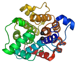
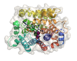
Poly [ADP-ribose] polymerase 1 (PARP-1) also known as NAD+ ADP-ribosyltransferase 1 or poly[ADP-ribose] synthase 1 is an enzyme that in humans is encoded by the PARP1 gene. It is the most abundant of the PARP family of enzymes, accounting for 90% of the NAD+ used by the family. PARP1 is mostly present in cell nucleus, but cytosolic fraction of this protein was also reported.

<span class="mw-page-title-main">ADP-ribose diphosphatase</span>

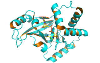
ADP-ribose diphosphatase (EC 3.6.1.13) is an enzyme that catalyzes a hydrolysis reaction in which water nucleophilically attacks ADP-ribose to produce AMP and D-ribose 5-phosphate. Enzyme hydrolysis occurs by the breakage of a phosphoanhydride bond and is dependent on Mg2+ ions that are held in complex by the enzyme.

<span class="mw-page-title-main">ADP-ribosyl-(dinitrogen reductase) hydrolase</span>

In enzymology, an ADP-ribosyl-[dinitrogen reductase] hydrolase is an enzyme that catalyzes the chemical reaction

In enzymology, a NAD+ glycohydrolase (EC 3.2.2.5) is an enzyme that catalyzes the chemical reaction

<span class="mw-page-title-main">ADP-ribosyl cyclase</span>

In enzymology, a ADP-ribosyl cyclase/cyclic ADP-ribose hydrolase (EC 3.2.2.6) is a bifunctional enzyme that catalyzes the chemical reaction

<span class="mw-page-title-main">NAD(P)(+)—protein-arginine ADP-ribosyltransferase</span>

In enzymology, a NAD(P)+-protein-arginine ADP-ribosyltransferase (EC 2.4.2.31) is an enzyme that catalyzes the chemical reaction using nicotinamide adenine dinucleotide

In enzymology, a NAD+-dinitrogen-reductase ADP-D-ribosyltransferase (EC 2.4.2.37) is an enzyme that catalyzes the chemical reaction

<span class="mw-page-title-main">Tankyrase</span> Enzyme

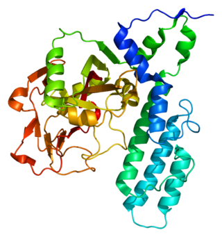
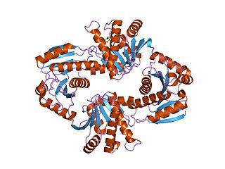
Tankyrase, also known as tankyrase 1, is an enzyme that in humans is encoded by the TNKS gene. It inhibits the binding of TERF1 to telomeric DNA. Tankyrase attracts substantial interest in cancer research through its interaction with AXIN1 and AXIN2, which are negative regulators of pro-oncogenic β-catenin signaling. Importantly, activity in the β-catenin destruction complex can be increased by tankyrase inhibitors and thus such inhibitors are a potential therapeutic option to reduce the growth of β-catenin-dependent cancers.

<span class="mw-page-title-main">PARP4</span> Enzyme

Poly [ADP-ribose] polymerase 4 is an enzyme that in humans is encoded by the PARP4 gene.

<span class="mw-page-title-main">PARP3</span> Protein-coding gene in the species Homo sapiens

Poly [ADP-ribose] polymerase 3 is an enzyme that in humans is encoded by the PARP3 gene.

<span class="mw-page-title-main">PARP2</span> Protein-coding gene in the species Homo sapiens

Poly [ADP-ribose] polymerase 2 is an enzyme that in humans is encoded by the PARP2 gene. It is one of the PARP family of enzymes.

<span class="mw-page-title-main">ADP-ribosylhydrolase 3</span> Protein-coding gene in the species Homo sapiens

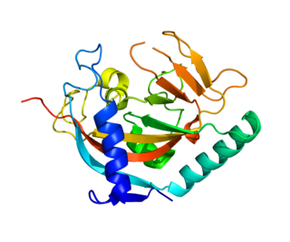
(ADP-ribosyl)hydrolase 3 (ARH3) is an enzyme that in humans is encoded by the ADPRHL2 gene (also called ADPRS). This enzyme reverses the proteins’ post-translational addition of ADP-ribose to serine residues as part of the DNA damage response The enzyme is also known to cleave poly(ADP-ribose) polymers, 1''-O-acetyl-ADP-ribose and alpha-NAD+

<span class="mw-page-title-main">Macro domain</span>

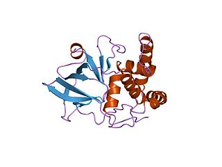
In molecular biology, the Macro domain or A1pp domain is a module of about 180 amino acids which can bind ADP-ribose, an NAD metabolite, or related ligands. Binding to ADP-ribose can be either covalent or non-covalent: in certain cases it is believed to bind non-covalently, while in other cases it appears to bind both non-covalently through a zinc finger motif, and covalently through a separate region of the protein.

<span class="mw-page-title-main">Clostridium botulinum C3 toxin</span>

Clostridium botulinum C3 exoenzyme is a toxin that causes the addition of one or more ADP-ribose moieties to Rho-like proteins. Many bacterial toxins nucleotide-binding modify by ADP-ribosylation proteins involved in essential cell functions, leading to their toxic effects.

<span class="mw-page-title-main">PARG</span> Protein-coding gene in the species Homo sapiens

Poly (ADP-ribose) glycohydrolase is an enzyme that in humans is encoded by the PARG gene.

<span class="mw-page-title-main">ADP-ribosylhydrolase 1</span>

(ADP-ribosyl)hydrolase 1, also termed [Protein ADP-ribosylarginine] hydrolase and protein-Nω-(ADP-D-ribosyl)-L-arginine ADP-ribosylhydrolase (EC 3.2.2.19), is an enzyme that in humans is encoded by the ADPRH gene. This enzyme is a specific mono(ADP-ribosyl)hydrolase that catalyses the removal of an ADP-ribosyl modification from target arginine residues of protein substrates. The chemical reactions can formally be described as follows:

<span class="mw-page-title-main">ADP-ribosylhydrolase 2</span> Protein-coding gene in the species Homo sapiens

(ADP-ribosyl)hydrolase 2 (ARH2) is a protein that in humans is encoded by the ADPRHL1 gene.

References

  1. 1 2 Rack JG, Palazzo L, Ahel I (March 2020). "(ADP-ribosyl)hydrolases: structure, function, and biology". Genes & Development. 34 (5–6): 263–284. doi:10.1101/gad.334631.119. PMC   7050489 . PMID   32029451.
  2. Takada T, Iida K, Moss J (August 1993). "Cloning and site-directed mutagenesis of human ADP-ribosylarginine hydrolase". The Journal of Biological Chemistry. 268 (24): 17837–43. doi: 10.1016/S0021-9258(17)46780-9 . PMID   8349667.
  3. Smith SJ, Towers N, Saldanha JW, Shang CA, Mahmood SR, Taylor WR, Mohun TJ (August 2016). "The cardiac-restricted protein ADP-ribosylhydrolase-like 1 is essential for heart chamber outgrowth and acts on muscle actin filament assembly". Developmental Biology. 416 (2): 373–88. doi:10.1016/j.ydbio.2016.05.006. PMC   4990356 . PMID   27217161.
  4. Fontana P, Bonfiglio JJ, Palazzo L, Bartlett E, Matic I, Ahel I (June 2017). "Serine ADP-ribosylation reversal by the hydrolase ARH3". eLife. 6: e28533. doi:10.7554/eLife.28533. PMC   5552275 . PMID   28650317.
  5. Stevens LA, Kato J, Kasamatsu A, Oda H, Lee DY, Moss J (December 2019). "The ARH and Macrodomain Families of α-ADP-ribose-acceptor Hydrolases Catalyze α-NAD + Hydrolysis". ACS Chemical Biology. 14 (12): 2576–2584. doi:10.1021/acschembio.9b00429. PMC   8388552 . PMID   31599159.
  6. Ono T, Kasamatsu A, Oka S, Moss J (November 2006). "The 39-kDa poly(ADP-ribose) glycohydrolase ARH3 hydrolyzes O-acetyl-ADP-ribose, a product of the Sir2 family of acetyl-histone deacetylases". Proceedings of the National Academy of Sciences of the United States of America. 103 (45): 16687–91. doi: 10.1073/pnas.0607911103 . PMC   1636516 . PMID   17075046.
  7. Oka S, Kato J, Moss J (January 2006). "Identification and characterization of a mammalian 39-kDa poly(ADP-ribose) glycohydrolase". The Journal of Biological Chemistry. 281 (2): 705–13. doi: 10.1074/jbc.M510290200 . PMID   16278211. S2CID   19256217.
  8. 1 2 Fitzmaurice WP, Saari LL, Lowery RG, Ludden PW, Roberts GP (August 1989). "Genes coding for the reversible ADP-ribosylation system of dinitrogenase reductase from Rhodospirillum rubrum". Molecular & General Genetics. 218 (2): 340–7. doi:10.1007/BF00331287. PMID   2506427. S2CID   35664554.
  9. 1 2 Piatigorsky J, Horwitz J, Norman BL (June 1993). "J1-crystallins of the cubomedusan jellyfish lens constitute a novel family encoded in at least three intronless genes". The Journal of Biological Chemistry. 268 (16): 11894–901. doi: 10.1016/S0021-9258(19)50284-8 . PMID   8505315.
  10. Castellano S, Lobanov AV, Chapple C, Novoselov SV, Albrecht M, Hua D, et al. (November 2005). "Diversity and functional plasticity of eukaryotic selenoproteins: identification and characterization of the SelJ family". Proceedings of the National Academy of Sciences of the United States of America. 102 (45): 16188–93. doi: 10.1073/pnas.0505146102 . PMC   1283428 . PMID   16260744.
This article incorporates text from the public domain Pfam and InterPro: IPR005502