Related Research Articles

An ulcer is a sore on the skin or a mucous membrane, accompanied by the disintegration of tissue. Ulcers can result in complete loss of the epidermis and often portions of the dermis and even subcutaneous fat. Ulcers are most common on the skin of the lower extremities and in the gastrointestinal tract. An ulcer that appears on the skin is often visible as an inflamed tissue with an area of reddened skin. A skin ulcer is often visible in the event of exposure to heat or cold, irritation, or a problem with blood circulation.

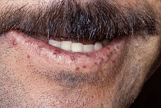
Hereditary hemorrhagic telangiectasia (HHT), also known as Osler–Weber–Rendu disease and Osler–Weber–Rendu syndrome, is a rare autosomal dominant genetic disorder that leads to abnormal blood vessel formation in the skin, mucous membranes, and often in organs such as the lungs, liver, and brain.
A skin condition, also known as cutaneous condition, is any medical condition that affects the integumentary system—the organ system that encloses the body and includes skin, hair, nails, and related muscle and glands. The major function of this system is as a barrier against the external environment.

Lichen planus (LP) is a chronic inflammatory and immune mediated disease that affects the skin, nails, hair, and mucous membranes. It is characterized by polygonal, flat-topped, violaceous papules and plaques with overlying, reticulated, fine white scale, commonly affecting dorsal hands, flexural wrists and forearms, trunk, anterior lower legs and oral mucosa. Although there is a broad clinical range of LP manifestations, the skin and oral cavity remain as the major sites of involvement. The cause is unknown, but it is thought to be the result of an autoimmune process with an unknown initial trigger. There is no cure, but many different medications and procedures have been used in efforts to control the symptoms.
Stasis dermatitis refers to the skin changes that occur in the leg as a result of "stasis" or blood pooling from insufficient venous return; the alternative name of varicose eczema comes from a common cause of this being varicose veins.

Chromoblastomycosis is a long-term fungal infection of the skin and subcutaneous tissue. The infection occurs most commonly in tropical or subtropical climates, often in rural areas. It can be caused by many different types of fungi which become implanted under the skin, often by thorns or splinters. Chromoblastomycosis spreads very slowly; it is rarely fatal and usually has a good prognosis, but it can be very difficult to cure. The several treatment options include medication and surgery.

Venous ulcers are wounds that are thought to occur due to improper functioning of venous valves, usually of the legs. They are the major occurrence of chronic wounds, occurring in 70% to 90% of leg ulcer cases. Venous ulcers develop mostly along the medial distal leg, and can be painful with negative effects on quality of life.

Cutis marmorata telangiectatica congenita is a rare congenital vascular disorder that usually manifests in affecting the blood vessels of the skin. The condition was first recognised and described in 1922 by Cato van Lohuizen, a Dutch pediatrician whose name was later adopted in the other common name used to describe the condition – Van Lohuizen Syndrome. CMTC is also used synonymously with congenital generalized phlebectasia, nevus vascularis reticularis, congenital phlebectasia, livedo telangiectatica, congenital livedo reticularis and Van Lohuizen syndrome.
Papular mucinosis is a rare skin disease. Localized and disseminated cases are called papular mucinosis or lichen myxedematosus while generalized, confluent papular forms with sclerosis are called scleromyxedema. Frequently, all three forms are regarded as papular mucinosis. However, some authors restrict it to only mild cases. Another form, acral persistent papular mucinosis is regarded as a separate entity.

Malakoplakia is a rare inflammatory condition which makes its presence known as a papule, plaque or ulceration that usually affects the genitourinary tract. However, it may also be associated with other bodily organs. It was initially described in the early 20th century as soft yellowish plaques found on the mucosa of the urinary bladder. Microscopically it is characterized by the presence of foamy histiocytes(called von Hansemann cells) with basophilic inclusions called Michaelis–Gutmann bodies.

Chronic venous insufficiency (CVI) is a medical condition in which blood pools in the veins, straining the walls of the vein. The most common cause of CVI is superficial venous reflux which is a treatable condition. As functional venous valves are required to provide for efficient blood return from the lower extremities, this condition typically affects the legs. If the impaired vein function causes significant symptoms, such as swelling and ulcer formation, it is referred to as chronic venous disease. It is sometimes called chronic peripheral venous insufficiency and should not be confused with post-thrombotic syndrome in which the deep veins have been damaged by previous deep vein thrombosis.
Actinic prurigo is a rare sunlight-induced, pruritic, papular or nodular skin eruption. Some medical experts use the term actinic prurigo to denote a rare photodermatosis that develops in childhood and is chronic and persistent; this rare photodermatosis, associated with the human leukocyte antigen HLA-DR4, is often called "Familial polymorphous light eruption of American Indians" or "Hereditary polymorphous light eruption of American Indians" but some experts consider it to be a variant of the syndrome known as polymorphous light eruption (PMLE). Some experts use the term actinic prurigo for Hutchinson's summer prurigo and several other photodermatoses that might, or might not, be distinct clinical entities.
Lipodermatosclerosis is a skin and connective tissue disease. It is a form of lower extremity panniculitis, an inflammation of the layer of fat under the epidermis.

Alternariosis is an infection by Alternaria, presenting cutaneously as focal, ulcerated papules and plaques.

Majocchi's granuloma is a skin condition characterized by deep, pustular plaques, and is a form of tinea corporis. It is a localized form of fungal folliculitis. Lesions often have a pink and scaly central component with pustules or folliculocentric papules at the periphery. The name comes from Professor Domenico Majocchi, who discovered the disorder in 1883. Majocchi was a professor of dermatology at the University of Parma and later the University of Bologna. The most common dermatophyte is called Trichophyton rubrum.

Klippel–Trénaunay syndrome formerly Klippel–Trénaunay–Weber syndrome and sometimes angioosteohypertrophy syndrome and hemangiectatic hypertrophy, is a rare congenital medical condition in which blood vessels and/or lymph vessels fail to form properly. The three main features are nevus flammeus, venous and lymphatic malformations, and soft-tissue hypertrophy of the affected limb. It is similar to, though distinctly separate from, the less common Parkes-Weber syndrome.

Blue rubber bleb nevus syndrome is a rare disorder that consists mainly of abnormal blood vessels affecting the skin or internal organs – usually the gastrointestinal tract. The disease is characterized by the presence of fluid-filled blisters (blebs) as visible, circumscribed, chronic lesions (nevus).
A vascular anomaly is any of a range of lesions from a simple birthmark to a large tumor that may be disfiguring. They are caused by a disorder of the vascular system. A vascular anomaly is a localized defect in blood or lymph vessels. These defects are characterized by an increased number of vessels, and vessels that are both enlarged and sinuous. Some vascular anomalies are congenital, others appear within weeks to years after birth, and others are acquired by trauma or during pregnancy. Inherited vascular anomalies are also described and often present with a number of lesions that increase with age. Vascular anomalies can also be a part of a syndrome.
Stasis papillomatosis is a disease characterized by chronic congestion of the extremities, with blood circulation interrupted in a specific area of the body. A consequence of this congestion and inflammation is long-term lymphatic obstruction. It is also typically characterized by the appearance of numerous papules. Injuries can range from small to large plates composed of brown or pink, smooth or hyperkeratotic papules. The most typical areas where injuries occur are the back of the feet, the toes, the legs, and the area around a venous ulcer formed in the extremities, although the latter is the rarest of all. These injuries include pachydermia, lymphedema, lymphomastic verrucusis and elephantosis verracosa. The disease can be either localized or generalized; the localized form makes up 78% of cases. Treatment includes surgical and pharmaceutical intervention; indications for partial removal include advanced fibrotic lymphedema and elephantiasis. Despite the existence of these treatments, chronic venous edema, which is a derivation of stasis papillomatosis, is only partially reversible. The skin is also affected and its partial removal may mean that the skin and the subcutaneous tissue are excised. A side effect of the procedure is the destruction of existing cutaneous lymphatic vessels. It also risks papillomatosis, skin necrosis and edema exacerbation.
Sarcoidosis, an inflammatory disease, involves the skin in about 25% of patients. The most common lesions are erythema nodosum, plaques, maculopapular eruptions, subcutaneous nodules, and lupus pernio. Treatment is not required, since the lesions usually resolve spontaneously in two to four weeks. Although it may be disfiguring, cutaneous sarcoidosis rarely causes major problems.
References
- 1 2 Rapini, Ronald P.; Bolognia, Jean L.; Jorizzo, Joseph L. (2007). Dermatology: 2-Volume Set. St. Louis: Mosby. ISBN 978-1-4160-2999-1.
- ↑ Scholz S, Schuller-Petrovic S, Kerl H (March 2005). "Mali acroangiodermatitis in homozygous activated protein C resistance". Arch Dermatol. 141 (3): 396–7. doi:10.1001/archderm.141.3.396. PMID 15781687.