Related Research Articles

Integrins are transmembrane receptors that help cell-cell and cell-extracellular matrix (ECM) adhesion. Upon ligand binding, integrins activate signal transduction pathways that mediate cellular signals such as regulation of the cell cycle, organization of the intracellular cytoskeleton, and movement of new receptors to the cell membrane. The presence of integrins allows rapid and flexible responses to events at the cell surface.

Cell adhesion is the process by which cells interact and attach to neighbouring cells through specialised molecules of the cell surface. This process can occur either through direct contact between cell surfaces such as cell junctions or indirect interaction, where cells attach to surrounding extracellular matrix, a gel-like structure containing molecules released by cells into spaces between them. Cells adhesion occurs from the interactions between cell-adhesion molecules (CAMs), transmembrane proteins located on the cell surface. Cell adhesion links cells in different ways and can be involved in signal transduction for cells to detect and respond to changes in the surroundings. Other cellular processes regulated by cell adhesion include cell migration and tissue development in multicellular organisms. Alterations in cell adhesion can disrupt important cellular processes and lead to a variety of diseases, including cancer and arthritis. Cell adhesion is also essential for infectious organisms, such as bacteria or viruses, to cause diseases.
Cell adhesion molecules (CAMs) are a subset of cell surface proteins that are involved in the binding of cells with other cells or with the extracellular matrix (ECM), in a process called cell adhesion. In essence, CAMs help cells stick to each other and to their surroundings. CAMs are crucial components in maintaining tissue structure and function. In fully developed animals, these molecules play an integral role in generating force and movement and consequently ensuring that organs are able to execute their functions normally. In addition to serving as "molecular glue", CAMs play important roles in the cellular mechanisms of growth, contact inhibition, and apoptosis. Aberrant expression of CAMs may result in a wide range of pathologies, ranging from frostbite to cancer.

Cell junctions or junctional complexes, are a class of cellular structures consisting of multiprotein complexes that provide contact or adhesion between neighboring cells or between a cell and the extracellular matrix in animals. They also maintain the paracellular barrier of epithelia and control paracellular transport. Cell junctions are especially abundant in epithelial tissues. Combined with cell adhesion molecules and extracellular matrix, cell junctions help hold animal cells together.

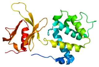
In mammalian cells, vinculin is a membrane-cytoskeletal protein in focal adhesion plaques that is involved in linkage of integrin adhesion molecules to the actin cytoskeleton. Vinculin is a cytoskeletal protein associated with cell-cell and cell-matrix junctions, where it is thought to function as one of several interacting proteins involved in anchoring F-actin to the membrane.

In cell biology, focal adhesions are large macromolecular assemblies through which mechanical force and regulatory signals are transmitted between the extracellular matrix (ECM) and an interacting cell. More precisely, focal adhesions are the sub-cellular structures that mediate the regulatory effects of a cell in response to ECM adhesion.

Integrin-linked kinase is an enzyme that in humans is encoded by the ILK gene involved with integrin-mediated signal transduction. Mutations in ILK are associated with cardiomyopathies. It is a 59kDa protein originally identified in a yeast-two hybrid screen with integrin β1 as the bait protein. Since its discovery, ILK has been associated with multiple cellular functions including cell migration, proliferation, and adhesion.

Integrin beta-1 (ITGB1), also known as CD29, is a cell surface receptor that in humans is encoded by the ITGB1 gene. This integrin associates with integrin alpha 1 and integrin alpha 2 to form integrin complexes which function as collagen receptors. It also forms dimers with integrin alpha 3 to form integrin receptors for netrin 1 and reelin. These and other integrin beta 1 complexes have been historically known as very late activation (VLA) antigens.
Talin is a high-molecular-weight cytoskeletal protein concentrated at regions of cell–substratum contact and, in lymphocytes, at cell–cell contacts. Discovered in 1983 by Keith Burridge and colleagues, talin is a ubiquitous cytosolic protein that is found in high concentrations in focal adhesions. It is capable of linking integrins to the actin cytoskeleton either directly or indirectly by interacting with vinculin and α-actinin.

Cadherin-2 also known as Neural cadherin (N-cadherin), is a protein that in humans is encoded by the CDH2 gene. CDH2 has also been designated as CD325 . Cadherin-2 is a transmembrane protein expressed in multiple tissues and functions to mediate cell–cell adhesion. In cardiac muscle, Cadherin-2 is an integral component in adherens junctions residing at intercalated discs, which function to mechanically and electrically couple adjacent cardiomyocytes. Alterations in expression and integrity of Cadherin-2 has been observed in various forms of disease, including human dilated cardiomyopathy. Variants in CDH2 have also been identified to cause a syndromic neurodevelopmental disorder.

Integrin beta-7 is an integrin protein that in humans is encoded by the ITGB7 gene. It can pair with ITGA4 (CD49d) to form the heterodimeric integrin receptor α4β7, or with ITGAE (CD103) to form αEβ7.

Alpha-7 integrin is a protein that in humans is encoded by the ITGA7 gene. Alpha-7 integrin is critical for modulating cell-matrix interactions. Alpha-7 integrin is highly expressed in cardiac muscle, skeletal muscle and smooth muscle cells, and localizes to Z-disc and costamere structures. Mutations in ITGA7 have been associated with congenital myopathies and noncompaction cardiomyopathy, and altered expression levels of alpha-7 integrin have been identified in various forms of muscular dystrophy.

Myosin X, also known as MYO10, is a protein that in humans is encoded by the MYO10 gene.

Stress fibers are contractile actin bundles found in non-muscle cells. They are composed of actin (microfilaments) and non-muscle myosin II (NMMII), and also contain various crosslinking proteins, such as α-actinin, to form a highly regulated actomyosin structure within non-muscle cells. Stress fibers have been shown to play an important role in cellular contractility, providing force for a number of functions such as cell adhesion, migration and morphogenesis.

Cadherin-1 or Epithelial cadherin(E-cadherin), is a protein that in humans is encoded by the CDH1 gene. Mutations are correlated with gastric, breast, colorectal, thyroid, and ovarian cancers. CDH1 has also been designated as CD324. It is a tumor suppressor gene.
Talin-1 is a protein that in humans is encoded by the TLN1 gene. Talin-1 is ubiquitously expressed, and is localized to costamere structures in cardiac and skeletal muscle cells, and to focal adhesions in smooth muscle and non-muscle cells. Talin-1 functions to mediate cell-cell adhesion via the linkage of integrins to the actin cytoskeleton and in the activation of integrins. Altered expression of talin-1 has been observed in patients with heart failure, however no mutations in TLN1 have been linked with specific diseases.

Keith Burridge is a British researcher and Kenan distinguished Professor at the University of North Carolina at Chapel Hill. His research on focal adhesions includes the discovery of many adhesion proteins including vinculin, talin and paxillin, and ranks him in top 1% of the most cited scientist in the field of molecular biology and genetics. Burridge has published more than 200 peer reviewed articles.

Fermitin family homolog 3) (FERMT3), also known as kindlin-3 (KIND3), MIG2-like protein (MIG2B), or unc-112-related protein 2 (URP2) is a protein that in humans is encoded by the FERMT3 gene. The kindlin family of proteins, member of the B4.1 superfamily, comprises three conserved protein homologues, kindlin 1, 2, and 3. They each contain a bipartite FERM domain comprising four subdomains F0, F1, F2, and F3 that show homology with the FERM head (H) domain of the cytoskeletal Talin protein. Kindlins have been linked to Kindler syndrome, leukocyte adhesion deficiency, cancer and other acquired human diseases. They are essential in the organisation of focal adhesions that mediate cell-extracellular matrix junctions and are involved in other cellular compartments that control cell-cell contacts and nucleus functioning. Therefore, they are responsible for cell to cell crosstalk via cell-cell contacts and integrin mediated cell adhesion through focal adhesion proteins and as specialised adhesion structures of hematopoietic cells they are also present in podosome's F actin surrounding ring structure. Isoform 2 may act as a repressor of NF-kappa-B and apoptosis
The secretome is the set of proteins expressed by an organism and secreted into the extracellular space. In humans, this subset of the proteome encompasses 13-20% of all proteins, including cytokines, growth factors, extracellular matrix proteins and regulators, and shed receptors. The secretome of a specific tissue can be measured by mass spectrometry and its analysis constitutes a type of proteomics known as secretomics.

Giantin or Golgin subfamily B member 1 is a protein that in humans is encoded by the GOLGB1 gene. Giantin is located at the cis-medial rims of the Golgi apparatus and is part of the Golgi matrix that is responsible for membrane trafficking in secretory pathway of proteins. This function is key for proper localisation of proteins at the plasma membrane and outside the cell which is important for cell function that is dependent on for example receptors and the extracellular matrix function. Recent animal model knockout studies of GOLGB1 in mice, rat, and zebrafish have shown that phenotypes are different between species ranging from mild to severe craniofacial defects in the rodent models to just minor size defects in zebrafish. However, in adult zebrafish a tumoral calcinosis-like phenotype was observed, and in humans such phenotype has been linked to defective glycosyltransferase function.
References
- ↑ Whittaker, Charles A.; Bergeron, Karl-Frederik; Whittle, James; Brandhorst, Bruce P.; Burke, Robert D.; Hynes, Richard O. (December 2006). "The echinoderm adhesome". Developmental Biology. 300 (1): 252–266. doi:10.1016/j.ydbio.2006.07.044. PMC 3565218 . PMID 16950242.
- ↑ Zaidel-Bar, Ronen; Itzkovitz, Shalev; Ma'ayan, Avi; Iyengar, Ravi; Geiger, Benjamin (2007). "Functional atlas of the integrin adhesome". Nature Cell Biology. 9 (8): 858–867. doi:10.1038/ncb0807-858. PMC 2735470 . PMID 17671451.
- 1 2 Zaidel-Bar, Ronen; Geiger, Benjamin (2010-05-01). "The switchable integrin adhesome". J Cell Sci. 123 (9): 1385–1388. doi:10.1242/jcs.066183. ISSN 0021-9533. PMC 2858016 . PMID 20410370.
- 1 2 3 Winograd-Katz, Sabina E.; Fässler, Reinhard; Geiger, Benjamin; Legate, Kyle R. (2014). "The integrin adhesome: from genes and proteins to human disease". Nature Reviews Molecular Cell Biology. 15 (4): 273–288. doi:10.1038/nrm3769. PMID 24651544. S2CID 5528372.
- 1 2 Zaidel-Bar, Ronen (2013-01-15). "Cadherin adhesome at a glance". J Cell Sci. 126 (2): 373–378. doi: 10.1242/jcs.111559 . ISSN 0021-9533. PMID 23547085.
- ↑ "Adhesome- Project Description". www.adhesome.org. Retrieved 2015-12-24.
- ↑ Byron, Adam; Humphries, Jonathan D.; Bass, Mark D.; Knight, David; Humphries, Martin J. (2011-04-05). "Proteomic Analysis of Integrin Adhesion Complexes". Sci. Signal. 4 (167): pt2. doi:10.1126/scisignal.2001827. ISSN 1945-0877. PMID 21467297. S2CID 25555835.
- 1 2 Schiller, Herbert B; Fässler, Reinhard (2013-06-01). "Mechanosensitivity and compositional dynamics of cell–matrix adhesions". EMBO Reports. 14 (6): 509–519. doi:10.1038/embor.2013.49. PMC 3674437 . PMID 23681438.
- 1 2 Kuo, Jean-Cheng; Han, Xuemei; Hsiao, Cheng-Te; III, John R. Yates; Waterman, Clare M. (2011). "Analysis of the myosin-II-responsive focal adhesion proteome reveals a role for β-Pix in negative regulation of focal adhesion maturation". Nature Cell Biology. 13 (4): 383–393. doi:10.1038/ncb2216. PMC 3279191 . PMID 21423176.
- ↑ Horton, Edward R.; Byron, Adam; Askari, Janet A.; Ng, Daniel H. J.; Millon-Frémillon, Angélique; Robertson, Joseph; Koper, Ewa J.; Paul, Nikki R.; Warwood, Stacey (2015). "Definition of a consensus integrin adhesome and its dynamics during adhesion complex assembly and disassembly". Nature Cell Biology. 17 (12): 1577–1587. doi:10.1038/ncb3257. PMC 4663675 . PMID 26479319.
- ↑ Kuo, J. C.; Han, X.; Yates Jr, 3rd; Waterman, C. M. (2012-01-01). Shimaoka, Motomu (ed.). Isolation of Focal Adhesion Proteins for Biochemical and Proteomic Analysis - Springer. Methods in Molecular Biology. Vol. 757. Humana Press. pp. 297–323. doi:10.1007/978-1-61779-166-6_19. ISBN 978-1-61779-165-9. PMC 4158431 . PMID 21909920.
- ↑ Roux, Kyle J.; Kim, Dae In; Raida, Manfred; Burke, Brian (2012-03-19). "A promiscuous biotin ligase fusion protein identifies proximal and interacting proteins in mammalian cells". The Journal of Cell Biology. 196 (6): 801–810. doi:10.1083/jcb.201112098. ISSN 0021-9525. PMC 3308701 . PMID 22412018.
- 1 2 Guo, Zhenhuan; Neilson, Lisa J.; Zhong, Hang; Murray, Paul S.; Zanivan, Sara; Zaidel-Bar, Ronen (2014-12-02). "E-cadherin interactome complexity and robustness resolved by quantitative proteomics". Sci. Signal. 7 (354): rs7. doi:10.1126/scisignal.2005473. ISSN 1945-0877. PMC 4972397 . PMID 25468996.
- ↑ Itallie, Christina M. Van; Tietgens, Amber Jean; Aponte, Angel; Fredriksson, Karin; Fanning, Alan S.; Gucek, Marjan; Anderson, James M. (2014-02-15). "Biotin ligase tagging identifies proteins proximal to E-cadherin, including lipoma preferred partner, a regulator of epithelial cell–cell and cell–substrate adhesion". J Cell Sci. 127 (4): 885–895. doi:10.1242/jcs.140475. ISSN 0021-9533. PMC 3924204 . PMID 24338363.
- ↑ Schiller, Herbert B.; Hermann, Michaela-Rosemarie; Polleux, Julien; Vignaud, Timothée; Zanivan, Sara; Friedel, Caroline C.; Sun, Zhiqi; Raducanu, Aurelia; Gottschalk, Kay-E. (2013). "β1- and αv-class integrins cooperate to regulate myosin II during rigidity sensing of fibronectin-based microenvironments". Nature Cell Biology. 15 (6): 625–636. doi:10.1038/ncb2747. PMID 23708002. S2CID 26767077.
- 1 2 Systems Biomedicine: Concepts and Perspectives ed. E.T. Liu and D.A. Lauffenburger. Oxford: Academic Press. 2009. pp. 139–152.
- ↑ Zaidel-Bar, Ronen (2009-08-10). "Evolution of complexity in the integrin adhesome". The Journal of Cell Biology. 186 (3): 317–321. doi:10.1083/jcb.200811067. ISSN 0021-9525. PMC 2728394 . PMID 19667126.
- 1 2 Murray, Paul S.; Zaidel-Bar, Ronen (2014-12-15). "Pre-metazoan origins and evolution of the cadherin adhesome". Biology Open. 3 (12): 1183–1195. doi:10.1242/bio.20149761. ISSN 2046-6390. PMC 4265756 . PMID 25395670.
- ↑ Padmanabhan, Anup; Rao, Megha Vaman; Wu, Yao; Zaidel-Bar, Ronen (2015). "Jack of all trades: functional modularity in the adherens junction". Current Opinion in Cell Biology. 36: 32–40. doi:10.1016/j.ceb.2015.06.008. PMID 26189061.