Amygdalotomy

Last updated

Amygdalotomy is a form of psychosurgery which involves the surgical removal or destruction of the amygdala, or parts of the amygdala. It is usually a last-resort treatment for severe aggressive behavioral disorders and similar behaviors including hyperexcitability, violent outbursts, and self-mutilation. [1] [2] [3] [4] The practice of medical amygdalotomy typically involves the administration of general anesthesia and is achieved through the application of cranial stereotactic surgery to target regions of the amygdala for surgical destruction. [3] While some studies have found stereotactic amygdalotomy in humans to be an effective treatment for severe cases of intractable aggressive behavior that has not responded to standard treatment methods, [5] [6] [7] [8] other studies remain inconclusive. [9] [10] In most cases of amygdalectomy in humans, there is no substantial evidence of impairment in overall cognitive function, including intelligence and working memory, however, deficits in specific areas of memory have been noted pertaining to the recognition and emotional interpretation of facial stimuli. This is because there are specialized cells in the amygdala which attend to facial stimuli.

Contents

Background

The amygdala is considered to be an important underlying structure in the fight-or-flight response, playing a mediating role in aggression in both humans and animals. [3] Clinical studies have revealed that the stimulation of the amygdala produces or accentuates rageful behavior in animals. [4] Research has also revealed that lesions of the amygdala in both humans and animals produces a calming effect on aggressive behavior. [3] Based on these findings, amygdalotomy was developed as a neurosurgical procedure to ameliorate aggression by reducing arousal levels in the amygdala. [3] [11]

The location of the amygdala in the human brain Amygdala.jpg
The location of the amygdala in the human brain

Medical use

Since the early 1900s there has been an accumulation of experimental evidence to demonstrate the role of the limbic system, specifically the amygdala complex in mediating emotional expressions of fear and anger. [11] [3] Early primate studies have revealed that chemical and electrical stimulation of the amygdala region accentuates aggressive behavior. Conversely, destruction of the amygdala nucleus results in a taming effect of normal anger and fearing responses in primate behavior. Similarly, clinical studies in humans have revealed the close etiological role of temporal lobe structures, particularly the limbic system and the amygdala in mediating fear and rageful behavior. [3] These findings have been instrumental in the development of clinical amygdalotomy as a form of neurosurgery to produce placating effects on abnormal aggressive behaviors. [11] [3] Procedural amygdalotomy is used as a last recourse treatment for severe intractable aggression when other options including pharmacological treatments have been exhausted. The psychopathology of patients with severe aggressive behavior in clinical cases of amygdalotomy over the past 20th century vary, including epileptics with violent convulsions, psychotics with violent outbursts, individuals with unmanageable conduct disorder, and patients with self-mutilative tendencies. The clinical practice of amygdalotomy in humans is commonly implemented under the stereotactic frame, with varying techniques used to destroy the amygdala, ranging from radiofrequency, mechanical destruction and the injection of oil, wax, and alcohol. [3] The preferred target zone of the amygdala also varies from basal and lateral nuclei, to the medial region, the cortico-medial group of nuclei and the bed of the stria terminalis. [3] The size of the lesion differs from one-third to one-half, to three-quarters, to the entire amygdalar region. [3] In spite of these methodological differences, most published accounts of human amygdalotomy have indicated beneficial outcomes in reducing the intensity and frequency of aggressive behaviors. [3] [11]

Historical evolution

Animal studies

Amongst some of the earliest studies conducted on the removal of the amygdala were animal and primate studies. [11] [3] In the early 1890s, Friedrich Goltz conducted experiments on temporal lobectomy in dogs including the removal of the amygdala and found that dogs post-surgery experienced a taming effects on aggressive behaviors. [11] Deep brain stimulation studies in animals revealed that the temporal lobe is involved in mediating expressions of rage and aggression. [11] A more detailed analysis of specific regions of the temporal lobe in animals revealed that the limbic system, specifically the amygdala complex, is involved in mediating fear and aggression. [11] Some of the earliest primate studies on amygdalotomy were carried out on rhesus monkeys by Kluver and Bucy in the late 1930s. [11] Data collected from these studies revealed that bilateral destruction of the amygdala resulted in a reduction in the intensity and frequency of fear and aggression behaviors.

Clinical trials

The stereotaxic apparatus used for clinical amygdalotomy The stereotaxic apparatus. Wellcome M0016268.jpg
The stereotaxic apparatus used for clinical amygdalotomy

The human counterpart of the role of the amygdala was then observed in the 20th century at the height of psychosurgery. [11] Professor Hirotaro Narabayashi and his colleagues were the first researchers to carry out stereotactic amygdalotomy for the treatment of abnormal aggression and hyperexcitability in a series of 60 patients with psychological disturbances. [3] [11] The procedure was performed under a stereotactic frame devised by Professor Narabayashi and involved the administration 0.6-0.8ml mixture of oil-wax to destroy the lateral groups of the amygdala nucleus, localized via pneumoencephalography. [11] The clinical results revealed a marked reduction in emotional disturbances amongst 85% of the cases. Following Narabayashi's study, there have been over 1000 cases of amygdalotomy reported in clinical trials as a last-resort treatment for severe intractable aggressive disorders. [3] Around the same time, Hatai Chitanondh utilized a slightly different technique of stereotactic amygdalotomy using an injection of an olive oil mixture to induce lesions to mechanically block signals in the amygdala. The results revealed an improvement in social adaptability of all seven patients. [11] In the late 1960s and 1970s, Balasubramaniam and Ramamurthi investigated the largest clinical patient series to undergo stereotactic amygdalotomy for aggression behaviours. The procedure was performed via high-frequency current generating electrodes inserted stereotactically to induce several small thermal lesions, creating a total lesion volume of 1800mm, a size that is larger than the amygdala. [11] The improvement in maladaptive behavior in patients, including hyperexcitability, rebellious behavior, and destructive behavior, ranged from moderate to high. [11] The development of MRI technology in the recent 20th century has enabled a more accurate and efficient process of amygdalotomy, with easier localization of amygdala regions during neuro-navigation as well as the use of advanced radiofrequency generating electrode to induce surgical lesions. [11] Despite these recent advances in technology, there has been a decline in clinical cases of amygdalotomy for treatment of maladaptive behavior, with growing skepticism in the medical community of the cost-benefits of the procedure and partly due to a greater reliance on pharmacological treatments [11] [12]

Clinical effectiveness

Short-term results

There has been a general consensus amongst many researchers on the general effectiveness of amygdalotomy in reducing aggression amongst patients with psychosis, violent epilepsy, and self-mutilative behavior. [8] [7] [5] A diverse study [8] on stereotactic amygdalotomy used to treat 25 patients primarily for aggressive behavior and violent epilepsy, found that behavioral abnormalities were eliminated in 2 of the 20 patients, with a significant improvement in another 9 of the 20 patients with aggressive behavior manifests. Convulsions were also eliminated in 4 of the 21 patients, whilst 12 of the patients experienced a significant decrease in the number of epileptic convulsions. Patient rehabilitation was also effective with 2 of the 12 patients committed to mental institutions being discharged, whilst 5 of 8 patients awaiting institutionalization were no longer being considered. A multi-disciplinary project carried out on amygdalotomy amongst epileptics with violent outbursts found that amygdalotomy showed promising results, with a decline in violent, aggressive and anti-social behavior as well as a reduction amongst patients and an improvement in the occupational functioning of some of the patients. [4] The researchers, however, concluded that the results cannot be generalized to non-epileptics. Other studies [5] [7] conducted on patients with conduct disorder, personality disorder, self-mutilation, and schizophrenics with violent hallucinations found that these maladaptive behaviors also improved across these groups of patients. Using reliable and objective methods of evaluation, Heimburger and colleagues found that in patients who did not respond to non-surgical therapy, amygdalotomy was effective, with both conditions of uncontrolled conduct disorder and seizures seeming improved after surgery. [7] Stereotaxic amygdalotomy conducted on 12 patients with schizophrenia and frequent self-mutilations found that, in 11 of the 12 patients, amygdalotomy resulted in elimination and or marked reduction of aggressive episodes. [5] In two of patients with frequent self-mutilative episodes and reactive psychotic hallucinations, however, these symptoms disappeared only after an additional basofrontal tractotomy had been performed.

Long-term results

There is a scarcity of long-term studies on the follow-up effects of clinical amygdalotomy in humans. [3] Amongst the few follow-up studies, includes a research study [6] which compared the results of clinical amygdalotomy in 58 patients pre and post-surgery over an average of 6 years using objective analysis such as psychiatric interviews, neuropsychological tests and EEG analysis and found no indication of worsening of symptoms. Additionally, the researchers found some evidence for the retention of positive outcomes in one-third of the patients, which were not limited to improvements in rageful behaviour but also included a decrease in the overall frequency of seizures. Another follow-up study [13] by Professor Narabayashi and colleagues observed the clinical effects of amygdalotomy in 40 cases from 3 to 5 years and found 27 of the cases had continued a satisfactory improvement in calming and taming effects on what was previously uncontrollable aggression including destructive and violent behaviour.

Risks and side-effects

In most cases of amygdalotomy in humans, there is no substantial evidence of impairment in overall cognitive function, including intelligence and working memory. [10] [9] However, deficits in specific areas of memory have been noted, particularly areas of memory pertaining to the recognition and emotional interpretation of facial stimuli. [10] These findings of face recognition impairment after amygdalotomy are of particular importance due to the neurophysiological data collected on the importance of cells in the amygdala that specifically attend to facial stimuli in both humans and primates. [10] A detailed case study of a patient who had undergone a bilateral amygdalotomy found incidences where the patient showed poor learning of new faces and impaired recognition of familiar faces, particularly troubles with naming faces. [10] Additionally the patient also revealed further deficits in the emotional processing of facial stimuli, demonstrating difficulty in identifying and matching a range of facial expressions. Another study of 15 patients showed no reduction in general intelligence, but there was a similar pattern of changes in attention and memory involving facial stimuli. This link between the amygdala and social disturbances pertaining to the processing of facial stimuli has been investigated as a possible side-effect of amygdalotomy in some patients [10] [9]

Related Research Articles

<span class="mw-page-title-main">Neurosurgery</span> Medical specialty of disorders which affect any portion of the nervous system.

Neurosurgery or neurological surgery, known in common parlance as brain surgery, is the medical specialty concerned with the surgical treatment of disorders which affect any portion of the nervous system including the brain, spinal cord and peripheral nervous system.

Psychosurgery, also called neurosurgery for mental disorder (NMD), is the neurosurgical treatment of mental disorder. Psychosurgery has always been a controversial medical field. The modern history of psychosurgery begins in the 1880s under the Swiss psychiatrist Gottlieb Burckhardt. The first significant foray into psychosurgery in the 20th century was conducted by the Portuguese neurologist Egas Moniz who during the mid-1930s developed the operation known as leucotomy. The practice was enthusiastically taken up in the United States by the neuropsychiatrist Walter Freeman and the neurosurgeon James W. Watts who devised what became the standard prefrontal procedure and named their operative technique lobotomy, although the operation was called leucotomy in the United Kingdom. In spite of the award of the Nobel prize to Moniz in 1949, the use of psychosurgery declined during the 1950s. By the 1970s the standard Freeman-Watts type of operation was very rare, but other forms of psychosurgery, although used on a much smaller scale, survived. Some countries have abandoned psychosurgery altogether; in others, for example the US and the UK, it is only used in a few centres on small numbers of people with depression or obsessive-compulsive disorder (OCD). In some countries it is also used in the treatment of schizophrenia and other disorders.

Bilateral cingulotomy is a form of psychosurgery, introduced in 1948 as an alternative to lobotomy. Today, it is mainly used in the treatment of depression and obsessive-compulsive disorder. In the early years of the twenty-first century, it was used in Russia to treat addiction. It is also used in the treatment of chronic pain. The objective of this procedure is the severing of the supracallosal fibres of the cingulum bundle, which pass through the anterior cingulate gyrus.

<span class="mw-page-title-main">Aggression</span> Social interaction aiming at inflicting harm or unpleasantness

Aggression is a behavior aimed at opposing or attacking something or someone. Though often done with the intent to cause harm, it can be channeled into creative and practical outlets for some. It may occur either reactively or without provocation. In humans, aggression can be caused by various triggers. For example, built-up frustration due to blocked goals or perceived disrespect. Human aggression can be classified into direct and indirect aggression; whilst the former is characterized by physical or verbal behavior intended to cause harm to someone, the latter is characterized by behavior intended to harm the social relations of an individual or group.

<span class="mw-page-title-main">Amygdala</span> Each of two small structures deep within the temporal lobe of complex vertebrates

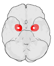
The amygdala is a paired nuclear complex present in the cerebral hemispheres of vertebrates. It is considered part of the limbic system. In primates, it is located medially within the temporal lobes. It consists of many nuclei, each made up of further subnuclei. The subdivision most commonly made is into the basolateral, central, cortical, and medial nuclei together with the intercalated cell clusters. The amygdala has a primary role in the processing of memory, decision-making, and emotional responses. The amygdala was first identified and named by Karl Friedrich Burdach in 1822.

<span class="mw-page-title-main">Deep brain stimulation</span> Neurosurgical treatment involving implantation of a brain pacemaker

Deep brain stimulation (DBS) is a surgical procedure that implants a neurostimulator and electrodes which sends electrical impulses to specified targets in the brain responsible for movement control. The treatment is designed for a range of movement disorders such as Parkinson's disease, essential tremor, and dystonia, as well as for certain neuropsychiatric conditions like obsessive-compulsive disorder (OCD) and epilepsy. The exact mechanisms of DBS are complex and not entirely clear, but it is known to modify brain activity in a structured way.
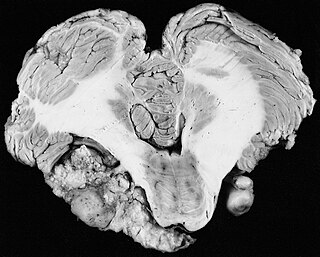
Lars Leksell (1907–1986) was a Swedish physician and Professor of Neurosurgery at the Karolinska Institute in Stockholm, Sweden. He was the inventor of radiosurgery.

<span class="mw-page-title-main">Radiosurgery</span> Surgical Specialty

Radiosurgery is surgery using radiation, that is, the destruction of precisely selected areas of tissue using ionizing radiation rather than excision with a blade. Like other forms of radiation therapy, it is usually used to treat cancer. Radiosurgery was originally defined by the Swedish neurosurgeon Lars Leksell as "a single high dose fraction of radiation, stereotactically directed to an intracranial region of interest".

<span class="mw-page-title-main">Stereotactic surgery</span> Medical procedure

Stereotactic surgery is a minimally invasive form of surgical intervention that makes use of a three-dimensional coordinate system to locate small targets inside the body and to perform on them some action such as ablation, biopsy, lesion, injection, stimulation, implantation, radiosurgery (SRS), etc.

<span class="mw-page-title-main">Intermittent explosive disorder</span> Behavioral disorder

Intermittent explosive disorder is a behavioral disorder characterized by explosive outbursts of anger and/or violence, often to the point of rage, that are disproportionate to the situation at hand. Impulsive aggression is not premeditated, and is defined by a disproportionate reaction to any provocation, real or perceived. Some individuals have reported affective changes prior to an outburst, such as tension, mood changes, energy changes, etc.

Corpus callosotomy is a palliative surgical procedure for the treatment of medically refractory epilepsy. In this procedure the corpus callosum is cut through, in an effort to limit the spread of epileptic activity between the two halves of the brain.

Reduced affect display, sometimes referred to as emotional blunting or emotional numbing, is a condition of reduced emotional reactivity in an individual. It manifests as a failure to express feelings either verbally or nonverbally, especially when talking about issues that would normally be expected to engage emotions. In this condition, expressive gestures are rare and there is little animation in facial expression or vocal inflection. Additionally, reduced affect can be symptomatic of autism, schizophrenia, depression, post-traumatic stress disorder, depersonalization disorder, schizoid personality disorder or brain damage. It may also be a side effect of certain medications.

<span class="mw-page-title-main">Inferior temporal gyrus</span> One of three gyri of the temporal lobe of the brain

The inferior temporal gyrus is one of three gyri of the temporal lobe and is located below the middle temporal gyrus, connected behind with the inferior occipital gyrus; it also extends around the infero-lateral border on to the inferior surface of the temporal lobe, where it is limited by the inferior sulcus. This region is one of the higher levels of the ventral stream of visual processing, associated with the representation of objects, places, faces, and colors. It may also be involved in face perception, and in the recognition of numbers and words.

Hirotaro Narabayashi was a prominent Japanese neurosurgeon.

Psychopathy is a mental health condition characterized by persistent antisocial behavior, impaired empathy and remorse, and bold, disinhibited, and egoistic traits masked by superficial charm and the outward presence of apparent normalcy. Different conceptions of psychopathy have been used throughout history that are only partly overlapping and may sometimes be contradictory.

Patient registration is used to correlate the reference position of a virtual 3D dataset gathered by computer medical imaging with the reference position of the patient. This procedure is crucial in computer assisted surgery, in order to insure the reproducitibility of the preoperative registration and the clinical situation during surgery. The use of the term "patient registration" out of this context can lead to a confusion with the procedure of registering a patient into the files of a medical institution.

Ablative brain surgery is the surgical ablation by various methods of brain tissue to treat neurological or psychological disorders. The word "Ablation" stems from the Latin word Ablatus meaning "carried away". In most cases, however, ablative brain surgery does not involve removing brain tissue, but rather destroying tissue and leaving it in place. The lesions it causes are irreversible. There are some target nuclei for ablative surgery and deep brain stimulation. Those nuclei are the motor thalamus, the globus pallidus, and the subthalamic nucleus.

Emotional lateralization is the asymmetrical representation of emotional control and processing in the brain. There is evidence for the lateralization of other brain functions as well.

Psychosurgery, also called neurosurgery for mental disorder or functional neurosurgery, is surgery in which brain tissue is destroyed with the aim of alleviating the symptoms of mental disorder. It was first used in modern times by Gottlieb Burckhardt in 1891, but only in a few isolated instances, not becoming more widely used until the 1930s following the work of Portuguese neurologist António Egas Moniz. The 1940s was the decade when psychosurgery was most popular, largely due to the efforts of American neurologist Walter Freeman; its use has been declining since then. Freeman's particular form of psychosurgery, the lobotomy, was last used in the 1970s, but other forms of psychosurgery, such as the cingulotomy and capsulotomy have survived.

<span class="mw-page-title-main">Konstantin Slavin</span>

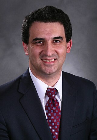
Konstantin Slavin is a Professor and Head of the Department of Stereotactic and functional neurosurgery at the University of Illinois College of Medicine. He is a former president of the American Society for Stereotactic and functional neurosurgery and current vice-president of the World Society for Stereotactic and Functional Neurosurgery. His specialties include Aneurysm, Brain surgery, Brain Tumor, Cerebrovascular Disorders, Craniotomy, Dystonia, Essential Tremor, Facial Nerve Pain, Facial Pain, Glioblastoma, Headache disorders, Laminectomy, Lower back pain, Movement Disorders, Multiple Sclerosis, Neck Pain, Neurosurgery, Neurosurgical Procedures, Pain, Parkinson Disease, Spinal Cord Injuries, and Stroke.

References

  1. Wang, W.; Li, P.; Jiang, S.; Zhou, P.; Zhang, S. (January 2017). "Bilateral anterior capsulotomy and amygdalotomy for mental retardation with psychiatric symptoms and aggression". Medicine. 96 (1): e5840. doi:10.1097/MD.0000000000005840. PMC   5228703 . PMID   28072743.
  2. "Violence, mental illness, and the brain – A brief history of psychosurgery: Part 3 – From deep brain stimulation to amygdalotomy for violent behavior, seizures, and pathological aggression in humans". Surgical Neurology International. Surgical Neurology International . Retrieved 2019-06-19.
  3. 1 2 3 4 5 6 7 8 9 10 11 12 13 14 15 16 Lee, G.P; Bechara, A; Adolphs, Meador, R; Arena, J; Meador, K.J; Loring, D.W; Smith, J.R (1998). "Clinical and physiological effects of stereotaxic bilateral amygdalotomy for intractable aggression". The Journal of Neuropsychiatry and Clinical Neurosciences. 10 (4): 413–420. doi:10.1176/jnp.10.4.413. PMID   9813786.
  4. 1 2 3 Hitchcock, E; Cairns, V (1973). "Amygdalotomy". Postgraduate Medical Journal. 49 (578): 894–904. doi:10.1136/pgmj.49.578.894. PMC   2495458 . PMID   4806269.
  5. 1 2 3 4 Vaernet, K; Madsen, A (1970). "Stereotaxic amygdalotomy and basofrontal tractotomy in psychotics with aggressive behaviour". Journal of Neurology, Neurosurgery & Psychiatry. 33 (6): 853–863. doi:10.1136/jnnp.33.6.858. PMC   493604 . PMID   5531905.
  6. 1 2 Small, I.F; Heimburger, R.F; Small, J.G; Milstein, V; Moore, D.F (1977). "Follow-up of stereotaxic amygdalotomy for seizure and behavior disorders". Biological Psychiatry. 12 (3): 401–411. PMID   326309.
  7. 1 2 3 4 Heimburger, R. F., R.F; Small, I.F; Small, J.G; Milstein, V; Moore, D (1978). "Stereotactic amygdalotomy for convulsive and behavioral disorders". Stereotactic and Functional Neurosurgery. 41 (4): 43–51. doi:10.1159/000102399. PMID   365101.
  8. 1 2 3 Heimburger, R.F; Whitlock, C.C; Kalsbeck, J.E (1966). "Stereotaxic amygdalotomy for epilepsy with aggressive behavior". JAMA. 198 (7): 741–745. doi:10.1001/jama.1966.03110200097026. PMID   5332043.
  9. 1 2 3 Anderson, R (1978). "Cognitive changes after amygdalotomy". Neuropsychologia. 16 (4): 439–451. doi:10.1016/0028-3932(78)90067-2. PMID   692856. S2CID   38237289.
  10. 1 2 3 4 5 6 Young, A.W; Agleton, J.P; Hellawell, D.J; Johnson, M; Broks, P; Hanley, J.R (1995). "Face processing impairments after amygdalotomy". Brain. 118 (1): 15–24. doi:10.1093/brain/118.1.15. PMID   7895001.
  11. 1 2 3 4 5 6 7 8 9 10 11 12 13 14 15 16 17 Fountas, K.N; Smith, J.R (2007). "Historical evolution of stereotactic amygdalotomy for the management of severe aggression". Journal of Neurosurgery. 106 (4): 710–713. doi:10.3171/jns.2007.106.4.710. PMID   17432727.
  12. Mpakopoulou, M; Gatos, H; Brotis, A; Paterakis, K.N; Fountas, K.N (2008). "Stereotactic amygdalotomy in the management of severe aggressive behavioral disorders". Neurosurgical Focus. 25 (1): E6. doi: 10.3171/FOC/2008/25/7/E6 . PMID   18590383.
  13. Narabayashi, H; Uno, M (1966). "Long range results of stereotaxic amygdalotomy for behavior disorders". Stereotactic and Functional Neurosurgery. 27 (1–3): 168–171. doi:10.1159/000103950. PMID   5334008.