The Asx turn[1][2][3][4][5][6][7] is a structural feature in proteins and polypeptides. It consists of three amino acid residues (labeled i, i+1 and i+2) in which residue i is an aspartate (Asp) or asparagine (Asn) that forms a hydrogen bond from its sidechain CO group to the mainchain NH group of residue i+2. About 14% of Asx residues present in proteins belong to Asx turns.
The name "Asx" is used here to represent either of the amino acids aspartate (Asp) or asparagine (Asn).
Types
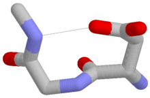
An Asx turn with an aspartate at residue i. One of the sidechain oxygens of the aspartate forms a hydrogen bond (dotted line) with the mainchain NH group of residue i+2. Colors: red, oxygen; grey, carbon; blue, nitrogen. Hydrogen atoms are omitted.
Four types of Asx turn can be distinguished:[8] types I, I’, II and II’. These categories correspond to those of the better-known hydrogen-bonded beta turns, which have four residues and a hydrogen bond between the CO of residue i and the NH of residue i+3. Asx turns and beta turns have structurally similar hydrogen-bonded loops and exhibit sidechain-mainchain mimicry in the sense that the sidechain of residue i of the Asx turn mimics the mainchain of residue i of the beta turn. Regarding their occurrence in proteins, they differ in that type I is the commonest of the four beta turns while type II’ is the commonest of the Asx turns.
Similar motifs occur with serine or threonine as residue i, which are called ST turns.[13] In spite of serine and threonine having one less sidechain atom, such that the sidechain-mainchain mimicry of β turns is imperfect, these features occur in proteins as the four types in numbers approaching those of Asx turns. They also exhibit a tendency to substitute each other over evolutionary time.[14]
A proportion of Asx turns are accompanied by a mainchain–mainchain hydrogen bond that qualifies them as Asx motifs.
↑ Rees, DC; Lewis M (1983). "Refined crystal structure of carboxypeptidase a at 1.54 Å resolution". Journal of Molecular Biology. 168 (2): 367–387. doi:10.1016/S0022-2836(83)80024-2. PMID6887246.
↑ Thakur, AK; Kishore R (2006). "Characterization of β-turn and asx-turns mimicry in a model peptide: Stabilization via C-H•••O interaction". Biopolymers. 81 (6): 440–449. doi:10.1002/bip.20441. PMID16411188. S2CID27091571.
↑ Wan, W-Y; Milner-White EJ (2009). "A Recurring Two-Hydrogen-bond Motif Incorporating a Serine or Threonine Residue is found both at α-Helical N Termini and in Other Situations". Journal of Molecular Biology. 286 (5): 1651–1662. doi:10.1006/jmbi.1999.2551. PMID10064721.
This page is based on this Wikipedia article Text is available under the CC BY-SA 4.0 license; additional terms may apply. Images, videos and audio are available under their respective licenses.