Related Research Articles

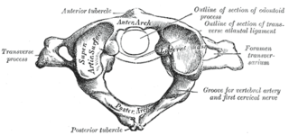
In anatomy, the atlas (C1) is the most superior (first) cervical vertebra of the spine and is located in the neck.

The neck is the part of the body on many vertebrates that connects the head with the torso. The neck supports the weight of the head and protects the nerves that carry sensory and motor information from the brain down to the rest of the body. In addition, the neck is highly flexible and allows the head to turn and flex in all directions. The structures of the human neck are anatomically grouped into four compartments: vertebral, visceral and two vascular compartments. Within these compartments, the neck houses the cervical vertebrae and cervical part of the spinal cord, upper parts of the respiratory and digestive tracts, endocrine glands, nerves, arteries and veins. Muscles of the neck are described separately from the compartments. They bound the neck triangles.

Whiplash, whose formal term is whiplash associated disorders (WAD), is a range of injuries to the neck caused by or related to a sudden distortion of the neck associated with extension, although the exact injury mechanisms remain unknown. The term "whiplash" is a colloquialism. "Cervical acceleration–deceleration" (CAD) describes the mechanism of the injury, while WAD describes the subsequent injuries and symptoms.

Back injuries result from damage, wear, or trauma to the bones, muscles, or other tissues of the back. Common back injuries include sprains and strains, herniated discs, and fractured vertebrae. The lumbar spine is often the site of back pain. The area is susceptible because of its flexibility and the amount of body weight it regularly bears. It is estimated that low-back pain may affect as much as 80 to 90 percent of the general population in the United States.

In tetrapods, cervical vertebrae are the vertebrae of the neck, immediately below the skull. Truncal vertebrae lie caudal of cervical vertebrae. In sauropsid species, the cervical vertebrae bear cervical ribs. In lizards and saurischian dinosaurs, the cervical ribs are large; in birds, they are small and completely fused to the vertebrae. The vertebral transverse processes of mammals are homologous to the cervical ribs of other amniotes. Most mammals have seven cervical vertebrae, with the only three known exceptions being the manatee with six, the two-toed sloth with five or six, and the three-toed sloth with nine.

Spinal adjustment and chiropractic adjustment are terms used by chiropractors to describe their approaches to spinal manipulation, as well as some osteopaths, who use the term adjustment. Despite anecdotal success, there is no scientific evidence that spinal adjustment is effective against disease.

In anatomy, the alar ligaments are ligaments which connect the dens to tubercles on the medial side of the occipital condyle.
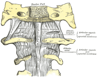
The atlanto-axial joint is a joint in the upper part of the neck between the atlas bone and the axis bone, which are the first and second cervical vertebrae. It is a pivot joint.

Hangman's fracture is the colloquial name given to a fracture of both pedicles, or partes interarticulares, of the axis vertebra (C2).

A spinal disc herniation is an injury to the intervertebral disc between two spinal vertebrae, usually caused by excessive strain or trauma to the spine. It may result in back pain, pain or sensation in different parts of the body, and physical disability. The most conclusive diagnostic tool for disc herniation is MRI, and treatments may range from painkillers to surgery. Protection from disc herniation is best provided by core strength and an awareness of body mechanics including good posture.
Atlanto-occipital dislocation, orthopedic decapitation, or internal decapitation describes ligamentous separation of the spinal column from the skull base. It is possible for a human to survive such an injury; however, 70% of cases result in immediate death. It should not be confused with atlanto-axial dislocation, which describes ligamentous separation between the first and second cervical vertebra.
The cruciate ligament of the atlas is a cross-shaped ligament in the neck forming part of the atlanto-axial joint. It consists of the transverse ligament of atlas, a superior longitudinal band, and an inferior longitudinal band.
A Jefferson fracture is a bone fracture of the anterior and posterior arches of the C1 vertebra, though it may also appear as a three- or two-part fracture. The fracture may result from an axial load on the back of the head or hyperextension of the neck, causing a posterior break, and may be accompanied by a break in other parts of the cervical spine.

Spinal disease refers to a condition impairing the backbone. These include various diseases of the back or spine ("dorso-"), such as kyphosis. Dorsalgia refers to back pain. Some other spinal diseases include spinal muscular atrophy, ankylosing spondylitis, scoliosis, lumbar spinal stenosis, spina bifida, spinal tumors, osteoporosis and cauda equina syndrome.
A spinal fracture, also called a vertebral fracture or a broken back, is a fracture affecting the vertebrae of the spinal column. Most types of spinal fracture confer a significant risk of spinal cord injury. After the immediate trauma, there is a risk of spinal cord injury if the fracture is unstable, that is, likely to change alignment without internal or external fixation.

The vertebral column, also known as the spinal column, spine or backbone, is the core part of the axial skeleton in vertebrate animals. The vertebral column is the defining and eponymous characteristic of the vertebrate endoskeleton, where the notochord found in all chordates has been replaced by a segmented series of mineralized irregular bones called vertebrae, separated by fibrocartilaginous intervertebral discs. The dorsal portion of the vertebral column houses the spinal canal, an elongated cavity formed by alignment of the vertebral neural arches that encloses and protects the spinal cord, with spinal nerves exiting via the intervertebral foramina to innervate each body segments.

Each vertebra is an irregular bone with a complex structure composed of bone and some hyaline cartilage, that make up the vertebral column or spine, of vertebrates. The proportions of the vertebrae differ according to their spinal segment and the particular species.

Forward head posture (FHP) is an excessively kyphotic (hunched) thoracic spine. It is clinically recognized as a form of repetitive strain injury. The posture can occur in dentists, surgeons, and hairdressers, or people who spend time on electronic devices. It is one of the most common postural issues. There is a correlation between forward head posture and neck pain in adults, but not adolescents.

Cervicocranial syndrome or is a combination of symptoms that are caused by an abnormality in the cervical vertebrae leading to improper function of cervical spinal nerves. Cervicocranial syndrome is either congenital or acquired. Cervicocranial syndrome may be caused by Chiari disease, Klippel-Feil malformation, osteoarthritis, and physical trauma. Treatment options include neck braces, pain medication and surgery. The quality of life for individuals suffering from Cervicocranial syndrome can improve through surgery.
The myodural bridge or miodural ligament is a bridge of connective tissue that extends between the suboccipital muscles and the cervical spinal dura mater, the outer membrane that envelops the spinal cord. It provides a physical connection between the musculoskeletal and nervous systems, and the circulation of cerebrospinal fluid. Its importance has been highlighted by various authors.
References
- ↑ Yang, King H. (2018-01-01), Yang, King-Hay (ed.), "Chapter 1 - Introduction", Basic Finite Element Method as Applied to Injury Biomechanics, Academic Press, pp. 3–49, doi:10.1016/b978-0-12-809831-8.00001-5, ISBN 978-0-12-809831-8 , retrieved 2024-06-11
- ↑ "axial loading". TheFreeDictionary.com. Retrieved 2024-06-11.
- ↑ Raniga, Sameer B.; Menon, Venugopal; Al Muzahmi, Khamis S.; Butt, Sajid (June 2014). "MDCT of acute subaxial cervical spine trauma: a mechanism-based approach". Insights into Imaging. 5 (3): 321–338. doi:10.1007/s13244-014-0311-y. ISSN 1869-4101. PMC 4035495 . PMID 24554380.
- ↑ Dave, Bharat R.; Krishnan, Ajay; Rai, Ravi Ranjan; Degulmadi, Devanand; Mayi, Shivanand (2020-03-30). "The Effect of Head Loading on Cervical Spine in Manual Laborers". Asian Spine Journal. 15 (1): 17–22. doi:10.31616/asj.2019.0221. ISSN 1976-1902. PMC 7904483 . PMID 32213796.
| | This biophysics-related article is a stub. You can help Wikipedia by expanding it. |