The striatum, or corpus striatum, is a nucleus in the subcortical basal ganglia of the forebrain. The striatum is a critical component of the motor and reward systems; receives glutamatergic and dopaminergic inputs from different sources; and serves as the primary input to the rest of the basal ganglia.

Tetanus toxin (TeNT) is an extremely potent neurotoxin produced by the vegetative cell of Clostridium tetani in anaerobic conditions, causing tetanus. It has no known function for clostridia in the soil environment where they are normally encountered. It is also called spasmogenic toxin, tentoxilysin, tetanospasmin or, tetanus neurotoxin. The LD50 of this toxin has been measured to be approximately 2.5–3 ng/kg, making it second only to the related botulinum toxin (LD50 2 ng/kg) as the deadliest toxin in the world. However, these tests are conducted solely on mice, which may react to the toxin differently from humans and other animals.

The olfactory bulb is a neural structure of the vertebrate forebrain involved in olfaction, the sense of smell. It sends olfactory information to be further processed in the amygdala, the orbitofrontal cortex (OFC) and the hippocampus where it plays a role in emotion, memory and learning. The bulb is divided into two distinct structures: the main olfactory bulb and the accessory olfactory bulb. The main olfactory bulb connects to the amygdala via the piriform cortex of the primary olfactory cortex and directly projects from the main olfactory bulb to specific amygdala areas. The accessory olfactory bulb resides on the dorsal-posterior region of the main olfactory bulb and forms a parallel pathway. Destruction of the olfactory bulb results in ipsilateral anosmia, while irritative lesions of the uncus can result in olfactory and gustatory hallucinations.

The barrel cortex is a region of the somatosensory cortex that is identifiable in some species of rodents and species of at least two other orders and contains the barrel field. The 'barrels' of the barrel field are regions within cortical layer IV that are visibly darker when stained to reveal the presence of cytochrome c oxidase and are separated from each other by lighter areas called septa. These dark-staining regions are a major target for somatosensory inputs from the thalamus, and each barrel corresponds to a region of the body. Due to this distinctive cellular structure, organisation, and functional significance, the barrel cortex is a useful tool to understand cortical processing and has played an important role in neuroscience. The majority of what is known about corticothalamic processing comes from studying the barrel cortex, and researchers have intensively studied the barrel cortex as a model of neocortical column.

Stellate cells are neurons in the central nervous system, named for their star-like shape formed by dendritic processes radiating from the cell body. Many stellate cells are GABAergic and are located in the molecular layer of the cerebellum. Stellate cells are derived from dividing progenitor cells in the white matter of postnatal cerebellum. Dendritic trees can vary between neurons. There are two types of dendritic trees in the cerebral cortex, which include pyramidal cells, which are pyramid shaped and stellate cells which are star shaped. Dendrites can also aid neuron classification. Dendrites with spines are classified as spiny, those without spines are classified as aspinous. Stellate cells can be spiny or aspinous, while pyramidal cells are always spiny. Most common stellate cells are the inhibitory interneurons found within the upper half of the molecular layer in the cerebellum. Cerebellar stellate cells synapse onto the dendritic trees of Purkinje cells and send inhibitory signals. Stellate neurons are sometimes found in other locations in the central nervous system; cortical spiny stellate cells are found in layer IVC of the primary visual cortex. In the somatosensory barrel cortex of mice and rats, glutamatergic (excitatory) spiny stellate cells are organized in the barrels of layer 4. They receive excitatory synaptic fibres from the thalamus and process feed forward excitation to 2/3 layer of the primary visual cortex to pyramidal cells. Cortical spiny stellate cells have a 'regular' firing pattern. Stellate cells are chromophobes, that is cells that does not stain readily, and thus appears relatively pale under the microscope.
Mriganka Sur is the Newton Professor of Neuroscience and Director of the Simons Center for the Social Brain at the Massachusetts Institute of Technology. He is also a Visiting Faculty Member in the Department of Computer Science and Engineering at the Indian Institute of Technology Madras and N.R. Narayana Murthy Distinguished Chair in Computational Brain Research at the Centre for Computational Brain Research, IIT Madras. He was on the Life Sciences jury for the Infosys Prize in 2010 and has been serving as Jury Chair from 2018.

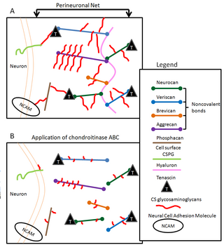
Perineuronal nets (PNNs) are specialized extracellular matrix structures responsible for synaptic stabilization in the adult brain. PNNs are found around certain neuron cell bodies and proximal neurites in the central nervous system. PNNs play a critical role in the closure of the childhood critical period, and their digestion can cause restored critical period-like synaptic plasticity in the adult brain. They are largely negatively charged and composed of chondroitin sulfate proteoglycans, molecules that play a key role in development and plasticity during postnatal development and in the adult.
Michael Greenberg is an American neuroscientist who specializes in molecular neurobiology. He served as the Chair of the Department of Neurobiology at Harvard Medical School from 2008 to 2022.

In neuroanatomy, pallium refers to the layers of grey and white matter that cover the upper surface of the cerebrum in vertebrates. The non-pallial part of the telencephalon builds the subpallium. In basal vertebrates the pallium is a relatively simple three-layered structure, encompassing 3–4 histogenetically distinct domains, plus the olfactory bulb.
Neurogenesis is the process by which nervous system cells, the neurons, are produced by neural stem cells (NSCs). In short, it is brain growth in relation to its organization. This occurs in all species of animals except the porifera (sponges) and placozoans. Types of NSCs include neuroepithelial cells (NECs), radial glial cells (RGCs), basal progenitors (BPs), intermediate neuronal precursors (INPs), subventricular zone astrocytes, and subgranular zone radial astrocytes, among others.

Madeline Lancaster is an American developmental biologist studying neurological development and diseases of the brain. Lancaster is a group leader at the Medical Research Council (MRC) Laboratory of Molecular Biology in Cambridge, UK.

Margarita Behrens is a neuroscientist and biochemist. She is currently an associate professor at the Salk Institute for Biological Studies where her lab studies the impact of oxidative stress on the post-natal brain through probing the biology of fast-spiking parvalbumin interneurons in models of schizophrenia.

Brenda Bloodgood is an American neuroscientist and associate professor of neurobiology at the University of California, San Diego. Bloodgood studies the molecular and cellular basis of brain circuitry changes in response to an animal's interactions with the environment.
Lisa Giocomo is an American neuroscientist who is a Professor in the Department of Neurobiology at Stanford University School of Medicine. Giocomo probes the molecular and cellular mechanisms underlying cortical neural circuits involved in spatial navigation and memory.
Nilay Yapici is a Turkish neuroscientist at Cornell University in Ithaca, New York, where she is the Nancy and Peter Meinig Family Investigator in the Life Sciences and Adelson Sesquicentennial Fellow in the Department of Neurobiology and Behavior. Yapici studies the neural circuits underlying decision making and feeding behavior in fruit fly models.
Jessica Cardin is an American neuroscientist who is an associate professor of neuroscience at Yale University School of Medicine. Cardin's lab studies local circuits within the primary visual cortex to understand how cellular and synaptic interactions flexibly adapt to different behavioral states and contexts to give rise to visual perceptions and drive motivated behaviors. Cardin's lab applies their knowledge of adaptive cortical circuit regulation to probe how circuit dysfunction manifests in disease models.
Corey C. Harwell is an American neuroscientist who is an assistant professor in the Department of Neurobiology at Harvard Medical School.

Eberhard Erich Fetz is an American neuroscientist, academic and researcher. He is a Professor of Physiology and Biophysics and DXARTS at the University of Washington.
Oscar Marín Parra FMedSci FRS is a Spanish and British neuroscientist. He is married to neuroscientist Beatriz Rico.
An assembloid is an in vitro model that combines two or more organoids, spheroids, or cultured cell types to recapitulate structural and functional properties of an organ. They are typically derived from induced pluripotent stem cells. Assembloids have been used to study cell migration, neural circuit assembly, neuro-immune interactions, metastasis, and other complex tissue processes. The term "assembloid" was coined by Sergiu P. Pașca's lab in 2017.


