
In optics, chromatic aberration (CA), also called chromatic distortion, color aberration, color fringing, or purple fringing, is a failure of a lens to focus all colors to the same point. It is caused by dispersion: the refractive index of the lens elements varies with the wavelength of light. The refractive index of most transparent materials decreases with increasing wavelength. Since the focal length of a lens depends on the refractive index, this variation in refractive index affects focusing. Since the focal length of the lens varies with the color of the light different colors of light are brought to focus at different distances from the lens or with different levels of magnification. Chromatic aberration manifests itself as "fringes" of color along boundaries that separate dark and bright parts of the image.

The optical microscope, also referred to as a light microscope, is a type of microscope that commonly uses visible light and a system of lenses to generate magnified images of small objects. Optical microscopes are the oldest design of microscope and were possibly invented in their present compound form in the 17th century. Basic optical microscopes can be very simple, although many complex designs aim to improve resolution and sample contrast.

In optical engineering, an objective is an optical element that gathers light from an object being observed and focuses the light rays from it to produce a real image of the object. Objectives can be a single lens or mirror, or combinations of several optical elements. They are used in microscopes, binoculars, telescopes, cameras, slide projectors, CD players and many other optical instruments. Objectives are also called object lenses, object glasses, or objective glasses.
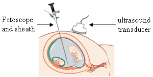
An endoscope is an inspection instrument composed of image sensor, optical lens, light source and mechanical device, which is used to look deep into the body by way of openings such as the mouth or anus. A typical endoscope applies several modern technologies including optics, ergonomics, precision mechanics, electronics, and software engineering. With an endoscope, it is possible to observe lesions that cannot be detected by X-ray, making it useful in medical diagnosis. An endoscope uses tubes only a few millimeters thick to transfer illumination in one direction and high-resolution video in the other, allowing minimally invasive surgeries. It is used to examine the internal organs like the throat or esophagus. Specialized instruments are named after their target organ. Examples include the cystoscope (bladder), nephroscope (kidney), bronchoscope (bronchus), arthroscope (joints) and colonoscope (colon), and laparoscope. They can be used to examine visually and diagnose, or assist in surgery such as an arthroscopy.

The Newtonian telescope, also called the Newtonian reflector or just a Newtonian, is a type of reflecting telescope invented by the English scientist Sir Isaac Newton, using a concave primary mirror and a flat diagonal secondary mirror. Newton's first reflecting telescope was completed in 1668 and is the earliest known functional reflecting telescope. The Newtonian telescope's simple design has made it very popular with amateur telescope makers.

Magnification is the process of enlarging the apparent size, not physical size, of something. This enlargement is quantified by a size ratio called optical magnification. When this number is less than one, it refers to a reduction in size, sometimes called de-magnification.

A fiberscope is a flexible optical fiber bundle with a lens on one end and an eyepiece or camera on the other. It is used to examine and inspect small, difficult-to-reach places such as the insides of machines, locks, and the human body.

An eyepiece, or ocular lens, is a type of lens that is attached to a variety of optical devices such as telescopes and microscopes. It is named because it is usually the lens that is closest to the eye when someone looks through an optical device to observe an object or sample. The objective lens or mirror collects light from an object or sample and brings it to focus creating an image of the object. The eyepiece is placed near the focal point of the objective to magnify this image to the eyes. The amount of magnification depends on the focal length of the eyepiece.

The Barlow lens, named after Peter Barlow, is a diverging lens which, used in series with other optics in an optical system, increases the effective focal length of an optical system as perceived by all components that are after it in the system. The practical result is that inserting a Barlow lens magnifies the image. A real Barlow lens is not a single glass element, because that would generate chromatic aberration, and spherical aberration if the lens is not aspheric. More common configurations use three or more elements for achromatic correction or apochromatic correction and higher image quality.

An optical fiber, or optical fibre, is a flexible glass or plastic fiber that can transmit light from one end to the other. Such fibers find wide usage in fiber-optic communications, where they permit transmission over longer distances and at higher bandwidths than electrical cables. Fibers are used instead of metal wires because signals travel along them with less loss and are immune to electromagnetic interference. Fibers are also used for illumination and imaging, and are often wrapped in bundles so they may be used to carry light into, or images out of confined spaces, as in the case of a fiberscope. Specially designed fibers are also used for a variety of other applications, such as fiber optic sensors and fiber lasers.
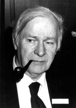
Harold Horace Hopkins FRS was a British physicist. His Wave Theory of Aberrations,, is central to all modern optical design and provides the mathematical analysis which enables the use of computers to create the highest quality lenses. In addition to his theoretical work, his many inventions are in daily use throughout the world. These include zoom lenses, coherent fibre-optics and more recently the rod-lens endoscopes which 'opened the door' to modern key-hole surgery. He was the recipient of many of the world's most prestigious awards and was twice nominated for a Nobel Prize. His citation on receiving the Rumford Medal from the Royal Society in 1984 stated: "In recognition of his many contributions to the theory and design of optical instruments, especially of a wide variety of important new medical instruments which have made a major contribution to clinical diagnosis and surgery."

A digital microscope is a variation of a traditional optical microscope that uses optics and a digital camera to output an image to a monitor, sometimes by means of software running on a computer. A digital microscope often has its own in-built LED light source, and differs from an optical microscope in that there is no provision to observe the sample directly through an eyepiece. Since the image is focused on the digital circuit, the entire system is designed for the monitor image. The optics for the human eye are omitted.

The stereo, stereoscopic or dissecting microscope is an optical microscope variant designed for low magnification observation of a sample, typically using light reflected from the surface of an object rather than transmitted through it. The instrument uses two separate optical paths with two objectives and eyepieces to provide slightly different viewing angles to the left and right eyes. This arrangement produces a three-dimensional visualization of the sample being examined. Stereomicroscopy overlaps macrophotography for recording and examining solid samples with complex surface topography, where a three-dimensional view is needed for analyzing the detail.
Remote Visual Inspection or Remote Digital Video Inspection, also known as RVI or RDVI, is a form of visual inspection which uses visual aids including video technology to allow an inspector to look at objects and materials from a distance because the objects are inaccessible or are in dangerous environments. RVI is also a specialty branch of nondestructive testing (NDT).

Afocal photography, also called afocal imaging or afocal projection is a method of photography where the camera with its lens attached is mounted over the eyepiece of another image forming system such as an optical telescope or optical microscope, with the camera lens taking the place of the human eye.

A USB microscope is a low-powered digital microscope which connects to a computer's USB port. Microscopes essentially the same as USB models are also available with other interfaces either in addition to or instead of USB, such as via WiFi. They are widely available at low cost for use at home or in commerce. Their cost varies in the range of tens to thousands of dollars. In essence, a USB microscope is a webcam with a high-powered macro lens, and generally uses reflected rather than transmitted light, using built-in LED light sources surrounding the lens. The camera is usually sensitive enough not to need additional illumination beyond normal ambient lighting. The camera attaches directly to the USB port of a computer without the need for an eyepiece, and the images are shown directly on the computer's display.
In optics, a relay lens is a lens or a group of lenses that receives the image from the objective lens and relays it to the eyepiece. Relay lenses are found in refracting telescopes, endoscopes, and periscopes to optically manipulate the light path, extend the length of the whole optical system, and usually serve the purpose of inverting the image. They may be made of one or more conventional lenses or achromatic doublets, or a long cylindrical gradient-index of refraction lens.
The term power assembly refers to an Electro-Motive Diesel (EMD) engine sub-assembly designed to be "easily" removed and replaced in order to restore engine performance lost to wear or engine failure. Typical of heavy-duty internal combustion engines used in industrial applications, EMD engines are designed to allow the cylinder liners, pistons, piston rings and connecting rods to be replaced at overhaul without removing the entire engine assembly from its application location. This increases engine value, reduces downtime and allows the engine to be returned to true new engine performance. Other terms such as cylinder pack, liner pack, cylinder assembly and cylinder kit are used in the engine industry to describe similar assemblies. In the large-engine industry, the term "power assembly" has also become generic and is often used to refer to the assemblies used in non-EMD engines where "power pack" may be the preferred term, although both terms are functionally the same.
Architectural endoscopy or architectural envisioning is used to photograph and film models of new buildings' exterior and interior in the planning stage. An architectural model of a new building in a 1:500 scale is thus correctly visualized from the perspective of a pedestrian walking by in the street. An endoscope connected to a video camera allows for the creation of walkthroughs, allowing the architect to develop the first draft further, and the public to share and critique the architect's vision of proposed buildings and cities.
A scanning fiber endoscope is a technology that uses a flexible, small peripheral or coronary catheter to provide wide-field, high-quality, full-color, laser-based video imaging. These differences distinguish SFE applications from current imaging approaches such as IVUS and Intracoronary OCT. Applications for the device, are expected to include medical diagnosis and support in determining interventional treatments such as surgery or biopsy. Providing both full-color images and a wide-field, real-time surgical view into the inner depths of arteries, enables physicians to circumnavigate hard to reach internal tissues to assess for potential disease.


