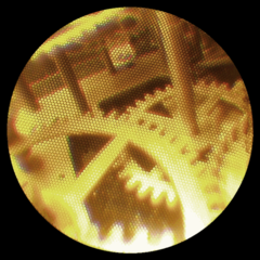
A fiberscope is a flexible optical fiber bundle with a lens on one end and an eyepiece or camera on the other. It is used to examine and inspect small, difficult-to-reach places such as the insides of machines, locks, and the human body.

A fiberscope is a flexible optical fiber bundle with a lens on one end and an eyepiece or camera on the other. It is used to examine and inspect small, difficult-to-reach places such as the insides of machines, locks, and the human body.
Guiding of light by refraction, the principle that makes fiber optics possible, was first demonstrated by Daniel Colladon and Jacques Babinet in Paris in the early 1840s. Then in 1930, Heinrich Lamm, a German medical student, became the first person to put together a bundle of optical fibers to carry an image. These discoveries led to the invention of endoscopes and fiberscopes. [1] In the 1960s the endoscope was upgraded with glass fiber, a flexible material that allowed light to transmit, even when bent. While this provided users with the capability of real-time observation, it did not provide them with the ability to take photographs. In 1964 the fiberscope, the first gastro camera, was invented. It was the first time an endoscope had a camera that could take pictures. This innovation led to more careful observations, and more accurate diagnoses. [2]
Fiberscopes work by utilizing the science of fiber-optic bundles, which consist of numerous fiber-optic cables. Fiber-optic cables are made of optically pure glass and are as thin as a human’s hair. The three main components of a fiber-optic cable are:
The following are the two different types of fiber-optic bundles in a fiberscope:
Fiber-optic cables use total internal reflection to carry information. When light travels from one medium to another it is refracted. If the light is traveling from a less dense medium to a dense medium it is refracted away from the normal. The opposite applies if the light is traveling from a dense medium to a less dense medium. In optic cables, light travels through the dense glass core (high refractive index) by constantly reflecting from the less dense cladding (lower refractive index). This happens because the surface of the core acts like a perfect mirror and the angle of the light is always larger than the critical angle. [4]
Fiberscopes are used in the medical field as a tool to help doctors and surgeons examine problems in a patient’s body without having to make large incisions. This procedure is called an endoscopy. Doctors use this when they suspect that a patient’s organ is infected, damaged, or cancerous. There are numerous types based on the area of the body being examined. They include:
Although any medical technique has its potential risks, using a fiberscope for endoscopy has a very low risk of causing infection and blood loss.
Locksmiths use fiberscopes to check the position of pins. Technicians and inspectors use fiberscopes to look at the inside of machines without having to disassemble them. Fiberscopes can also be used in a military or police application to check beneath doors or around corners, or otherwise perform surveillance or reconnaissance.

In optics, the numerical aperture (NA) of an optical system is a dimensionless number that characterizes the range of angles over which the system can accept or emit light. By incorporating index of refraction in its definition, NA has the property that it is constant for a beam as it goes from one material to another, provided there is no refractive power at the interface. The exact definition of the term varies slightly between different areas of optics. Numerical aperture is commonly used in microscopy to describe the acceptance cone of an objective, and in fiber optics, in which it describes the range of angles within which light that is incident on the fiber will be transmitted along it.

In fiber-optic communication, a single-mode optical fiber (SMF), also known as fundamental- or mono-mode, is an optical fiber designed to carry only a single mode of light - the transverse mode. Modes are the possible solutions of the Helmholtz equation for waves, which is obtained by combining Maxwell's equations and the boundary conditions. These modes define the way the wave travels through space, i.e. how the wave is distributed in space. Waves can have the same mode but have different frequencies. This is the case in single-mode fibers, where we can have waves with different frequencies, but of the same mode, which means that they are distributed in space in the same way, and that gives us a single ray of light. Although the ray travels parallel to the length of the fiber, it is often called transverse mode since its electromagnetic oscillations occur perpendicular (transverse) to the length of the fiber. The 2009 Nobel Prize in Physics was awarded to Charles K. Kao for his theoretical work on the single-mode optical fiber. The standards G.652 and G.657 define the most widely used forms of single-mode optical fiber.
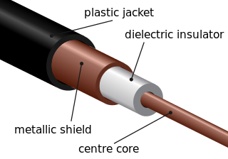
A transmission medium is a system or substance that can mediate the propagation of signals for the purposes of telecommunication. Signals are typically imposed on a wave of some kind suitable for the chosen medium. For example, data can modulate sound, and a transmission medium for sounds may be air, but solids and liquids may also act as the transmission medium. Vacuum or air constitutes a good transmission medium for electromagnetic waves such as light and radio waves. While a material substance is not required for electromagnetic waves to propagate, such waves are usually affected by the transmission media they pass through, for instance, by absorption or reflection or refraction at the interfaces between media. Technical devices can therefore be employed to transmit or guide waves. Thus, an optical fiber or a copper cable is used as transmission media.

The optical microscope, also referred to as a light microscope, is a type of microscope that commonly uses visible light and a system of lenses to generate magnified images of small objects. Optical microscopes are the oldest design of microscope and were possibly invented in their present compound form in the 17th century. Basic optical microscopes can be very simple, although many complex designs aim to improve resolution and sample contrast.
Optics is the branch of physics which involves the behavior and properties of light, including its interactions with matter and the construction of instruments that use or detect it. Optics usually describes the behavior of visible, ultraviolet, and infrared light. Because light is an electromagnetic wave, other forms of electromagnetic radiation such as X-rays, microwaves, and radio waves exhibit similar properties.
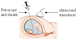
An endoscope is an inspection instrument composed of image sensor, optical lens, light source and mechanical device, which is used to look deep into the body by way of openings such as the mouth or anus. A typical endoscope applies several modern technologies including optics, ergonomics, precision mechanics, electronics, and software engineering. With an endoscope, it is possible to observe lesions that cannot be detected by X-ray, making it useful in medical diagnosis. Endoscopes use tubes which are only a few millimeters thick to transfer illumination in one direction and high-resolution images in real time in the other direction, resulting in minimally invasive surgeries. It is used to examine the internal organs like the throat or esophagus. Specialized instruments are named after their target organ. Examples include the cystoscope (bladder), nephroscope (kidney), bronchoscope (bronchus), arthroscope (joints) and colonoscope (colon), and laparoscope. They can be used to examine visually and diagnose, or assist in surgery such as an arthroscopy.

A borescope is an optical instrument designed to assist visual inspection of narrow, difficult-to-reach cavities, consisting of a rigid or flexible tube with an eyepiece or display on one end, an objective lens or camera on the other, linked together by an optical or electrical system in between. The optical system in some instances is accompanied by illumination to enhance brightness and contrast. An internal image of the illuminated object is formed by the objective lens and magnified by the eyepiece which presents it to the viewer's eye.

Gradient-index (GRIN) optics is the branch of optics covering optical effects produced by a gradient of the refractive index of a material. Such gradual variation can be used to produce lenses with flat surfaces, or lenses that do not have the aberrations typical of traditional spherical lenses. Gradient-index lenses may have a refraction gradient that is spherical, axial, or radial.

A flexible Videoscope or Video Borescope is an advanced type of borescope that houses a very small image sensor embedded into the tip of the scope. The video image is relayed from the distal tip and focusable lens assembly back to the display via internal wiring. This is unlike a traditional rigid borescope and flexible fiberscope. Rigid borescopes use hard optical relay components to transfer the image from the tip to an eyepiece and flexible fiberscopes use coherent image fiber optics to relay the image to one's eye through an eyepiece. The image quality of a videoscope is superior to that of a fiberscope and can be compared to that of an intermediate camcorder.
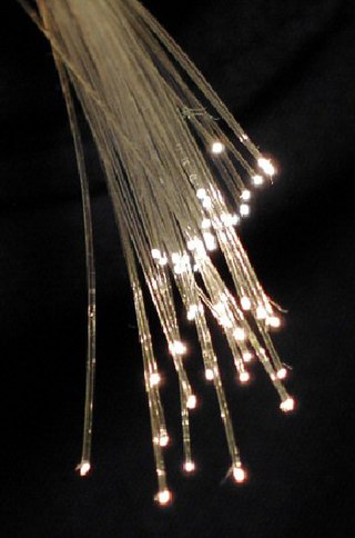
An optical fiber, or optical fibre, is a flexible glass or plastic fiber that can transmit light from one end to the other. Such fibers find wide usage in fiber-optic communications, where they permit transmission over longer distances and at higher bandwidths than electrical cables. Fibers are used instead of metal wires because signals travel along them with less loss and are immune to electromagnetic interference. Fibers are also used for illumination and imaging, and are often wrapped in bundles so they may be used to carry light into, or images out of confined spaces, as in the case of a fiberscope. Specially designed fibers are also used for a variety of other applications, such as fiber optic sensors and fiber lasers.

A fiber-optic cable, also known as an optical-fiber cable, is an assembly similar to an electrical cable but containing one or more optical fibers that are used to carry light. The optical fiber elements are typically individually coated with plastic layers and contained in a protective tube suitable for the environment where the cable is used. Different types of cable are used for fiber-optic communication in different applications, for example long-distance telecommunication or providing a high-speed data connection between different parts of a building.

A microlens is a small lens, generally with a diameter less than a millimetre (mm) and often as small as 10 micrometres (μm). The small sizes of the lenses means that a simple design can give good optical quality but sometimes unwanted effects arise due to optical diffraction at the small features. A typical microlens may be a single element with one plane surface and one spherical convex surface to refract the light. Because micro-lenses are so small, the substrate that supports them is usually thicker than the lens and this has to be taken into account in the design. More sophisticated lenses may use aspherical surfaces and others may use several layers of optical material to achieve their design performance.

Harold Horace Hopkins FRS was a British physicist. His Wave Theory of Aberrations,, is central to all modern optical design and provides the mathematical analysis which enables the use of computers to create the highest quality lenses. In addition to his theoretical work, his many inventions are in daily use throughout the world. These include zoom lenses, coherent fibre-optics and more recently the rod-lens endoscopes which 'opened the door' to modern key-hole surgery. He was the recipient of many of the world's most prestigious awards and was twice nominated for a Nobel Prize. His citation on receiving the Rumford Medal from the Royal Society in 1984 stated: "In recognition of his many contributions to the theory and design of optical instruments, especially of a wide variety of important new medical instruments which have made a major contribution to clinical diagnosis and surgery."

The stereo, stereoscopic or dissecting microscope is an optical microscope variant designed for low magnification observation of a sample, typically using light reflected from the surface of an object rather than transmitted through it. The instrument uses two separate optical paths with two objectives and eyepieces to provide slightly different viewing angles to the left and right eyes. This arrangement produces a three-dimensional visualization of the sample being examined. Stereomicroscopy overlaps macrophotography for recording and examining solid samples with complex surface topography, where a three-dimensional view is needed for analyzing the detail.

A USB microscope is a low-powered digital microscope which connects to a computer's USB port. Microscopes essentially the same as USB models are also available with other interfaces either in addition to or instead of USB, such as via WiFi. They are widely available at low cost for use at home or in commerce. Their cost varies in the range of tens to thousands of dollars. In essence, a USB microscope is a webcam with a high-powered macro lens, and generally uses reflected rather than transmitted light, using built-in LED light sources surrounding the lens. The camera is usually sensitive enough not to need additional illumination beyond normal ambient lighting. The camera attaches directly to the USB port of a computer without the need for an eyepiece, and the images are shown directly on the computer's display.
In optics, a relay lens is a lens or a group of lenses that receives the image from the objective lens and relays it to the eyepiece. Relay lenses are found in refracting telescopes, endoscopes, and periscopes to optically manipulate the light path, extend the length of the whole optical system, and usually serve the purpose of inverting the image. They may be made of one or more conventional lenses or achromatic doublets, or a long cylindrical gradient-index of refraction lens.
Cladding in optical fibers is one or more layers of materials of lower refractive index in intimate contact with a core material of higher refractive index.
Endomicroscopy is a technique for obtaining histology-like images from inside the human body in real-time, a process known as ‘optical biopsy’. It generally refers to fluorescence confocal microscopy, although multi-photon microscopy and optical coherence tomography have also been adapted for endoscopic use. Commercially available clinical and pre-clinical endomicroscopes can achieve a resolution on the order of a micrometre, have a field-of-view of several hundred μm, and are compatible with fluorophores which are excitable using 488 nm laser light. The main clinical applications are currently in imaging of the tumour margins of the brain and gastro-intestinal tract, particularly for the diagnosis and characterisation of Barrett’s Esophagus, pancreatic cysts and colorectal lesions. A number of pre-clinical and transnational applications have been developed for endomicroscopy as it enables researchers to perform live animal imaging. Major pre-clinical applications are in gastro-intestinal tract, toumour margin detection, uterine complications, ischaemia, live imaging of cartilage and tendon and organoid imaging.
A scanning fiber endoscope is a technology that uses a flexible, small peripheral or coronary catheter to provide wide-field, high-quality, full-color, laser-based video imaging. These differences distinguish SFE applications from current imaging approaches such as IVUS and Intracoronary OCT. Applications for the device, are expected to include medical diagnosis and support in determining interventional treatments such as surgery or biopsy. Providing both full-color images and a wide-field, real-time surgical view into the inner depths of arteries, enables physicians to circumnavigate hard to reach internal tissues to assess for potential disease.

A ball lens is an optical lens in the shape of a sphere. Formally, it is a bi-convex spherical lens with the same radius of curvature on both sides, and diameter equal to twice the radius of curvature. The same optical laws may be applied to analyze its imaging characteristics as for other lenses.