Related Research Articles

A urethral stricture is a narrowing of the urethra, the tube connected to the bladder that allows the passing of urine. The narrowing reduces the flow of urine and makes it more difficult or even painful to empty the bladder.

Hypospadias is a common variation in fetal development of the penis in which the urethra does not open from its usual location on the head of the penis. It is the second-most common birth abnormality of the male reproductive system, affecting about one of every 250 males at birth. Roughly 90% of cases are the less serious distal hypospadias, in which the urethral opening is on or near the head of the penis (glans). The remainder have proximal hypospadias, in which the meatus is all the way back on the shaft of the penis, near or within the scrotum. Shiny tissue that typically forms the urethra instead extends from the meatus to the tip of the glans; this tissue is called the urethral plate.

The penile artery is the artery that serves blood to the penis.

Penile fracture is rupture of one or both of the tunica albuginea, the fibrous coverings that envelop the penis's corpora cavernosa. It is caused by rapid blunt force to an erect penis, usually during vaginal intercourse, or aggressive masturbation. It sometimes also involves partial or complete rupture of the urethra or injury to the dorsal nerves, veins and arteries.

Hydronephrosis describes hydrostatic dilation of the renal pelvis and calyces as a result of obstruction to urine flow downstream. Alternatively, hydroureter describes the dilation of the ureter, and hydronephroureter describes the dilation of the entire upper urinary tract.

A suprapubic cystostomy or suprapubic catheter (SPC) is a surgically created connection between the urinary bladder and the skin used to drain urine from the bladder in individuals with obstruction of normal urinary flow. The connection does not go through the abdominal cavity.
A urethrotomy is an operation which involves incision of the urethra, especially for relief of a stricture. It is most often performed in the outpatient setting, with the patient (usually) being discharged from the hospital or surgery center within six hours from the procedure's inception.

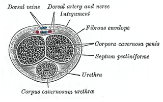
A corpus cavernosum penis (singular) is one of a pair of sponge-like regions of erectile tissue, which contain most of the blood in the penis during an erection.

Prostaglandin E1 (PGE1) is a naturally occurring prostaglandin and is also used as a medication (alprostadil).
Urethroplasty is the surgical repair of an injury or defect within the walls of the urethra. Trauma, iatrogenic injury and infections are the most common causes of urethral injury/defect requiring repair. Urethroplasty is regarded as the gold standard treatment for urethral strictures and offers better outcomes in terms of recurrence rates than dilatations and urethrotomies. It is probably the only useful modality of treatment for long and complex strictures though recurrence rates are higher for this difficult treatment group.

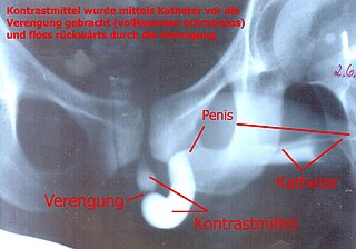
A retrograde urethrography is a routine radiologic procedure used to image the integrity of the urethra. Hence a retrograde urethrogram is essential for diagnosis of urethral injury, or urethral stricture.

Urethrostomy is a surgical procedure that creates a permanent opening in the urethra, commonly to remove obstructions to urine flow. The procedure is most often performed in male cats, where the opening is made in the perineum.

Scrotalultrasound is a medical ultrasound examination of the scrotum. It is used in the evaluation of testicular pain, and can help identify solid masses.

Ultrasonography of suspected or previously confirmed chronic venous insufficiency of leg veins is a risk-free, non-invasive procedure. It gives information about the anatomy, physiology and pathology of mainly superficial veins. As with heart ultrasound (echocardiography) studies, venous ultrasonography requires an understanding of hemodynamics in order to give useful examination reports. In chronic venous insufficiency, sonographic examination is of most benefit; in confirming varicose disease, making an assessment of the hemodynamics, and charting the progression of the disease and its response to treatment. It has become the reference standard for examining the condition and hemodynamics of the lower limb veins. Particular veins of the deep venous system (DVS), and the superficial venous system (SVS) are looked at. The great saphenous vein (GSV), and the small saphenous vein (SSV) are superficial veins which drain into respectively, the common femoral vein and the popliteal vein. These veins are deep veins. Perforator veins drain superficial veins into the deep veins. Three anatomic compartments are described, (N1) containing the deep veins, (N2) containing the perforator veins, and (N3) containing the superficial veins, known as the saphenous compartment. This compartmentalisation makes it easier for the examiner to systematize and map. The GSV can be located in the saphenous compartment where together with the Giacomini vein and the accessory saphenous vein (ASV) an image resembling an eye, known as the 'eye sign' can be seen. The ASV which is often responsible for varicose veins, can be located at the 'alignment sign', where it is seen to align with the femoral vessels.

Doppler ultrasonography is medical ultrasonography that employs the Doppler effect to perform imaging of the movement of tissues and body fluids, and their relative velocity to the probe. By calculating the frequency shift of a particular sample volume, for example, flow in an artery or a jet of blood flow over a heart valve, its speed and direction can be determined and visualized.

Penile Artery Shunt Syndrome (PASS) is an iatrogenic clinical phenomenon first described by Tariq Hakky, Christopher Yang, Jonathan Pavlinec, Kamal Massis, and Rafael Carrion within the Sexual Medicine Program in the Department of Urology, at the University of South Florida, and Ricardo Munarriz, of Boston University School of Medicine Department of Urology in 2013. It may be a cause of refractory Erectile Dysfunction in patients who have undergone Penile Revascularization Surgery.
A penile injury is a medical emergency that afflicts the penis. Common injuries include fracture, avulsion injury, strangulation, entrapment, and amputation.
Rupture of the urethra is an uncommon result of penile injury, incorrect catheter insertion, straddle injury, or pelvic girdle fracture. The urethra, the muscular tube that allows for urination, may be damaged by trauma. When urethral rupture occurs, urine may extravasate (escape) into the surrounding tissues. The membranous urethra is most likely to be injured in pelvic fractures, allowing urine and blood to enter the deep perineal space and subperitoneal spaces via the genital hiatus. The spongy urethra is most likely to be injured with a catheter or in a straddle injury, allowing urine and blood to escape into the scrotum, the penis, and the superficial peritoneal space. Urethral rupture may be diagnosed with a cystourethrogram. Due to the tight adherence of the fascia lata, urine from a urethral rupture cannot spread into the thighs.
Penile ulltrasonography is medical ultrasonography of the penis. Ultrasound is an excellent method for the study of the penis, such as indicated in trauma, priapism, erectile dysfunction or suspected Peyronie's disease.

Glans insufficiency syndrome, also known as the soft glans, cold glans, or glans insufficiency, is a medical condition that affects male individuals. This condition is characterized by the persistent inability of the glans penis to achieve and maintain an erect or turgid state during sexual arousal, remaining soft and cold. This condition can have an impact on a person's sexual function, including decreased sensitivity, difficulty in maintaining an erection, and overall quality of life.
References
- ↑ Bhagat SK, Gopalakrishnan G, Kumar S, Devasia A, Kekre NS. Redo-urethroplasty in pelvic fracture urethral distraction defect: an audit. World J Urol. 2011:1:97–101.
- ↑ Chen L, Hu B, Feng C, Sun XJ. Predictive value of penile dynamic colour duplex Doppler ultrasound parameters in patients with posttraumatic urethral stricture. J Int Med Res. 2011:39:1513–9.