Related Research Articles
The term abdominal surgery broadly covers surgical procedures that involve opening the abdomen (laparotomy). Surgery of each abdominal organ is dealt with separately in connection with the description of that organ Diseases affecting the abdominal cavity are dealt with generally under their own names.

The peritoneum is the serous membrane forming the lining of the abdominal cavity or coelom in amniotes and some invertebrates, such as annelids. It covers most of the intra-abdominal organs, and is composed of a layer of mesothelium supported by a thin layer of connective tissue. This peritoneal lining of the cavity supports many of the abdominal organs and serves as a conduit for their blood vessels, lymphatic vessels, and nerves.
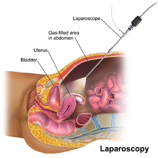
Laparoscopy is an operation performed in the abdomen or pelvis using small incisions with the aid of a camera. The laparoscope aids diagnosis or therapeutic interventions with a few small cuts in the abdomen.

Appendicitis is inflammation of the appendix. Symptoms commonly include right lower abdominal pain, nausea, vomiting, and decreased appetite. However, approximately 40% of people do not have these typical symptoms. Severe complications of a ruptured appendix include widespread, painful inflammation of the inner lining of the abdominal wall and sepsis.

Peritonitis is inflammation of the localized or generalized peritoneum, the lining of the inner wall of the abdomen and cover of the abdominal organs. Symptoms may include severe pain, swelling of the abdomen, fever, or weight loss. One part or the entire abdomen may be tender. Complications may include shock and acute respiratory distress syndrome.

The mesothelium is a membrane composed of simple squamous epithelial cells of mesodermal origin, which forms the lining of several body cavities: the pleura, peritoneum and pericardium.

The thoracic diaphragm, or simply the diaphragm, is a sheet of internal skeletal muscle in humans and other mammals that extends across the bottom of the thoracic cavity. The diaphragm is the most important muscle of respiration, and separates the thoracic cavity, containing the heart and lungs, from the abdominal cavity: as the diaphragm contracts, the volume of the thoracic cavity increases, creating a negative pressure there, which draws air into the lungs. Its high oxygen consumption is noted by the many mitochondria and capillaries present; more than in any other skeletal muscle.
A laparotomy is a surgical procedure involving a surgical incision through the abdominal wall to gain access into the abdominal cavity. It is also known as a celiotomy.

An injury is any physiological damage to living tissue caused by immediate physical stress. An injury can occur intentionally or unintentionally and may be caused by blunt trauma, penetrating trauma, burning, toxic exposure, asphyxiation, or overexertion. Injuries can occur in any part of the body, and different symptoms are associated with different injuries.

The mesentery is an organ that attaches the intestines to the posterior abdominal wall in humans and is formed by the double fold of peritoneum. It helps in storing fat and allowing blood vessels, lymphatics, and nerves to supply the intestines, among other functions.
Peritoneal dialysis (PD) is a type of dialysis which uses the peritoneum in a person's abdomen as the membrane through which fluid and dissolved substances are exchanged with the blood. It is used to remove excess fluid, correct electrolyte problems, and remove toxins in those with kidney failure. Peritoneal dialysis has better outcomes than hemodialysis during the first couple of years. Other benefits include greater flexibility and better tolerability in those with significant heart disease.
The peritoneal cavity is a potential space between the parietal peritoneum and visceral peritoneum. The parietal and visceral peritonea are layers of the peritoneum named depending on their function/location. It is one of the spaces derived from the coelomic cavity of the embryo, the others being the pleural cavities around the lungs and the pericardial cavity around the heart.

The patellar tendon is the distal portion of the common tendon of the quadriceps femoris, which is continued from the patella to the tibial tuberosity. It is also sometimes called the patellar ligament as it forms a bone to bone connection when the patella is fully ossified.

Adhesions are fibrous bands that form between tissues and organs, often as a result of injury during surgery. They may be thought of as internal scar tissue that connects tissues not normally connected.
An adhesion barrier is a medical implant that can be used to reduce abnormal internal scarring (adhesions) following surgery by separating the internal tissues and organs while they heal.
Ovarian drilling, also known as multiperforation or laparoscopic ovarian diathermy, is a surgical technique of puncturing the membranes surrounding the ovary with a laser beam or a surgical needle using minimally invasive laparoscopic procedures. It differs from ovarian wedge resection, which involves the cutting of tissue. Minimally invasive ovarian drilling procedures have replaced wedge resections. Ovarian drilling is preferred to wedge resection because cutting into the ovary can cause adhesions which may complicate postoperative outcomes. Ovarian drilling and ovarian wedge resection are treatment options to reduce the amount of androgen producing tissue in women with polycystic ovarian syndrome (PCOS). PCOS is the primary cause of anovulation, which results in female infertility. The induction of mono-ovulatory cycles can restore fertility.

Intraperitoneal hyperthermic chemoperfusion is a type of hyperthermia therapy used in combination with surgery in the treatment of advanced abdominal cancers. In this procedure, warmed anti-cancer medications are infused and circulated in the peritoneal cavity (abdomen) for a short period of time. The chemotherapeutic agents generally infused during IPHC are mitomycin-C and cisplatin.
Surgical humidification is the conditioning of insufflation gas with water vapour (humidity) and heat during surgery. Surgical humidification is used to reduce the risk of tissue drying and evaporative cooling.
The development of the digestive system in the human embryo concerns the epithelium of the digestive system and the parenchyma of its derivatives, which originate from the endoderm. Connective tissue, muscular components, and peritoneal components originate in the mesoderm. Different regions of the gut tube such as the esophagus, stomach, duodenum, etc. are specified by a retinoic acid gradient that causes transcription factors unique to each region to be expressed. Differentiation of the gut and its derivatives depends upon reciprocal interactions between the gut endoderm and its surrounding mesoderm. Hox genes in the mesoderm are induced by a Hedgehog signaling pathway secreted by gut endoderm and regulate the craniocaudal organization of the gut and its derivatives. The gut system extends from the oropharyngeal membrane to the cloacal membrane and is divided into the foregut, midgut, and hindgut.
Local anesthetic nerve block is a short-term nerve block involving the injection of local anesthetic as close to the nerve as possible for pain relief. The local anesthetic bathes the nerve and numbs the area of the body that is supplied by that nerve. The goal of the nerve block is to prevent pain by blocking the transmission of pain signals from the affected area. Nerve blocks have a number of uses including treating headache disorders and providing anesthesia during surgery. The pain relief provided by the block is present during the surgery and continues to last after the procedure. This can lead to a reduction in the amount of opiates needed for pain control. The advantages of nerve blocks over general anesthesia include faster recovery, monitored anesthesia care vs. intubation with an airway tube, and much less postoperative pain.
References
- ↑ Huang, Yi-Ting; Su, Yu-Ying; Wu, Kai-Yun; Huang, Hui-Yu; Lin, Yu-Shan; Weng, Cindy Hsuan; Yang, Lan-Yan; Pan, Yu-Bin; Wang, Chin-Jung (2020-10-06). "Learning curve analysis of applying Seprafilm hyaluronic acid/carboxymethylcellulose membrane during laparoscopic hysterectomy". Scientific Reports. 10 (1): 16592. doi:10.1038/s41598-020-73692-y. ISSN 2045-2322. PMC 7538582 . PMID 33024214.
- ↑ Craig, A. B.; Ellis, A. G. (June 1905). "I. An Experimental and Histological Study of Cargile Membrane: With Reference to (1) Its Efficacy in Preventing Adhesion in the Abdominal and Cranial Cavities and Around Nerves and Tendons, and (2) Its Ultimate Fate in the Tissues". Annals of Surgery. 41 (6): 801–822. doi:10.1097/00000658-190506000-00001. ISSN 0003-4932. PMC 1426045 . PMID 17861646.
- ↑ Some uses of Cargile membrane – Red Cross Notes. Johnson & Johnson. 1904. pp. 70–.
- ↑ "Find A Grave". Archived from the original on 2016-12-21. Retrieved 2016-02-26.