Chemorepulsion

Last updated

Chemorepulsion is the directional movement of a cell away from a substance. Of the two directional varieties of chemotaxis, chemoattraction has been studied to a much greater extent. Only recently have the key components of the chemorepulsive pathway been elucidated. [1] The exact mechanism is still being investigated, and its constituents are currently being explored as likely candidates for immunotherapies. [2]

Contents

Cell Migration Glossary
Chemotaxis Cellular response to an environmental substance with a directional movement.
Chemokinesis Cellular response to an environmental substance with a random, non-vectorial movement.
Chemoattraction Directional cell movement towards a substance
Chemorepulsion Directional cell movement away from a substance
Chemokines Secreted cell-signaling proteins able to induce chemotaxis in nearby cells.
Immunorepulsion The active movement of immune cells away from a substance

History and etymology

Neutrophils being repelled from a chemokinetic agent Immunorepulsion 2.jpg
Neutrophils being repelled from a chemokinetic agent

The mechanism of the chemorepulsion of immune cells was first acknowledged by medical researchers at the Massachusetts General Hospital in Boston in early 2002. [1] The phenomenon was originally referred to as "reverse chemotaxis," and later, “fugetaxis” (derived from the Latin words fugere, to flee from; and taxis, movement). [1] For a time, the words were used interchangeably before being replaced almost exclusively by “chemorepulsion.” While "chemorepulsion" applies to all cell types, the term "immunorepulsion" is gaining momentum as a more specific term that only applies to hematopoietic blood cell types that are involved in immune responses. Different cell types to which the term "immunorepulsion" could potentially be applied include: Myeloid lineage cells (monocytes, macrophages, neutrophils, basophils, eosinophils, erythrocytes, platelets, dendritic cells) and Lymphoid lineage cells (T-cells, B-cells, NK-cells).

Role in physiological processes

The chemorepulsion of immune cells was first postulated a priori based on the established migratory behavior of cells evidenced in several naturally occurring physiological processes: the development of the Central Nervous System, the establishment of immune-privileged sites, and thymic emigration.

Central nervous system development

During the development of the Central Nervous System, chemokinetic agents influence the localization of neuronal cells by either attracting or repelling the growing axon. [3] This mechanism of context-dependent bidirectionality serves as a valuable model of chemorepulsion that can be studied in vivo. [1] Additionally, there is growing evidence that chemorepulsion is probably a key mechanism involved in regulating leukocyte motility. [4] Many of the chemorepellents that affect neuronal cell migration, including netrins, semaphorins, slit ligands, and ephrins have recently been implicated in the motility of immune cells. [1] For example, the Slit protein that mediates axonal chemorepulsion has also been shown to inhibit the directed migration of leukocytes in response to chemoattractants. [5] Other factors might also provide chemorepulsive effects on immune cells, and these inhibitory effects might be regulated by the tissue microenvironment.

Immune-privileged sites

Certain body tissues are able to tolerate antigens without an inflammatory immune response. [6] Immune privilege is thought to be an evolutionary adaptation to protect the most vital sensory organs and reproductive structures that would be otherwise severely impaired during an inflammatory response. [7] Although these locations are often physically isolated or segregated from access by immune cells, there are some functionally significant characteristics of such environments that are unique, and could potentially be replicated to keep immune cells away from targeted areas. Known immunologically privileged sites include the:

  1. Brain and central nervous system
  2. Eyes
  3. Placenta and fetus
  4. Testicles

Characteristics that are particular to immune-privileged sites should be seriously considered when investigating candidates for immunorepulsion therapy. These characteristics include:

  1. Low expression of Classical MHC Class IA molecules.
  2. Expression of immunoregulatory Nonclassical MHC Class IB molecules.
  3. Increased expression of surface molecules that inhibit complement activation.
  4. Local production of immunosuppressive cytokines, such as TGF-β
  5. Presence of neuropeptides.
  6. Expression of Fas ligand that controls the entry of Fas-expressing lymphoid cells.

Thymic emigration

T-cells are one of the most critical constituents of the adaptive immune system due to their ability to continue developing after activation. [8] To prevent premature instigation, it is necessary for T-cells to mature in an environment completely isolated from any potentially activating factors (antigens, cytokines, steroids, receptor antagonists, adhesion molecules, etc.). [9] As a result, T-cells are formed in the bone marrow and subsequently migrate to the cortex of the thymus where they can mature in an antigen-free environment. The thymus supports the differentiation of multiple distinct T cell subsets that play unique roles in the immune system. For example, T-helper, T-cytotoxic, T-memory, and T-suppressor cells all develop in the thymus and must leave it to provide their functions elsewhere in the body during an immune response. [10] In vitro models of the T-lymphopoiesis system have revealed that the emigration of mature T-cells occurs as a result of immunorepulsion away from a chemokinetic agent generated from within the thymic organ via a G-protein coupled receptor. [11]

Role in pathological processes

Viral and bacterial immune evasion

Pathogens have evolved various strategies of evasion to thwart the host’s mobilization of immune cells, some of which are relevant to immunrepulsion. [12] For example, some microbes actively seek out and infect immune-privileged tissues where the immune response is not active. [13] Others produce immunomodulatory proteins that interfere with the host’s normal immune system response. [14] These proteins function by modulating elements of the host:

  1. Complement system and inflammatory response [15]
  2. Cytokine network [16]
  3. Antigen processing and presentation pathway [17]

Historically, the active sites of immunomodulatory proteins have suggested relevant targets for conventional immunotherapies. [18] In the current paradigm, these targets also harbor potential for innovative immunorepulsion therapies. [1]

Cancer immune evasion

Cancer cells leverage the chemorepulsion of immune cells to evade recognition and destruction by immune cells. [19] Without a targeted immune response, the cancer cells can proliferate and even metastasize. Studies have been conducted to investigate which chemokines are secreted by tumors that allow them to evade response so diligently. [20] One study showed that high expression of SDF-1 was responsible for the down-regulation of MHC class I molecules, which significantly interferes with tumor antigen recognition. [21] Further investigations of high SDF-1 activity indicate that tumors eventually establish an immune privileged site through repulsion of tumor-specific lymphocytes. [22]

Potentially clinically relevant cancer chemokines include:

  1. IL-8: Many cancers have been found to produce and express IL-8. Binding of IL-8 to CXCR1 and CXCR2 receptors has been associated with tumor establishment. [23]
  2. SDF-1: Other cancers express high levels of SDF-1, which stimulates tumor growth and disrupts normal immune cell trafficking. [24]

Pharmacological relevance

Inflammation

Neutrophils are critical constituents of the innate immune system Neutrophil2.jpg
Neutrophils are critical constituents of the innate immune system

Inflammation is one of the first responses of the immune system to infection or irritation. The response is stimulated by chemical factors released by injured cells. These chemical factors induce all associated inflammatory symptoms by sensitizing pain receptors, causing vasodilation of the blood vessels at the scene, and attracting phagocytes. [25]

Neutrophils are the first to the scene, triggering other parts of the immune system by releasing factors to summon other leukocytes and lymphocytes. Other innate leukocytes include natural killer cells, mast cells, eosinophils, basophils, macrophages, and dendritic cells. These cells function in concert by identifying and eliminating pathogens that might cause infection. [25]

As first responders, the innate immune cells cannot afford to be specific, and must respond to foreign substances in a generic way. [26] Neutrophils, for example, contain toxic substances in their granules that kill or hinder the expansion of pathogens. The cells attack pathogens by releasing strong oxidizing agents including hydrogen peroxide, free oxygen radicals, and hypochlorite. [25] Although the attack is effective against bacteria and fungi, the response can inadvertently inflict severe damage to the surrounding host tissue. The misregulation of innate immune cells plays a key role in promulgating inflammatory conditions.

Chemorepulsion is currently being explored as a practicable therapy for the prevention or resolution of unwanted inflammatory responses. A chemorepellent functions by conveying chemical signals to immune cells that instruct them to leave or stay away from a targeted area or tissue in order to restore the tissue to a normal state.

Graft rejections

The objective of using chemorepulsion therapy in transplantation medicine is to procure sustainable, site-specific unresponsiveness for the prevention of graft rejection. [27] Current therapies achieve rejection control by indiscriminately suppressing the immune response. In this approach, any benefits achieved by immunosuppression are overcome by increasing the patient's risk of deadly, opportunistic infections. If attainable, constitutive expression of chemorepellents by the donor tissue would create an inducible immune-privileged site for the allograft, and would be an effective alternative treatment for graft rejection prevention. [28]

Mechanism

Along the PIP3 gradient, the signaling pathway is highly conserved between D.discoideum and human neutrophils Chemotaxis Mechanism.jpg
Along the PIP3 gradient, the signaling pathway is highly conserved between D.discoideum and human neutrophils

Chemorepulsion is enabled by the same gradient-sensing capability that governs chemotaxis. The gradient signal of the chemokinetic agent is received through specific receptors on the cell surface and is transduced through intracellular machinery to generate the directional response. The cell moves up a gradient of a chemoattractant or down a gradient of a chemorepellent. In addition to axon growth cones, the model organism Dictyostelium discoideum has been instrumental in determining the mechanisms that mediate chemorepulsion and immunorepulsion. [29] The mechanisms of gradient-sensing and cell polarization in D. discoideum are remarkably conserved in human neutrophils. [30]

Bidirectional decisions

Directional Decision Making Mechanism in a Human Neutrophil Il88.jpg
Directional Decision Making Mechanism in a Human Neutrophil

Leukocytes can exhibit active chemorepulsion away from a factor that is normally considered to stimulate chemoattraction depending on the context. [31] For example, lymphocytes can migrate away from a high concentration of the chemokine SDF-1 rather than be attracted by lower concentrations of the same factor. Similar results have been reported for human neutrophils to the chemokine IL-8. [32]

• The directional decision to move towards or away from a chemokine appears to be determined by:
• Differential receptor occupancy
• Intracellular kinase activation
• Cyclic nucleotide concentrations

Signaling pathways

Abbreviations Legend
PI3K Phosphoinositide 3-kinase
PLC Phospholipase C
cAMP Cyclic adenosine monophosphate, a chemoattractant
8CPT-cAMP 8-para-chlorphenylthio, a chemorepellent
IP-3 Inositol trisphosphate
Pt dIns(3,4,5)P3 Phosphatidylinositol (3,4,5)-triphosphate
SDF-1 Stromal cell-derived factor 1

In both D. discoideum and human neutrophils, there is a reversal of polarity that occurs when converting from a chemoattraction to a chemorepulsion response. [33] Evidenced chemotaxis models have been observed using cAMP analogs. [34] During cAMP-mediated chemoattraction, the chemoattractant cAMP acti vates PI3K at the leading edge along with the localized activation of the small GTPases Rac and Cdc42. [35] This in turn activates PLC which leads to the generation of IP-3, which results in a loss of PtdIns(4,5)P2 at the leading edge. [36] The chemorepellent 8CPT-cAMP inhibits PLC activity and thereby increases Ptds(3,4,5)P3 accumulation and activation of PTEN. In this manner, the chemorepellant reverses the polarity of the PtdIns(3,4,5)P3 gradient and induces chemorepulsion. Recent evidence also implicates a role for PI5K and Rho signaling during directional decision making and migration. [37]

Inhibitors

Useful inhibitors have been investigated in T cells. For example, T cell chemoattraction to SDF-1 is inhibited by the tyrosine kinase inhibitors, genistein and herbimycin. [38]

Related Research Articles

<span class="mw-page-title-main">Chemotaxis</span> Movement of an organism or entity in response to a chemical stimulus

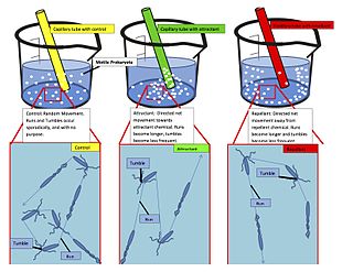
Chemotaxis is the movement of an organism or entity in response to a chemical stimulus. Somatic cells, bacteria, and other single-cell or multicellular organisms direct their movements according to certain chemicals in their environment. This is important for bacteria to find food by swimming toward the highest concentration of food molecules, or to flee from poisons. In multicellular organisms, chemotaxis is critical to early development and development as well as in normal function and health. In addition, it has been recognized that mechanisms that allow chemotaxis in animals can be subverted during cancer metastasis. The aberrant chemotaxis of leukocytes and lymphocytes also contribute to inflammatory diseases such as atherosclerosis, asthma, and arthritis. Sub-cellular components, such as the polarity patch generated by mating yeast, may also display chemotactic behavior.

<span class="mw-page-title-main">Immune system</span> Biological system protecting an organism against disease

The immune system is a network of biological systems that protects an organism from diseases. It detects and responds to a wide variety of pathogens, from viruses to parasitic worms, as well as cancer cells and objects such as wood splinters, distinguishing them from the organism's own healthy tissue. Many species have two major subsystems of the immune system. The innate immune system provides a preconfigured response to broad groups of situations and stimuli. The adaptive immune system provides a tailored response to each stimulus by learning to recognize molecules it has previously encountered. Both use molecules and cells to perform their functions.

<span class="mw-page-title-main">Inflammation</span> Physical effects resulting from activation of the immune system

Inflammation is part of the biological response of body tissues to harmful stimuli, such as pathogens, damaged cells, or irritants. It is a protective response involving immune cells, blood vessels, and molecular mediators. The function of inflammation is to eliminate the initial cause of cell injury, clear out damaged cells and tissues, and initiate tissue repair.

<span class="mw-page-title-main">Cytokine</span> Broad and loose category of small proteins important in cell signaling

Cytokines are a broad and loose category of small proteins important in cell signaling. Due to their size, cytokines cannot cross the lipid bilayer of cells to enter the cytoplasm and therefore typically exert their functions by interacting with specific cytokine receptors on the target cell surface. Cytokines have been shown to be involved in autocrine, paracrine and endocrine signaling as immunomodulating agents.

<span class="mw-page-title-main">Chemokine</span> Small cytokines or signaling proteins secreted by cells

Chemokines, or chemotactic cytokines, are a family of small cytokines or signaling proteins secreted by cells that induce directional movement of leukocytes, as well as other cell types, including endothelial and epithelial cells. In addition to playing a major role in the activation of host immune responses, chemokines are important for biological processes, including morphogenesis and wound healing, as well as in the pathogenesis of diseases like cancers.

Haptotaxis is the directional motility or outgrowth of cells, e.g. in the case of axonal outgrowth, usually up a gradient of cellular adhesion sites or substrate-bound chemoattractants. These gradients are naturally present in the extracellular matrix (ECM) of the body during processes such as angiogenesis or artificially present in biomaterials where gradients are established by altering the concentration of adhesion sites on a polymer substrate.

<span class="mw-page-title-main">CXCR4</span> Protein

C-X-C chemokine receptor type 4 (CXCR-4) also known as fusin or CD184 is a protein that in humans is encoded by the CXCR4 gene. The protein is a CXC chemokine receptor.

<span class="mw-page-title-main">Interleukin 8</span> Mammalian protein found in Homo sapiens

Interleukin 8 is a chemokine produced by macrophages and other cell types such as epithelial cells, airway smooth muscle cells and endothelial cells. Endothelial cells store IL-8 in their storage vesicles, the Weibel-Palade bodies. In humans, the interleukin-8 protein is encoded by the CXCL8 gene. IL-8 is initially produced as a precursor peptide of 99 amino acids which then undergoes cleavage to create several active IL-8 isoforms. In culture, a 72 amino acid peptide is the major form secreted by macrophages.

<span class="mw-page-title-main">L-selectin</span> Mammalian protein found in Homo sapiens

L-selectin, also known as CD62L, is a cell adhesion molecule found on the cell surface of leukocytes, and the blastocyst. It is coded for in the human by the SELL gene. L-selectin belongs to the selectin family of proteins, which recognize sialylated carbohydrate groups containing a Sialyl LewisX (sLeX) determinant. L-selectin plays an important role in both the innate and adaptive immune responses by facilitating leukocyte-endothelial cell adhesion events. These tethering interactions are essential for the trafficking of monocytes and neutrophils into inflamed tissue as well as the homing of lymphocytes to secondary lymphoid organs. L-selectin is also expressed by lymphoid primed hematopoietic stem cells and may participate in the migration of these stem cells to the primary lymphoid organs. In addition to its function in the immune response, L-selectin is expressed on embryonic cells and facilitates the attachment of the blastocyst to the endometrial endothelium during human embryo implantation.

Chemokine ligands 4 previously known as macrophage inflammatory protein (MIP-1β), is a protein which in humans is encoded by the CCL4 gene. CCL4 belongs to a cluster of genes located on 17q11-q21 of the chromosomal region. Identification and localization of the gene on the chromosome 17 was in 1990 although the discovery of MIP-1 was initiated in 1988 with the purification of a protein doublet corresponding to inflammatory activity from supernatant of endotoxin-stimulated murine macrophages. At that time, it was also named as "macrophage inflammatory protein-1" (MIP-1) due to its inflammatory properties.

Chemokine ligand 1 (CCL1) is also known as small inducible cytokine A1 and I-309 in humans. CCL1 is a small glycoprotein that belongs to the CC chemokine family.

<span class="mw-page-title-main">CCL7</span> Mammalian protein found in Homo sapiens

Chemokine ligand 7 (CCL7) is a small cytokine that was previously called monocyte-chemotactic protein 3 (MCP3). CCL7 is a small protein that belongs to the CC chemokine family and is most closely related to CCL2.

<span class="mw-page-title-main">CCL21</span> Mammalian protein found in Homo sapiens

Chemokine ligand 21 (CCL21) is a small cytokine belonging to the CC chemokine family. This chemokine is also known as 6Ckine, exodus-2, and secondary lymphoid-tissue chemokine (SLC). CCL21 elicits its effects by binding to a cell surface chemokine receptor known as CCR7. The main function of CCL21 is to guide CCR7 expressing leukocytes to the secondary lymphoid organs, such as lymph nodes and Peyer´s patches.

<span class="mw-page-title-main">CXCL9</span> Mammalian protein found in Homo sapiens

Chemokine ligand 9 (CXCL9) is a small cytokine belonging to the CXC chemokine family that is also known as monokine induced by gamma interferon (MIG). The CXCL9 is one of the chemokine which plays role to induce chemotaxis, promote differentiation and multiplication of leukocytes, and cause tissue extravasation.

<span class="mw-page-title-main">CXCL1</span> Mammalian protein found in Homo sapiens

The chemokine ligand 1 (CXCL1) is a small peptide belonging to the CXC chemokine family that acts as a chemoattractant for several immune cells, especially neutrophils or other non-hematopoietic cells to the site of injury or infection and plays an important role in regulation of immune and inflammatory responses. It was previously called GRO1 oncogene, GROα, neutrophil-activating protein 3 (NAP-3) and melanoma growth stimulating activity, alpha (MGSA-α). CXCL1 was first cloned from a cDNA library of genes induced by platelet-derived growth factor (PDGF) stimulation of BALB/c-3T3 murine embryonic fibroblasts and named "KC" for its location in the nitrocellulose colony hybridization assay. This designation is sometimes erroneously believed to be an acronym and defined as "keratinocytes-derived chemokine". Rat CXCL1 was first reported when NRK-52E cells were stimulated with interleukin-1β (IL-1β) and lipopolysaccharide (LPS) to generate a cytokine that was chemotactic for rat neutrophils, cytokine-induced neutrophil chemoattractant (CINC). In humans, this protein is encoded by the gene Cxcl1 and is located on human chromosome 4 among genes for other CXC chemokines.

<span class="mw-page-title-main">CXCL5</span> Mammalian protein found in Homo sapiens

C-X-C motif chemokine 5 is a protein that in humans is encoded by the CXCL5 gene.

<span class="mw-page-title-main">CD137</span> Member of the tumor necrosis factor (TNF) receptor family

CD137, a member of the tumor necrosis factor (TNF) receptor family, is a type 1 transmembrane protein, expressed on surfaces of leukocytes and non-immune cells. Its alternative names are tumor necrosis factor receptor superfamily member 9 (TNFRSF9), 4-1BB, and induced by lymphocyte activation (ILA). It is of interest to immunologists as a co-stimulatory immune checkpoint molecule, and as a potential target in cancer immunotherapy.

<span class="mw-page-title-main">Leukocyte extravasation</span>

Leukocyte extravasation is the movement of leukocytes out of the circulatory system and towards the site of tissue damage or infection. This process forms part of the innate immune response, involving the recruitment of non-specific leukocytes. Monocytes also use this process in the absence of infection or tissue damage during their development into macrophages.

<span class="mw-page-title-main">C-C chemokine receptor type 7</span> Protein-coding gene in the species Homo sapiens

C-C chemokine receptor type 7 is a protein that in humans is encoded by the CCR7 gene. Two ligands have been identified for this receptor: the chemokines ligand 19 (CCL19/ELC) and ligand 21 (CCL21). The ligands have similar affinity for the receptor, though CCL19 has been shown to induce internalisation of CCR7 and desensitisation of the cell to CCL19/CCL21 signals. CCR7 is a transmembrane protein with 7 transmembrane domains, which is coupled with heterotrimeric G proteins, which transduce the signal downstream through various signalling cascades. The main function of the receptor is to guide immune cells to immune organs by detecting specific chemokines, which these tissues secrete.

<span class="mw-page-title-main">Dedicator of cytokinesis protein 2</span> Protein found in humans

Dedicator of cytokinesis protein 2 (Dock2) is a protein encoded in the human by the DOCK2 gene. Dock2 is a large protein involved in intracellular signalling networks. It is a member of the DOCK-A subfamily of the DOCK family of guanine nucleotide exchange factors (GEFs) which function as activators of small G-proteins. Dock2 specifically activates isoforms of the small G protein Rac.

References

  1. 1 2 3 4 5 6 Vianello, F., E. Righi, et al. (2010). Methods for Quantitation of Leukocyte Chemotaxis and Fugetaxis. T-Cell Trafficking. F. M. Marelli-Berg and S. Nourshargh, Humana Press. 616: 115-124.
  2. IMMUNOLOGY - CHAPTER ONE > INNATE (NON-SPECIFIC) IMMUNITY Gene Mayer, Ph.D. Immunology Section of Microbiology and Immunology On-line. University of South Carolina.
  3. Wu, W; et al. (1999). "Directional guidance of neuronal migration in the olfactory system by the protein slit". Nature. 400 (6742): 331–336. Bibcode:1999Natur.400..331W. doi:10.1038/22477. PMC   2041931 . PMID   10432110.
  4. Yoshie, O (2000). "Role of chemokines in trafficking of lymphocytes and dendritic cells". Int. J. Hematol. 72 (4): 399–407. PMID   11197204.
  5. Wu, J.Y.; et al. (2001). "The neuronal repellent Slit inhibits leukocyte chemotaxis induced by chemotactic factors". Nature. 410 (6831): 948–952. Bibcode:2001Natur.410..948W. doi:10.1038/35073616. PMC   2072862 . PMID   11309622.
  6. Streilein, JW (1993). "Immune privilege as the result of local tissue barriers and immunosuppressive microenvironments". Current Opinion in Immunology. 5 (3): 428–432. doi:10.1016/0952-7915(93)90064-Y. PMID   8347303.
  7. Streilein, JW (1993). "Tissue barriers, immunosuppressive microenvironments, and privileged sites: the eye's point of view". Reg Immunol. 5 (5): 253–268. PMID   8148235.
  8. Metloubian, M.; et al. (2004). "Lymphocyte egress from thymus and peripheral lymphoid organs is dependent on S1P receptor 1". Nature. 427 (6972): 355–360. Bibcode:2004Natur.427..355M. doi:10.1038/nature02284. PMID   14737169. S2CID   4371877.
  9. Ueno, T.; et al. (2002). "Role for CCR7 ligands in the emigration of newly generated T lymphocytes from the neonatal thy mus". Immunity. 16 (2): 205–218. doi: 10.1016/S1074-7613(02)00267-4 . PMID   11869682.
  10. Chaffin, K.E.; Prelmutter, R.M (1991). "A pertussis toxin-sensitive process controls thymocyte emigration". Eur. J. Immunol. 21 (10): 2565–2573. doi:10.1002/eji.1830211038. PMID   1655469. S2CID   36339438.
  11. Vianello, F.; et al. (2005). "A CXCR4-dependent chemorepellent signal contributes to the emigration of mature single-positive CD4 cel.ls from the fetal thymus". Journal of Immunology. 175 (8): 5115–5125. doi: 10.4049/jimmunol.175.8.5115 . PMID   16210615.
  12. Howard, J; et al. (1998). "Molecular mimicry of the inflammation modulatory proteins (IMPs) of poxviruses: evasion of the inflammatory response to preserve viral habitat". Journal of Leukocyte Biology. 64 (1): 68–71. doi: 10.1002/jlb.64.1.68 . PMID   9665277.
  13. Farrell, HE; Degli-Esposti, Davis-Poynter NJ (1999). "Cytomegalovirus evasion of natural killer cell responses". Immunological Reviews. 168: 187–197. doi:10.1111/j.1600-065X.1999.tb01293.x. PMID   10399075. S2CID   38742701.
  14. Murphy, PM (1994). "Molecular piracy of chemokine receptors by herpesviruses". Infect Agents Dis. 3 (2): 137–164. PMID   7812652.
  15. Kotwal, GJ (2000). "Poxviral mimicry of complement and chemokine system components: what's the end game?". Immunol Today. 21 (5): 242–248. doi:10.1016/S0167-5699(00)01606-6. PMID   10782056.
  16. Moore, PS; et al. (1996). "Molecular mimicry of human cytokine and cytokine response pathway genes by KSHV". Science. 274 (5293): 1739–1744. Bibcode:1996Sci...274.1739M. doi:10.1126/science.274.5293.1739. PMID   8939871. S2CID   29713179.
  17. Fruh, K; et al. (1999). "A comparison of viral immune escape strategies targeting the MHC class I assembly pathway". Immunol Rev. 168: 157–166. doi:10.1111/j.1600-065X.1999.tb01290.x. PMID   10399072. S2CID   21017460.
  18. Milne, RS; et al. (2000). "RANTES binding and down-regulation by a novel human perpesvirus-6 beta chemokine receptor". Journal of Immunology. 164 (5): 2396–2404. doi: 10.4049/jimmunol.164.5.2396 . PMID   10679075.
  19. Nomura, T; et al. (2001). "Enhancement of anti-tumor immunity by tumor cells transfected with the secondary lymphoid tissue chemokine EBI-1-ligand chemokine and stromal cell-derived factor-1 alpha chemokine genes". Int J Cancer. 91 (5): 597–606. doi:10.1002/1097-0215(200002)9999:9999<::AID-IJC1107>3.0.CO;2-J. PMID   11267967.
  20. Rempel, SA; et al. (2000). "Identification and localization of the cytokine SDF1 and its receptor, CXC chemokine receptor 4, to regions of necrosis and angiogenesis in human glioblastoma". Clin Cancer Res. 6 (1): 102–111. PMID   10656438.
  21. Zou, W; et al. (2001). "Stromal-derived factor-1 in human tumors recruits and alters the function of plasmacytoid precursor dendritic cells". Nat Med. 7 (12): 1339–1346. doi:10.1038/nm1201-1339. PMID   11726975. S2CID   24369357.
  22. Barbero, S; et al. (2003). "Stromal cell-derived factor 1-alpha stimulates human gliblastoma cell growth through the activation of both extracellular signal-regulated kinases 1/2 and Akt". Cancer Res. 63 (8): 1969–1974. PMID   12702590.
  23. Balkwill, F (2004). "Cancer and the chemokine network". Nature Reviews Cancer. 4 (7): 540–550. doi:10.1038/nrc1388. PMID   15229479. S2CID   205467365.
  24. Strieter, RM (2001). "Chemokines: not just leukocyte chemoattractants in the promotion of cancer". Nat Immunol. 2 (4): 285–286. doi: 10.1038/86286 . PMID   11276195.
  25. 1 2 3 Stvrtinová, Viera; Ján Jakubovský and Ivan Hulín (1995), "Inflammation and Fever from Pathophysiology: Principles of Disease", Computing Centre, Slovak Academy of Sciences: Academic Electronic Press
  26. Alberts, Bruce; Alexander Johnson, Julian Lewis, Martin Raff, Keith Roberts, and Peter Walters (2002), "Molecular Biology of the Cell; Fourth Edition. New York and London: Garland Science", " ISBN   0-8153-3218-1".
  27. Head, JR; Billingham RE (1985). "Immunologically privileged sites in transplantation immunology and oncology". Perspect Biol Med. 29 (1): 115–131. doi:10.1353/pbm.1985.0038. PMID   3906552. S2CID   35574942.
  28. Head, JR; Billingham RE (1985). "Immune privilege in the testis:evaluation of potential local factors for transplantation". Transplantation. 40 (3): 269–275. doi: 10.1097/00007890-198509000-00010 . PMID   3898493. S2CID   40237018.
  29. Manahan, C.L.; et al. (2004). "Chemoattractant signaling in Dictyostelium discoideum" (PDF). Annu. Rev. Cell Dev. Biol. 20: 223–253. doi:10.1146/annurev.cellbio.20.011303.132633. PMID   15473840. S2CID   4656628. Archived from the original (PDF) on 2019-02-20.
  30. Willard, SS.; Devreotes, PN (2006). "Signaling pathways mediating chemotaxis in the social amoeaba, Dictyostelium discoideum". Eur. J. Cell Biol. 85 (9–10): 897–904. doi:10.1016/j.ejcb.2006.06.003. PMID   16962888.
  31. Devreotes, P.; Janetopoulos, C. (2003). "Eukaryotic chemotaxis: distinctions between directional sensing and polarization". J. Biol. Chem. 278 (23): 20445–20448. doi: 10.1074/jbc.R300010200 . PMID   12672811.
  32. Tharp, W.G.; et al. (2006). "Neutrophil chemorepulsion in defined interleukin-8 gradients in vitro and in vivo". J. Leukoc. Biol. 79 (3): 539–554. doi:10.1189/jlb.0905516. PMID   16365152. S2CID   1708811.
  33. Bacon, K.B. (1997). "Analysis of signal transduction following lymphocyte activation by chemokines". Chemokine Receptors. Methods in Enzymology. Vol. 288. pp. 340–361. doi:10.1016/S0076-6879(97)88023-8. ISBN   978-0-12-182189-0. PMID   9357003.
  34. Dustin, M.L.; Chakrabortk, A.K. (2008). "Tug of war at the exit door". Immunity. 28 (1): 15–17. doi:10.1016/j.immuni.2008.01.001. PMC   2719829 . PMID   18199414.
  35. Heit, B.; et al. (2002). "An intracellular signaling hierarchy determines direction of migration in opposing chemotactic gradients". J. Cell Biol. 159 (1): 91–102. doi:10.1083/jcb.200202114. PMC   2173486 . PMID   12370241.
  36. Armengol, MP; et al. (2003). "Chemokines determine local lymphoneogenesis and a reduction of circulating CXCR4+ T and CCR7 B and T lymphocytes in thyroid autoimmune diseases". Journal of Immunology. 170 (12): 6320–6328. doi: 10.4049/jimmunol.170.12.6320 . PMID   12794165.
  37. Alblas, J.; et al. (2001). "Activation of RhoA and ROCK are essential for detachment of migrating leukocytes". Mol. Biol. Cell. 12 (7): 2137–2145. doi:10.1091/mbc.12.7.2137. PMC   55668 . PMID   11452009.
  38. Pham, !.H.; et al. (2008). "S1P1 receptor signaling overrides retention mediated G alpha i-coupled receptors to promote T cell egress". Immunity. 28 (1): 122–133. doi:10.1016/j.immuni.2007.11.017. PMC   2691390 . PMID   18164221.