| Chorangiosis | |
|---|---|
 | |
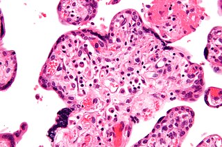
| Micrograph of a chorangiosis. H&E stain. | |
| Specialty | Pathology |
Chorangiosis is a placental pathology characterized by an abundance of blood vessels within the chorionic villi.
| Chorangiosis | |
|---|---|
| | |
| Micrograph of a chorangiosis. H&E stain. | |
| Specialty | Pathology |
Chorangiosis is a placental pathology characterized by an abundance of blood vessels within the chorionic villi.
It is associated with gestational diabetes, [1] smoking and high altitude.
It is diagnosed by a microscopic examination of the placenta.
Commonly used criteria from Altshuler [2] [3] are: "a minimum of 10 villi, each with 10 or more vascular channels, in 10 or more areas of 3 or more random, non-infarcted placental areas when using a ×10 ocular." The Altshuler criteria are not theoretically rigorous, as they do not define the area. Normal villi have up to five vascular channels. [3]


Gestational diabetes is a condition in which a woman without diabetes develops high blood sugar levels during pregnancy. Gestational diabetes generally results in few symptoms; however, it increases the risk of pre-eclampsia, depression, and of needing a Caesarean section. Babies born to mothers with poorly treated gestational diabetes are at increased risk of macrosomia, of having hypoglycemia after birth, and of jaundice. If untreated, diabetes can also result in stillbirth. Long term, children are at higher risk of being overweight and of developing type 2 diabetes.

Gestational hypertension or pregnancy-induced hypertension (PIH) is the development of new hypertension in a pregnant woman after 20 weeks' gestation without the presence of protein in the urine or other signs of pre-eclampsia. Gestational hypertension is defined as having a blood pressure greater than 140/90 on two occasions at least 6 hours apart.
Oligohydramnios is a medical condition in pregnancy characterized by a deficiency of amniotic fluid, the fluid that surrounds the fetus in the abdomen, in the amniotic sac. It is typically diagnosed by ultrasound when the amniotic fluid index (AFI) measures less than 5 cm or when the single deepest pocket (SDP) of amniotic fluid measures less than 2 cm. Amniotic fluid is necessary to allow for normal fetal movement, lung development, and cushioning from uterine compression. Low amniotic fluid can be attributed to a maternal, fetal, placental or idiopathic cause and can result in poor fetal outcomes including death. The prognosis of the fetus is dependent on the etiology, gestational age at diagnosis, and the severity of the oligohydramnios.

Hemangioendotheliomas are a family of vascular neoplasms of intermediate malignancy.

The decidua is the modified mucosal lining of the uterus that forms every month, in preparation for pregnancy. It is shed off each month when there is no fertilised egg to support. The decidua is under the influence of progesterone. Endometrial cells become highly characteristic. The decidua forms the maternal part of the placenta and remains for the duration of the pregnancy. After birth the decidua is shed together with the placenta.

Intrauterine hypoxia occurs when the fetus is deprived of an adequate supply of oxygen. It may be due to a variety of reasons such as prolapse or occlusion of the umbilical cord, placental infarction, maternal diabetes and maternal smoking. Intrauterine growth restriction may cause or be the result of hypoxia. Intrauterine hypoxia can cause cellular damage that occurs within the central nervous system. This results in an increased mortality rate, including an increased risk of sudden infant death syndrome (SIDS). Oxygen deprivation in the fetus and neonate have been implicated as either a primary or as a contributing risk factor in numerous neurological and neuropsychiatric disorders such as epilepsy, attention deficit hyperactivity disorder, eating disorders and cerebral palsy.
Placental insufficiency or utero-placental insufficiency is the failure of the placenta to deliver sufficient nutrients to the fetus during pregnancy, and is often a result of insufficient blood flow to the placenta. The term is also sometimes used to designate late decelerations of fetal heart rate as measured by cardiotocography or an NST, even if there is no other evidence of reduced blood flow to the placenta, normal uterine blood flow rate being 600mL/min.

A placental disease is any disease, disorder, or pathology of the placenta.

Placentitis is an inflammation of the placenta. The main forms of placentitis are:
In pathology, hypertrophic decidual vasculopathy, abbreviated HDV, is the histomorphologic correlate of gestational hypertension, as may be seen in intrauterine growth restriction (IUGR) and HELLP syndrome.

Intermediate trophoblast is a distinct subtype of trophoblastic tissue that arises from the cytotrophoblast.
Telepathology is the practice of pathology at a distance. It uses telecommunications technology to facilitate the transfer of image-rich pathology data between distant locations for the purposes of diagnosis, education, and research. Performance of telepathology requires that a pathologist selects the video images for analysis and the rendering of diagnoses. The use of "television microscopy", the forerunner of telepathology, did not require that a pathologist have physical or virtual "hands-on" involvement in the selection of microscopic fields-of-view for analysis and diagnosis.

Fetal thrombotic vasculopathy is a chronic disorder characterized by thrombosis in the fetus leading to vascular obliteration and hypoperfusion.
Villitis of unknown etiology (VUE), also known as chronic villitis, is a placental injury. VUE is an inflammatory condition involving the chorionic villi. VUE is a recurrent condition and can be associated with intrauterine growth restriction (IUGR). IUGR involves the poor growth of the foetus, stillbirth, miscarriage, and premature delivery. VUE recurs in about 1/3 of subsequent pregnancies.

Intranodal palisaded myofibroblastoma (IPM) is a rare primary tumour of lymph nodes, that classically presents as an inguinal mass.
Pleomorphic anaplastic neuroblastoma (PAN) is a striking aspect of neuroblastoma first described by Cozzutto and Carbone in 1988. Another case was thereafter reported by Cowan, et al. with cytogenetic and immunohistological analysis in a 28-year-old man. The case described by Navarro, et al. showed MYCN amplification and a 1p36 deletion as measured with FISH in 13% of cells. Additionally there was a main cell population with a DNA index of 2 indicating a tetraploid DNA content and a high expression of MIBI (Ki-67), bel 2, p53, and P-glycoprotein, either correlated with rapid progression of disease.

Placental villous immaturity is chorionic villous development that is inappropriate for the gestational age.
The Azzopardi phenomenon, or Azzopardi effect, is the presence of DNA in necrotic venules. It can occur in small cell carcinomas and in some high-grade malignant neoplasms. The effect is well known in diagnostic surgical pathology. The phenomenon is named after the pathologist, John G. Azzopardi.
John G. Azzopardi was a prominent pathologist, recognized for his contributions to diagnostic surgical pathology, particularly in breast pathology. His name is also eponymously connected with his elucidation of the Azzopardi phenomenon.
Extravillous trophoblasts(EVTs), are one form of differentiated trophoblast cells of the placenta. They are invasive mesenchymal cells which function to establish critical tissue connection in the developing placental-uterine interface. EVTs derive from progenitor cytotrophoblasts (CYTs), as does the other main trophoblast subtype, syncytiotrophoblast (SYN). They are sometimes called intermediate trophoblast.